
Carcinosarcomas are rare lung tumors belonging to the group of sarcomatoid carcinomas. They are a very rare histological type of lung tumor, representing less than 1% of all lung cancers and the definitive diagnosis is obtained, in many cases, by studying the resection piece.1,2
These tumors contain a mixture of malignant elements: epithelial (epidermoid or adenocarcinoma) and sarcomatous (rhabdomyosarcoma, osteosarcoma and chondrosarcoma).
Preoperative diagnosis is difficult because the histological samples are too small or necrotic to show both components and patients often have an erroneous diagnosis prior to surgery, as was the case with our patient.3
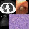
Our case is a 75-year-old man, a former smoker who quit 10 years ago, came to the emergency room for a cough with purulent sputum without fever. A chest scan was performed where pulmonary consolidation with a patchy distribution was detected in LSD, LM, LID and LII, and bilateral pneumonia was diagnosed (Fig. 1 A). The patient was admitted to the ward for control and treatment.

In subsequent controls, there was little radiological improvement, so a bronchoscopy was performed to take samples. An endobronchial mass in the intermediate bronchus was discovered. A biopsy of the lesion resulted in necrotic material (Fig. 1B).
An endobronchial ultrasound (EBUS) was then performed for lymph node staging, with a negative (N0) result with a new biopsy where, once again, the material was necrotic but the bronchialaspiration was positive for squamous cell carcinoma (Fig. 1C).
A cryobiopsy of the lesion was performed due to the absence of sufficient histological material and a tumor with an angiocentric growth pattern was identified, composed of pleomorphic cells growing on a lax stroma. Part of the tumor cellularity exhibit an enlarged nucleus of ovoid morphology with central nucleolus and clear eosinophilic cytoplasms. Other fields show a fusiform cellularity with clear cytoplasms that are arranged on a desmoplastic stroma. Histological and immunophenotypic findings compatible with carcinosarcoma (Fig. 1D).
In conclusion, pulmonary carcinosarcomas constitute a very rare histological type of tumor, difficult to diagnose anatomopathologically before surgery, requiring large samples to reach a correct diagnosis. Cryobiopsy is an easy and safe tool that allows us to obtain larger tissue samples in better condition for an anatomopathological study as tissue alterations are avoided because the sample is frozen before extraction. Likewise, it can be performed on an outpatient basis, with the patient requiring just a few hours of monitoring if there are no complications.
The authors thank Kimberly Fitler and Carlos Disdier for her advice during the preparation of this paper.


