
Primary pulmonary lymphomas are uncommon entities in respiratory medicine1. They present most often as mucosa-associated lymphoid tissue (MALT) lymphomas (80%) in asymptomatic middle-aged adults in the form of pulmonary nodules that in some cases may be associated with amyloidosis2,3,4. Diagnosis is not easy, and they may appear on a chest tomography (CT) as a pulmonary lesion with low attenuation in the interior. Lymphoid infiltration of the lung mucosa or a monoclonal tissue-level peak may be detected on bronchoscopy, but in the vast proportion of cases, surgical biopsy is required for confirmation. Located lesions can be treated with surgery or radiotherapy, and the rest can be treated with chemotherapy1.
A 62-year-old man with history of tuberculosis correctly treated at a younger age, with an active intake of 2–3 beers a day, was admitted to the hospital with a 1-week history of unproductive cough, asthenia and anorexia. No fever was reported at any time.
In the medical examination, the patient showed good general condition, and no adenopathy was reported. In pulmonary auscultation, crackles in the left lower lobe (LLL) were present, without any other pathological signs in the rest of the examination.
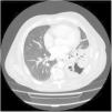
In the blood tests, the findings were as follows: leukocytosis with neutrophilia, raised C-reactive protein and procalcitonin, slight alteration of liver function tests, and B12 vitamin 991.70pg/mL. Chest radiography showed LLL opacity and urine antigens were negative for pneumococcus and legionella. All these findings were interpreted as LLL community-acquired pneumonia, and the patient received a 1-week course of levofloxacin 500mg. There was no clinical or radiological improvement, so the drug therapy was adjusted to moxifloxacin 400mg for 21 days. One month later, the patient attended our clinic. No changes were detected in the radiological image, so additional studies were implemented. A chest CT was performed (Fig. 1), showing a LLL opacity with air bronchogram, and multiple images of cavities with necrosis and bronchiectasis. The flexible bronchoscopy showed no endobronchial, bacterial or anatomopathological findings. Finally, we performed a CT-guided biopsy on the LLL lesion. Pathology results reported a low-grade B-small cell non-Hodgkin lymphoma, compatible with marginal MALT-type lymphoma associated with vascular and stromal lambda amyloidosis type A. This case was reviewed by the haematology department, who requested an abdominal scan, a positron emission tomography (PET) and an echocardiogram. Finally, the patient received radiotherapy to the lesion.
Funding
The authors have not received any type of funding.
Conflict of InterestThe authors declare no conflict of interests.


