In this study we analyzed the characteristics of patients with pleural effusion secondary to Streptococcus milleri studied retrospectively between January and March 2013 and found seven patients with a mean age of 60 years; 43% of them were smokers and 57% with a drinking habit. The most common associated factors were alcoholism, previous pneumonia and diabetes. Other bacteria were identified as Enterobacter aerogenes, Bacteroides and Prevotella intermedia capillosus in two patients. The mean duration of antibiotic therapy was 28 days; six patients underwent pleural drainage by chest tube and one patient needed surgery due to poor clinical progress. The mean duration of hospitalization was 30days with satisfactory outcome in all cases, despite some changes in residual function.
Se realiza un análisis retrospectivo de las características de los pacientes con derrame pleural secundario a Streptococcus milleri diagnosticados en nuestro hospital entre enero de 2011 y marzo 2013. Se diagnosticaron 7pacientes con una edad media de 60años, el 57% con hábito enólico importante y el 43% fumadores. Los factores asociados más frecuentemente fueron el alcoholismo, la existencia de neumonía previa y diabetes mellitus. En 2pacientes se identificaron otros gérmenes, como Enterobacter aerogenes, Bacteroides capillosus y Prevotella intermedia. La duración media del tratamiento antibiótico fue de 28días. En 6casos (86%) se realizó drenaje pleural con tubo de tórax, y un paciente precisó cirugía por evolución tórpida. La duración media de la hospitalización fue de 30días, con evolución satisfactoria en todos los casos, aunque con alteración funcional restrictiva residual.
The term Streptococcus milleri, traditionally classified under the viridans group, is a heterogeneous group of microorganisms that includes Streptococcus anginosus, intermedius and constellatus. They are oral, nasopharyngeal, gastrointestinal and genitourinary mucosa saprophytes and are pathological in patients with predisposing factors.1 In contrast to the patterns observed some decades ago, bacteria in this group are now the most common pathogens in the etiology of community-acquired pleural infections.1,2 The aim of our study is to analyze the clinical characteristics, associated factors, and clinical course of patients with S. milleri empyema.
Patients and MethodsThis was a retrospective study performed in patients with a diagnosis of pleural empyema in whom S. milleri was isolated in pleural fluid in our hospital between January 2011 and March 2013. The variables studied were age, sex, associated factors, smoking habit, consumption of alcohol, previous endoscopic procedures, symptoms, clinical laboratory and radiological changes, treatment and clinical course.
The SPSS 12.0 statistical software package was used for statistical analysis. A descriptive analysis was performed with qualitative variables expressed as simple frequencies and quantitative variables as mean and standard deviation.
ResultsA total of 7 patients were studied, of which 6 (86%) were male. Mean age was 60.2 years (55–67; SD: 3.7). Three were smokers (43%) and 2 were ex-smokers (27%). Most common associated factors were alcoholism in 4 patients (57%), previous pneumonia in 3 (43%) and diabetes mellitus in 3 (43%). None of the patients had neurological disease or swallowing disorders. The main symptoms were fever in 5 patients (71%), pleuritic pain in 4 (57%) and cough with purulent expectoration in 3 (43%). Elevated C-reactive protein (CRP) was found in all patients, with a mean level of 36mg/l (SD: 15). Only 2 patients (29%) had raised procalcitonin (PCT); the mean value was 0.8ng/ml (SD: 0.8). Empyema was primary with no evidence of consolidation in 3 patients (43%). Anaerobic bacteria were isolated in 2 patients (29%): Enterobacter aerogenes, Bacteroides capillosus and Prevotella intermedia. In 3 cases, S. milleri was resistant to erythromycin, clindamycin and amoxicillin. The mean duration of antibiotic treatment was 28 days (SD: 11). Pleural drainage by chest tube was required in 6 patients (86%) and 1 patient underwent ultrasound-guided thoracocentesis for evacuation of fluid. Three patients required fibrinolytics with urokinase for a mean of 6 days (SD: 2). Clinical course was satisfactory in 5 patients (71%). One patient required surgery due to poor clinical progress, undergoing pleural decortication and pulmonary debridement without complications. One patient was admitted to the intensive care unit due to septic shock associated with empyema and sub-diaphragmatic abscess secondary to perforated gangrenous appendicitis. Length of hospitalization was 30 days (8–72; SD: 23.7). None of the patients died. They all had minimal pleural thickening and 2 individuals (29%) had restrictive ventilatory changes.
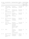
The main patient characteristics are described in Table 1. Fig. 1 shows the radiological characteristics of one of the patients.
Clinical, Radiological and Therapeutic Characteristics.
| Sex | Age (years) | Associated factors | Radiological features | Antibiotic used | Duration of antibiotic (days) | Drainage with chest tube |
| M | 67 | COPD. Diabetes. Alcoholism. Septic mouth. | Left massive multiloculated PE | Amoxicillin–clavulanate | 40 | Yes |
| M | 58 | Asthma. Crohn's disease. Immunosuppression. | Submassive non-loculated PE with abscess | Piperacillin–tazobactam | 29 | Yes |
| F | 59 | Previous pneumonia due to bronchoaspiration secondary to drug overdose | Submassive loculated PE with abscess. Consolidation. | Piperacillin–tazobactam | 16 | Yes |
| M | 62 | Previous pneumonia due to bronchoaspiration. Alcoholism. | Submassive loculated PE. Consolidation. | Piperacillin–tazobactam | 14 | No |
| M | 61 | Chronic liver disease. Alcoholism. Diabetes. | Submassive loculated PE. Lesion with residual inflammatory appearance in lateral segment of middle lobe. | Amoxicillin–clavulanate | 29 | Yes |
| M | 55 | DILD | Submassive loculated PE. Anterior hydropneumothorax | Piperacillin–tazobactam | 40 | Yes |
| M | 60 | Perforated appendix with subdiaphragmatic abscess. | Bilateral submassive loculated PE. Consolidation. | Imipenem. Amikacin. Linezolid | 40 | Yes |
PE: pleural effusion; DILD: diffuse interstitial lung disease; COPD: chronic obstructive pulmonary disease; F: female; M: male.
S. milleri is the most common causative agent in pyogenic infections, and most of which are located in the abdomen, the central nervous system and the chest.3 These infections usually occur in adults over 50 years of age and more often in patients with predisposing pathological factors. In our series, 86% of the patients had predisposing factors which could cause a certain degree of immunosuppression and their mean hospital stay was prolonged due to the need for pleural procedures and prolonged antimicrobial regimens.
S. milleri bacteremia is uncommon and generally associated with a septic intraabdominal focus,5 as was the case in one of our patients. The chest is involved in one fifth of S. milleri infections, the most common manifestations being empyema and less frequently mediastinitis and lung abscess.1,3–5 In our series, empyema was associated with lung abscess in 2 cases (29%), and 3 cases (43%) were primary empyema.
The pathogenic role of the co-existence of anaerobic bacteria and Streptococcus has been established. This concomitancy leads to synergy that increases virulence, lung tissue damage and the dissemination of the infection4: this was the case in 2 of our patients (29%).
Earlier studies in patients with S. milleri pleuropulmonary infection found that between 67% and 87% of cases required invasive procedures and antimicrobial treatment.5 Pleural fluid drainage may suffice in early stages, but advanced stages require video-assisted thoracic surgery (VATS) that according to most studies should be carried out between 3 and 7 days after failed drainage.5
This group of bacteria are very sensitive to ureidopenicillins, carbapenem and cephalosporins,3–5 although resistance has been reported in up to 33% of cases of S. intermedius.3 In our series, the mean antibiotic treatment time was 28 days, but this varies in other published series (10–65 days).5 Our conclusion is that S. milleri must be considered in community-acquired empyemas, and should be appropriately treated with antibiotics and early pleural drainage in order to avoid major surgical procedures and long periods of hospitalization and to improve prognosis.
Please cite this article as: Madrid-Carbajal CJ, Molinos L, García-Clemente M, Pando-Sandoval A, Fleites A, Casan-Clarà P. Descripción de casos de derrame pleural secundario a Streptococcus milleri. Arch Bronconeumol. 2014;50:404–406.


