Superior vena cava syndrome is a clear sign for clinicians of infiltrative mediastinal involvement, usually caused by neoplasms in this location, and it is an indicator of poor prognosis. However, other diseases of benign origin can also cause these alterations. We present the case of a 34-year-old patient who debuted with symptoms of superior vena cava syndrome due to idiopathic mediastinal fibrosis, which presented a torpid evolution and few therapeutic alternatives.
El síndrome de vena cava superior es para el clínico una señal inequívoca de afectación mediastínica por procesos infiltrativos, generalmente neoplasias, en esta localización, y es indicador de un mal pronóstico. Sin embargo, otras enfermedades de origen benigno pueden causar estas alteraciones. Presentamos el caso de un paciente de 34 años que empezó con un cuadro de síndrome de vena cava superior debido a fibrosis mediastínica de origen idiopático y que presentó una evolución tórpida con escasas alternativas terapéuticas.
Superior vena cava syndrome (SVCS) is a disease entity with notable signs and symptoms that cannot go unnoticed by clinicians. It is a relatively common complication of lung cancer, and may constitute one of the early manifestations of this disease.1 However, the pathogenesis of SVCS includes non-neoplastic causes that must also be considered within its differential diagnosis. Mediastinal fibrosis following thrombosis of intravascular devices (pacemaker leads) is the second most common cause of these benign entities that can affect the large vessels of the mediastinum.2
Clinical FindingsWe present the case of a 34-year-old male with a history of active smoking (15 packs/year) and sleep apnoea-hypopnoea syndrome on nocturnal continuous positive airway pressure (CPAP) therapy, with no other medical history of interest. He worked in excavations and had travelled to the Dominican Republic 5 years before the onset of symptoms.
The patient attended the clinic due to a 5-day history of dyspnoea on minimal exertion, not accompanied by cough, expectoration or fever. He described a two-year history of neck and face oedema with dilated neck veins, which was generally greater upon wakening and decreased over the day, and which had increased in the previous 2 weeks. He did not report asthenia, anorexia or weight loss.
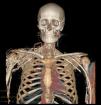
Physical examination revealed facial oedema, dilated neck veins and an increase in the number of collateral veins in the upper thorax, shoulder and right arm. Mediastinal widening and bilateral subpulmonary pleural effusion were observed on the chest radiograph. Chest computed tomography (CT) showed a widened mediastinum with fat trabeculation which caused a mass effect affecting the superior vena cava (Fig. 1). The 3D angiographic reconstruction showed an extensive network of collateral circulation due to obstruction of the superior vena cava that extended towards the chest wall, upper limbs and abdomen (Fig. 2). No thrombi were observed in the vena cava. Based on these findings, a diagnosis of SVCS was made. A phlebography was performed to assess the vascular involvement and to place an endovascular stent in order to re-permeabilise the venous circulation. However, this treatment was unsuccessful due to total obstruction of the subclavian veins, which made it difficult to access the vena cava. In view of the obstruction of the superior vena cava by a mediastinal infiltrative process, a mediastinotomy was performed to determine its cause. The biopsy showed adipose tissue with little cellularity; no granulomas, calcifications or malignant cells were identified. The tuberculin test did not reveal induration, and the bronchoaspirate and biopsy material cultures for mycobacteria and fungi were negative. The patient was diagnosed with SVCS due to mediastinal fibrosis of idiopathic origin.
Mediastinal fibrosis is a rare disease characterised by the proliferation of collagen tissue and establishment of fibrous tissue in the mediastinum.3 In most cases, the cause of this process is unknown, although in endemic zones it has been related with Histoplasma capsulatum infection, specifically with an abnormal inflammatory response to the antigens of this fungus and, less commonly, with other granulomatous diseases such as tuberculosis.4 There is an idiopathic form with a possible autoimmune component that may be associated with fibrosing processes in other locations, such as retroperitoneal fibrosis, pseudotumour of the orbit and Riedel's thyroiditis.5,6 It affects young patients, with a slight predominance in males, and its symptoms are insidious and progressive, with a variable natural history.5 The signs and symptoms depend on the mediastinal structures affected; thus, typical complications are the result of a compromised respiratory tract, heart and major vessels, or oesophagus. Obstruction of the superior vena cava is the most common complication in this disease: it generally develops slowly over a period of years, allowing the formation of an extensive network of collateral circulation that aims to prevent blood stasis and increased pressure in the tributaries of the superior vena cava.3 Diagnostic studies in patients with suspected mediastinal fibrosis may include chest radiograph, CT scanning and magnetic resonance imaging. CT angiography plays an essential role in the diagnosis of complications with evaluation of the vascular bed, and enables surgical measures to be planned and the disease monitored.7 Positron emission tomography (PET) combined with CT enables the anatomical study of mediastinal and pulmonary lesions, and allows their metabolic activity to be determined, which is particularly useful in lung cancer.8 However, benign processes with a notable inflammatory component (tuberculosis, histoplasmosis, aspergillosis, sarcoidosis) can show intense metabolic activity, which limits the value of this tool for the differential diagnosis of masses in the mediastinum.9 The histological study shows fibrous, paucicellular tissue and adipocytes, with the presence of mononuclear cells, calcifications and granulomas in cases related with infections (histoplasmosis, tuberculosis).5 Within the differential diagnosis, certain fibrosis-causing neoplasms must be considered, such as sclerosing non-Hodgkin's lymphoma and the sclerosing variant of Hodgkin's lymphoma, localised mesotheliomas, low grade sarcomas, thymomas and thymic carcinoids, which can show a fibrous inflammatory reaction.10
There is no curative treatment for this disease. Anti-fungal agents have been used in cases that may be related with histoplasmosis, although they have not been effective.11 The use of corticoids does not provide any benefit except in cases of autoimmune aetiology, in which there may be a response.6 Therefore, therapeutic measures will be aimed at relieving obstructive symptoms in the airways, major vessels and oesophagus. When there is involvement of the vena cava, the placement of endovascular stents to permeabilise the vessel is an option that produces a symptomatic improvement. Other techniques have been described, such as bypass surgery with saphenous vein grafts or bioprostheses.12,13
In the case of our patient, the symptoms began with SVCS, and after performing additional tests and a biopsy, the diagnosis of idiopathic mediastinal fibrosis was reached, having excluded other possible causes. Placement of a stent in the superior vena cava was attempted as palliative treatment but was unsuccessful, so bypass surgery was proposed; however this possibility was discarded due to the patient's poor distal bed, opting instead for indefinite anticoagulation and follow-up checks.
Please cite this article as: Novella Sánchez L, et al. Fibrosis mediastínica y síndrome de vena cava superior. Arch Bronconeumol. 2013;49:340–2.


