array:24 [
  "pii" => "S1579212913001122"
  "issn" => "15792129"
  "doi" => "10.1016/j.arbr.2013.06.007"
  "estado" => "S300"
  "fechaPublicacion" => "2013-08-01"
  "aid" => "775"
  "copyright" => "SEPAR"
  "copyrightAnyo" => "2013"
  "documento" => "simple-article"
  "crossmark" => 0
  "subdocumento" => "crp"
  "cita" => "Arch Bronconeumol. 2013;49:337-9"
  "abierto" => array:3 [
    "ES" => false
    "ES2" => false
    "LATM" => false
  ]
  "gratuito" => false
  "lecturas" => array:2 [
    "total" => 3352
    "formatos" => array:3 [
      "EPUB" => 130
      "HTML" => 2498
      "PDF" => 724
    ]
  ]
  "Traduccion" => array:1 [
    "es" => array:19 [
      "pii" => "S030028961300121X"
      "issn" => "03002896"
      "doi" => "10.1016/j.arbres.2013.04.005"
      "estado" => "S300"
      "fechaPublicacion" => "2013-08-01"
      "aid" => "775"
      "copyright" => "SEPAR"
      "documento" => "article"
      "crossmark" => 0
      "subdocumento" => "sco"
      "cita" => "Arch Bronconeumol. 2013;49:337-9"
      "abierto" => array:3 [
        "ES" => true
        "ES2" => true
        "LATM" => true
      ]
      "gratuito" => true
      "lecturas" => array:2 [
        "total" => 5827
        "formatos" => array:3 [
          "EPUB" => 102
          "HTML" => 4998
          "PDF" => 727
        ]
      ]
      "es" => array:13 [
        "idiomaDefecto" => true
        "cabecera" => "<span class="elsevierStyleTextfn">Nota cl&#237;nica</span>"
        "titulo" => "Diagn&#243;stico de lesiones paratraqueobronquiales no adenop&#225;ticas mediante ecobroncoscopia lineal"
        "tienePdf" => "es"
        "tieneTextoCompleto" => "es"
        "tieneResumen" => array:2 [
          0 => "es"
          1 => "en"
        ]
        "paginas" => array:1 [
          0 => array:2 [
            "paginaInicial" => "337"
            "paginaFinal" => "339"
          ]
        ]
        "titulosAlternativos" => array:1 [
          "en" => array:1 [
            "titulo" => "Diagnosis of Non-nodal Paratracheobronchial Lesions by Linear Endobronchial Ultrasound"
          ]
        ]
        "contieneResumen" => array:2 [
          "es" => true
          "en" => true
        ]
        "contieneTextoCompleto" => array:1 [
          "es" => true
        ]
        "contienePdf" => array:1 [
          "es" => true
        ]
        "resumenGrafico" => array:2 [
          "original" => 0
          "multimedia" => array:7 [
            "identificador" => "fig0005"
            "etiqueta" => "Figura 1"
            "tipo" => "MULTIMEDIAFIGURA"
            "mostrarFloat" => true
            "mostrarDisplay" => false
            "figura" => array:1 [
              0 => array:4 [
                "imagen" => "gr1.jpeg"
                "Alto" => 3747
                "Ancho" => 2167
                "Tamanyo" => 691344
              ]
            ]
            "descripcion" => array:1 [
              "es" => "<p id="spar0015" class="elsevierStyleSimplePara elsevierViewall">Ejemplos de im&#225;genes radiol&#243;gicas y sonogr&#225;ficas de 4 de los casos de la serie&#46;</p> <p id="spar0020" class="elsevierStyleSimplePara elsevierViewall">A y B&#58; Im&#225;genes de tomograf&#237;a computarizada &#40;TC&#41; y ecoendosc&#243;pica de una masa localizada en el l&#243;bulo superior derecho puncionada por ecobroncoscopia&#44; con resultado de adenocarcinoma de origen pulmonar&#46; C y D&#58; Im&#225;genes de TC y ecoendosc&#243;pica de un paciente con una lesi&#243;n pulmonar en l&#243;bulo inferior izquierdo y una adenopat&#237;a en la estaci&#243;n mediast&#237;nica 9 derecha&#44; puncionadas ambas por ecobroncoscopia a trav&#233;s del es&#243;fago con resultado en las 2 de met&#225;stasis de carcinoma de colon&#46; E y F&#58; Im&#225;genes de TC y ecoendosc&#243;pica de una masa hiliar izquierda que engloba la arteria pulmonar con punci&#243;n guiada por ecobroncoscopia diagn&#243;stica de adenocarcinoma de origen pulmonar&#46; G y H&#58; Im&#225;genes de TC y ecoendosc&#243;pica de una masa paratraqueoesof&#225;gica derecha de posible origen en el l&#243;bulo superior derecho con diagn&#243;stico tras punci&#243;n realizada a trav&#233;s del es&#243;fago de adenocarcinoma de origen pulmonar&#46; <span class="elsevierStyleSmallCaps">I</span> y J&#58; Im&#225;genes de TC y ecoendosc&#243;pica de una masa localizada en el l&#243;bulo superior derecho que infiltraba mediastino puncionada por ecobroncoscopia&#44; con resultado de adenocarcinoma de origen pulmonar&#46;</p>"
            ]
          ]
        ]
        "autores" => array:1 [
          0 => array:2 [
            "autoresLista" => "Tamara Lourido, Maribel Botana, Virginia Leiro, Manuel N&#250;&#241;ez, Alberto Fern&#225;ndez-Villar"
            "autores" => array:5 [
              0 => array:2 [
                "nombre" => "Tamara"
                "apellidos" => "Lourido"
              ]
              1 => array:2 [
                "nombre" => "Maribel"
                "apellidos" => "Botana"
              ]
              2 => array:2 [
                "nombre" => "Virginia"
                "apellidos" => "Leiro"
              ]
              3 => array:2 [
                "nombre" => "Manuel"
                "apellidos" => "N&#250;&#241;ez"
              ]
              4 => array:2 [
                "nombre" => "Alberto"
                "apellidos" => "Fern&#225;ndez-Villar"
              ]
            ]
          ]
        ]
      ]
      "idiomaDefecto" => "es"
      "Traduccion" => array:1 [
        "en" => array:9 [
          "pii" => "S1579212913001122"
          "doi" => "10.1016/j.arbr.2013.06.007"
          "estado" => "S300"
          "subdocumento" => ""
          "abierto" => array:3 [
            "ES" => false
            "ES2" => false
            "LATM" => false
          ]
          "gratuito" => false
          "lecturas" => array:1 [
            "total" => 0
          ]
          "idiomaDefecto" => "en"
          "EPUB" => "https://multimedia.elsevier.es/PublicationsMultimediaV1/item/epub/S1579212913001122?idApp=UINPBA00003Z"
        ]
      ]
      "EPUB" => "https://multimedia.elsevier.es/PublicationsMultimediaV1/item/epub/S030028961300121X?idApp=UINPBA00003Z"
      "url" => "/03002896/0000004900000008/v1_201308011253/S030028961300121X/v1_201308011253/es/main.assets"
    ]
  ]
  "itemSiguiente" => array:19 [
    "pii" => "S1579212913001109"
    "issn" => "15792129"
    "doi" => "10.1016/j.arbr.2013.06.006"
    "estado" => "S300"
    "fechaPublicacion" => "2013-08-01"
    "aid" => "719"
    "copyright" => "SEPAR"
    "documento" => "simple-article"
    "crossmark" => 0
    "subdocumento" => "crp"
    "cita" => "Arch Bronconeumol. 2013;49:340-2"
    "abierto" => array:3 [
      "ES" => false
      "ES2" => false
      "LATM" => false
    ]
    "gratuito" => false
    "lecturas" => array:2 [
      "total" => 7415
      "formatos" => array:3 [
        "EPUB" => 172
        "HTML" => 6058
        "PDF" => 1185
      ]
    ]
    "en" => array:13 [
      "idiomaDefecto" => true
      "cabecera" => "<span class="elsevierStyleTextfn">Case Report</span>"
      "titulo" => "Mediastinal Fibrosis and Superior Vena Cava Syndrome"
      "tienePdf" => "en"
      "tieneTextoCompleto" => "en"
      "tieneResumen" => array:2 [
        0 => "en"
        1 => "es"
      ]
      "paginas" => array:1 [
        0 => array:2 [
          "paginaInicial" => "340"
          "paginaFinal" => "342"
        ]
      ]
      "titulosAlternativos" => array:1 [
        "es" => array:1 [
          "titulo" => "Fibrosis mediast&#237;nica y s&#237;ndrome de vena cava superior"
        ]
      ]
      "contieneResumen" => array:2 [
        "en" => true
        "es" => true
      ]
      "contieneTextoCompleto" => array:1 [
        "en" => true
      ]
      "contienePdf" => array:1 [
        "en" => true
      ]
      "resumenGrafico" => array:2 [
        "original" => 0
        "multimedia" => array:7 [
          "identificador" => "fig0005"
          "etiqueta" => "Fig&#46; 1"
          "tipo" => "MULTIMEDIAFIGURA"
          "mostrarFloat" => true
          "mostrarDisplay" => false
          "figura" => array:1 [
            0 => array:4 [
              "imagen" => "gr1.jpeg"
              "Alto" => 983
              "Ancho" => 1500
              "Tamanyo" => 112975
            ]
          ]
          "descripcion" => array:1 [
            "en" => "<p id="spar0015" class="elsevierStyleSimplePara elsevierViewall">Mediastinal mass including and obliterating the superior vena cava&#44; reducing the passage of iodated contrast &#40;arrow&#41;&#46;</p>"
          ]
        ]
      ]
      "autores" => array:1 [
        0 => array:2 [
          "autoresLista" => "Laura Novella S&#225;nchez, Francisco Sanz Herrero, Javier Berraondo Fraile, Estrella Fern&#225;ndez Fabrellas"
          "autores" => array:4 [
            0 => array:2 [
              "nombre" => "Laura"
              "apellidos" => "Novella S&#225;nchez"
            ]
            1 => array:2 [
              "nombre" => "Francisco"
              "apellidos" => "Sanz Herrero"
            ]
            2 => array:2 [
              "nombre" => "Javier"
              "apellidos" => "Berraondo Fraile"
            ]
            3 => array:2 [
              "nombre" => "Estrella"
              "apellidos" => "Fern&#225;ndez Fabrellas"
            ]
          ]
        ]
      ]
    ]
    "idiomaDefecto" => "en"
    "Traduccion" => array:1 [
      "es" => array:9 [
        "pii" => "S0300289612003274"
        "doi" => "10.1016/j.arbres.2012.11.006"
        "estado" => "S300"
        "subdocumento" => ""
        "abierto" => array:3 [
          "ES" => true
          "ES2" => true
          "LATM" => true
        ]
        "gratuito" => true
        "lecturas" => array:1 [
          "total" => 0
        ]
        "idiomaDefecto" => "es"
        "EPUB" => "https://multimedia.elsevier.es/PublicationsMultimediaV1/item/epub/S0300289612003274?idApp=UINPBA00003Z"
      ]
    ]
    "EPUB" => "https://multimedia.elsevier.es/PublicationsMultimediaV1/item/epub/S1579212913001109?idApp=UINPBA00003Z"
    "url" => "/15792129/0000004900000008/v1_201307270144/S1579212913001109/v1_201307270144/en/main.assets"
  ]
  "itemAnterior" => array:19 [
    "pii" => "S1579212913001146"
    "issn" => "15792129"
    "doi" => "10.1016/j.arbr.2013.06.008"
    "estado" => "S300"
    "fechaPublicacion" => "2013-08-01"
    "aid" => "776"
    "copyright" => "SEPAR"
    "documento" => "article"
    "crossmark" => 0
    "subdocumento" => "fla"
    "cita" => "Arch Bronconeumol. 2013;49:330-6"
    "abierto" => array:3 [
      "ES" => false
      "ES2" => false
      "LATM" => false
    ]
    "gratuito" => false
    "lecturas" => array:2 [
      "total" => 3384
      "formatos" => array:3 [
        "EPUB" => 127
        "HTML" => 2525
        "PDF" => 732
      ]
    ]
    "en" => array:13 [
      "idiomaDefecto" => true
      "cabecera" => "<span class="elsevierStyleTextfn">Original Article</span>"
      "titulo" => "Evolution of the Use of Noninvasive Mechanical Ventilation in Chronic Obstructive Pulmonary Disease in a Spanish Region&#44; 1997&#8211;2010"
      "tienePdf" => "en"
      "tieneTextoCompleto" => "en"
      "tieneResumen" => array:2 [
        0 => "en"
        1 => "es"
      ]
      "paginas" => array:1 [
        0 => array:2 [
          "paginaInicial" => "330"
          "paginaFinal" => "336"
        ]
      ]
      "titulosAlternativos" => array:1 [
        "es" => array:1 [
          "titulo" => "Evoluci&#243;n del uso de la ventilaci&#243;n mec&#225;nica no invasiva en enfermedad pulmonar obstructiva cr&#243;nica en una regi&#243;n espa&#241;ola&#44; 1997-2010"
        ]
      ]
      "contieneResumen" => array:2 [
        "en" => true
        "es" => true
      ]
      "contieneTextoCompleto" => array:1 [
        "en" => true
      ]
      "contienePdf" => array:1 [
        "en" => true
      ]
      "resumenGrafico" => array:2 [
        "original" => 0
        "multimedia" => array:7 [
          "identificador" => "fig0005"
          "etiqueta" => "Fig&#46; 1"
          "tipo" => "MULTIMEDIAFIGURA"
          "mostrarFloat" => true
          "mostrarDisplay" => false
          "figura" => array:1 [
            0 => array:4 [
              "imagen" => "gr1.jpeg"
              "Alto" => 1791
              "Ancho" => 2925
              "Tamanyo" => 339188
            ]
          ]
          "descripcion" => array:1 [
            "en" => "<p id="spar0045" class="elsevierStyleSimplePara elsevierViewall">Hospital attendance and use of each ventilatory technique&#46; Annual rates per 100<span class="elsevierStyleHsp" style=""></span>000 population&#44; standardised by age&#46; Murcia region 1997&#8211;2010&#46; JoinPoint regression&#46;</p>"
          ]
        ]
      ]
      "autores" => array:1 [
        0 => array:2 [
          "autoresLista" => "Bienvenida Carpe-Carpe, Lauro Hernando-Arizaleta, M&#46; Carmen Ib&#225;&#241;ez-P&#233;rez, Joaqu&#237;n A&#46; Palomar-Rodr&#237;guez, Antonio M&#46; Esquinas-Rodr&#237;guez"
          "autores" => array:5 [
            0 => array:2 [
              "nombre" => "Bienvenida"
              "apellidos" => "Carpe-Carpe"
            ]
            1 => array:2 [
              "nombre" => "Lauro"
              "apellidos" => "Hernando-Arizaleta"
            ]
            2 => array:2 [
              "nombre" => "M&#46; Carmen"
              "apellidos" => "Ib&#225;&#241;ez-P&#233;rez"
            ]
            3 => array:2 [
              "nombre" => "Joaqu&#237;n A&#46;"
              "apellidos" => "Palomar-Rodr&#237;guez"
            ]
            4 => array:2 [
              "nombre" => "Antonio M&#46;"
              "apellidos" => "Esquinas-Rodr&#237;guez"
            ]
          ]
        ]
      ]
    ]
    "idiomaDefecto" => "en"
    "Traduccion" => array:1 [
      "es" => array:9 [
        "pii" => "S0300289613001221"
        "doi" => "10.1016/j.arbres.2013.04.006"
        "estado" => "S300"
        "subdocumento" => ""
        "abierto" => array:3 [
          "ES" => true
          "ES2" => true
          "LATM" => true
        ]
        "gratuito" => true
        "lecturas" => array:1 [
          "total" => 0
        ]
        "idiomaDefecto" => "es"
        "EPUB" => "https://multimedia.elsevier.es/PublicationsMultimediaV1/item/epub/S0300289613001221?idApp=UINPBA00003Z"
      ]
    ]
    "EPUB" => "https://multimedia.elsevier.es/PublicationsMultimediaV1/item/epub/S1579212913001146?idApp=UINPBA00003Z"
    "url" => "/15792129/0000004900000008/v1_201307270144/S1579212913001146/v1_201307270144/en/main.assets"
  ]
  "en" => array:20 [
    "idiomaDefecto" => true
    "cabecera" => "<span class="elsevierStyleTextfn">Case Report</span>"
    "titulo" => "Diagnosis of Non-nodal Paratracheobronchial Lesions by Linear Endobronchial Ultrasound"
    "tieneTextoCompleto" => true
    "paginas" => array:1 [
      0 => array:2 [
        "paginaInicial" => "337"
        "paginaFinal" => "339"
      ]
    ]
    "autores" => array:1 [
      0 => array:4 [
        "autoresLista" => "Tamara Lourido, Maribel Botana, Virginia Leiro, Manuel N&#250;&#241;ez, Alberto Fern&#225;ndez-Villar"
        "autores" => array:5 [
          0 => array:2 [
            "nombre" => "Tamara"
            "apellidos" => "Lourido"
          ]
          1 => array:2 [
            "nombre" => "Maribel"
            "apellidos" => "Botana"
          ]
          2 => array:2 [
            "nombre" => "Virginia"
            "apellidos" => "Leiro"
          ]
          3 => array:2 [
            "nombre" => "Manuel"
            "apellidos" => "N&#250;&#241;ez"
          ]
          4 => array:4 [
            "nombre" => "Alberto"
            "apellidos" => "Fern&#225;ndez-Villar"
            "email" => array:1 [
              0 => "alberto&#46;fernandez&#46;villar&#64;sergas&#46;es"
            ]
            "referencia" => array:1 [
              0 => array:2 [
                "etiqueta" => "<span class="elsevierStyleSup">¿</span>"
                "identificador" => "cor0005"
              ]
            ]
          ]
        ]
        "afiliaciones" => array:1 [
          0 => array:2 [
            "entidad" => "Servicio de Neumolog&#237;a&#44; Complexo Hospitalario Universitario de Vigo&#44; Vigo&#44; Spain"
            "identificador" => "aff0005"
          ]
        ]
        "correspondencia" => array:1 [
          0 => array:3 [
            "identificador" => "cor0005"
            "etiqueta" => "&#8270;"
            "correspondencia" => "Corresponding author&#46;"
          ]
        ]
      ]
    ]
    "titulosAlternativos" => array:1 [
      "es" => array:1 [
        "titulo" => "Diagn&#243;stico de lesiones paratraqueobronquiales no adenop&#225;ticas mediante ecobroncoscopia lineal"
      ]
    ]
    "resumenGrafico" => array:2 [
      "original" => 0
      "multimedia" => array:7 [
        "identificador" => "fig0005"
        "etiqueta" => "Fig&#46; 1"
        "tipo" => "MULTIMEDIAFIGURA"
        "mostrarFloat" => true
        "mostrarDisplay" => false
        "figura" => array:1 [
          0 => array:4 [
            "imagen" => "gr1.jpeg"
            "Alto" => 2246
            "Ancho" => 1301
            "Tamanyo" => 386280
          ]
        ]
        "descripcion" => array:1 [
          "en" => "<p id="spar0015" class="elsevierStyleSimplePara elsevierViewall">Examples of radiological and ultrasound images from four of the cases from the series&#46; &#40;A and B&#41; Computed tomography &#40;CT&#41; and endoscopic ultrasound images of a mass located in the right upper lobe&#44; aspirated using endobronchial ultrasound&#44; the result of which was adenocarcinoma of pulmonary origin&#46; &#40;C and D&#41; CT and endoscopic ultrasound images of a patient with a pulmonary lesion in the left lower lobe and lymphadenopathy in right mediastinal station 9&#46; Both were aspirated using endobronchial ultrasound via the oesophagus&#59; the result in both was colon cancer metastasis&#46; &#40;E and F&#41; CT and endoscopic ultrasound images of a left hilar mass surrounding the pulmonary artery&#44; with EBUS-guided needle aspiration diagnostic of adenocarcinoma of pulmonary origin&#46; &#40;G and H&#41; CT and endoscopic ultrasound images of a right paratracheoesophageal mass possibly originating in the right upper lobe&#44; with needle aspiration carried out via the oesophagus&#44; resulting in a diagnosis of adenocarcinoma of pulmonary origin&#46; &#40;I and J&#41; CT and endoscopic ultrasound images of a mass located in the right upper lobe infiltrating the mediastinum&#46; EBUS-guided fine needle aspiration was performed with a result of adenocarcinoma of pulmonary origin&#46;</p>"
        ]
      ]
    ]
    "textoCompleto" => "<span class="elsevierStyleSections"><span id="sec0005" class="elsevierStyleSection elsevierViewall"><span class="elsevierStyleSectionTitle" id="sect0025">Introduction</span><p id="par0005" class="elsevierStylePara elsevierViewall">Linear endobronchial ultrasound &#40;EBUS&#41; allows the non-invasive and safe sampling of any type of lesion close to the airways and digestive tract&#44; as it enables needle aspiration to be performed under visual control in real time &#40;EBUS-TBNA&#41;&#46;<a class="elsevierStyleCrossRef" href="#bib0005"><span class="elsevierStyleSup">1</span></a> The most widely-accepted indications for EBUS-TBNA are the diagnosis&#44; staging and restaging of lung carcinoma from mediastinal lymph node samples&#44; although its application in the study of other diseases such as lymphomas&#44; and granulomatous diseases like sarcoidosis and tuberculosis&#44; has also been described&#46;<a class="elsevierStyleCrossRef" href="#bib0005"><span class="elsevierStyleSup">1</span></a> However&#44; little evidence is available on the usefulness and safety of EBUS-TBNA in the diagnosis of other types of lung and mediastinal lesions adjacent to the airways or oesophagus&#46;<a class="elsevierStyleCrossRefs" href="#bib0010"><span class="elsevierStyleSup">2&#44;3</span></a></p></span><span id="sec0010" class="elsevierStyleSection elsevierViewall"><span class="elsevierStyleSectionTitle" id="sect0030">Clinical Observation &#40;Case Series&#41;</span><p id="par0010" class="elsevierStylePara elsevierViewall">Forty-three &#40;43&#41; lesions of possible non-nodal origin were selected from a database of 730 different lesions aspirated by EBUS-TBNA in 450 patients&#46; After a comprehensive review of the imaging studies and cytopathological findings&#44; 26 cases in which the authors were completely sure that the aspirated lesion was non-nodal were included in this analysis&#46; The other 17 lesions were hilar or paratracheal masses from which&#44; although suitable material was obtained for the diagnosis of malignancy in all cases&#44; with no histological data to suggest that a lymph gland had been aspirated&#44; there was some possibility&#44; due their location&#44; that they could have initially originated from metastatic hilar or paratracheal lymph node involvement&#46;</p><p id="par0015" class="elsevierStylePara elsevierViewall">The procedures were performed by four bronchoscopists in an outpatient setting&#44; with conscious sedation with midozalam and fentanyl&#46; The samples were examined immediately by a pathologist&#46; An Olympus UC180F endobronchial ultrasound scope and an Aloka Alpha 5 ultrasound system were used&#46; All patients signed an informed consent form&#46;</p><p id="par0020" class="elsevierStylePara elsevierViewall">The mean age of the 26 patients was 62 &#40;SD&#44; 12&#41; years and 18 &#40;69&#37;&#41; were male&#46; None had endobronchial lesions&#46; In 8 &#40;30&#46;8&#37;&#41; of the cases&#44; the lesion was exclusively intrapulmonary&#44; in 16 &#40;65&#46;4&#37;&#41; the lesions were pulmonary with mediastinal contact or infiltration and another 2 &#40;7&#46;6&#37;&#41; cases were paraesophageal lesions&#46; All were less than 10<span class="elsevierStyleHsp" style=""></span>mm from the airways or oesophagus&#59; the mean diameter was 31 &#40;SD&#44; 16&#41; mm&#44; the most common location was the right upper lobe &#40;61&#46;5&#37;&#41;&#44; the mean SUV on PET-CT was 11&#46;9 &#40;SD&#44; 4&#46;2&#41; and 2 &#40;SD&#44; 0&#46;9&#41; passes were made each time&#46; In 3 cases&#44; access was via the oesophagus and samples were obtained satisfactorily in all 3 cases&#46;</p><p id="par0025" class="elsevierStylePara elsevierViewall">Suitable material was obtained from 25 &#40;96&#46;1&#37;&#41; lesions&#46; Of these&#44; 16 &#40;61&#46;5&#37;&#41; were pulmonary adenocarcinomas and 6 &#40;23&#46;7&#37;&#41; other subtypes&#44; 2 &#40;7&#46;6&#37;&#41; were extrapulmonary carcinomas&#44; 1 &#40;3&#46;8&#37;&#41; a tubercular lesion and 1 &#40;3&#46;8&#37;&#41; a pneumonic process&#46; In the case of one patient with pulmonary adenocarcinoma&#44; insufficient material was obtained for a specific diagnosis and it was confirmed by surgery&#46;</p><p id="par0030" class="elsevierStylePara elsevierViewall"><a class="elsevierStyleCrossRef" href="#fig0005">Fig&#46; 1</a> shows some examples of the computed tomography &#40;CT&#41; and endoscopic ultrasound images obtained&#46;</p><elsevierMultimedia ident="fig0005"></elsevierMultimedia><p id="par0035" class="elsevierStylePara elsevierViewall">No complications related with the procedures were described&#46;</p></span><span id="sec0015" class="elsevierStyleSection elsevierViewall"><span class="elsevierStyleSectionTitle" id="sect0035">Discussion</span><p id="par0040" class="elsevierStylePara elsevierViewall">The diagnosis of paramediastinal or central pulmonary lesions with no endobronchial lesion is a clinical problem&#44; since conventional diagnostic techniques are not very effective for obtaining a cytohistological diagnosis&#44; and the results depend in large part on the size of the lesions&#44; presence or absence of the bronchus sign and the availability of new technologies&#46;<a class="elsevierStyleCrossRef" href="#bib0020"><span class="elsevierStyleSup">4</span></a> For lesions closer to the central airways or the digestive tract&#44; one possible option is the use of EBUS&#44; a technique that has been widely validated in the study of mediastinal lymphadenopathies&#44; although there is little published evidence in these types of lesions and few series address this indication exclusively&#46;<a class="elsevierStyleCrossRefs" href="#bib0010"><span class="elsevierStyleSup">2&#44;3&#44;5</span></a> In the study by Tournoy et al&#46;&#44;<a class="elsevierStyleCrossRef" href="#bib0010"><span class="elsevierStyleSup">2</span></a> all the lesions were intrapulmonary and of malignant aetiology&#44; while in the report published by Yang et al&#46;&#44;<a class="elsevierStyleCrossRef" href="#bib0015"><span class="elsevierStyleSup">3</span></a> as in ours&#44; lesions adjacent to the airways&#44; whether intrapulmonary or mediastinal&#44; and of benign aetiology &#40;such as tuberculosis and mediastinal cysts&#41; were also included&#46; We found only one series published in Spain in the form of an abstract&#46;<a class="elsevierStyleCrossRef" href="#bib0025"><span class="elsevierStyleSup">5</span></a></p><p id="par0045" class="elsevierStylePara elsevierViewall">The review of these cases confirms the value and safety of EBUS-TBNA in both the respiratory tract and the oesophagus in the diagnosis of non-endobronchial central lesions of different aetiologies&#46; One possible limitation of this study is that we could not clearly define if a central or paratracheal lesion was of nodal origin or if it was a lung lesion that had extended beyond the bronchi or towards the mediastinum&#46; This was the situation encountered in 17 of the 43 cases initially selected&#44; and the reason for excluding them from the analysis&#44; even though a diagnosis of malignancy could be made by EBUS-TBNA in all cases&#46; This serves to further reinforce the usefulness of this technique in these types of lesions&#46;</p></span></span>"
    "textoCompletoSecciones" => array:1 [
      "secciones" => array:8 [
        0 => array:2 [
          "identificador" => "xres232845"
          "titulo" => "Abstract"
        ]
        1 => array:2 [
          "identificador" => "xpalclavsec219439"
          "titulo" => "Keywords"
        ]
        2 => array:2 [
          "identificador" => "xres232844"
          "titulo" => "Resumen"
        ]
        3 => array:2 [
          "identificador" => "xpalclavsec219438"
          "titulo" => "Palabras clave"
        ]
        4 => array:2 [
          "identificador" => "sec0005"
          "titulo" => "Introduction"
        ]
        5 => array:2 [
          "identificador" => "sec0010"
          "titulo" => "Clinical Observation &#40;Case Series&#41;"
        ]
        6 => array:2 [
          "identificador" => "sec0015"
          "titulo" => "Discussion"
        ]
        7 => array:1 [
          "titulo" => "References"
        ]
      ]
    ]
    "pdfFichero" => "main.pdf"
    "tienePdf" => true
    "fechaRecibido" => "2013-02-13"
    "fechaAceptado" => "2013-04-10"
    "PalabrasClave" => array:2 [
      "en" => array:1 [
        0 => array:4 [
          "clase" => "keyword"
          "titulo" => "Keywords"
          "identificador" => "xpalclavsec219439"
          "palabras" => array:4 [
            0 => "Endoscopic ultrasound"
            1 => "Linear endobronchial ultrasound-guided fine needle aspiration"
            2 => "Lung cancer"
            3 => "Paratracheobronchial lesion"
          ]
        ]
      ]
      "es" => array:1 [
        0 => array:4 [
          "clase" => "keyword"
          "titulo" => "Palabras clave"
          "identificador" => "xpalclavsec219438"
          "palabras" => array:4 [
            0 => "Ultrasonograf&#237;a endosc&#243;pica"
            1 => "Punci&#243;n guiada por ultrasonograf&#237;a endobronquial lineal"
            2 => "C&#225;ncer de pulm&#243;n"
            3 => "Lesi&#243;n paratraqueobronquial"
          ]
        ]
      ]
    ]
    "tieneResumen" => true
    "resumen" => array:2 [
      "en" => array:2 [
        "titulo" => "Abstract"
        "resumen" => "<p id="spar0005" class="elsevierStyleSimplePara elsevierViewall">Linear endobronchial ultrasound &#40;EBUS&#41; allows samples of lesions close to the airways to be obtained&#44; as it enables aspiration to be performed under visual control in real time&#44; opening new possibilities for minimally invasive examination of the mediastinum&#46; While there are many publications on its usefulness in the study of mediastinal or hilar lymphadenopathies&#44; there are few that analyse the role of EBUS-guided transbronchial needle aspiration for the diagnosis of other lesions adjacent to the airways or digestive tract&#46; We describe the characteristics and results obtained in a series of 26 cases of non-nodal lesions of different aetiologies studied by EBUS-guided transbronchial needle aspiration through the airways or oesophagus&#44; demonstrating the usefulness and safety of this technique in the diagnosis of these types of lesions&#46;</p>"
      ]
      "es" => array:2 [
        "titulo" => "Resumen"
        "resumen" => "<p id="spar0010" class="elsevierStyleSimplePara elsevierViewall">La ultrasonograf&#237;a endobronquial lineal &#40;USEB&#41; facilita la obtenci&#243;n de muestras de lesiones pr&#243;ximas a la v&#237;a a&#233;rea&#44; ya que permite realizar la punci&#243;n bajo control visual en tiempo real&#44; abriendo nuevas posibilidades de exploraci&#243;n m&#237;nimamente invasiva del mediastino&#46; Mientras que existen multitud de publicaciones acerca de su utilidad en el estudio de adenopat&#237;as mediast&#237;nicas o hiliares&#44; son escasas aquellas que analizan el papel de la punci&#243;n guiada por USEB para el diagn&#243;stico de otras lesiones adyacentes a la v&#237;a &#225;rea o digestiva&#46; Describimos las caracter&#237;sticas y los resultados obtenidos en una serie de 26 casos de lesiones no adenop&#225;ticas de diferentes etiolog&#237;as estudiadas mediante punci&#243;n guiada por USEB a trav&#233;s de la v&#237;a a&#233;rea o esof&#225;gica&#44; demostrando la utilidad y seguridad de esta t&#233;cnica en el diagn&#243;stico de este tipo de lesiones&#46;</p>"
      ]
    ]
    "NotaPie" => array:1 [
      0 => array:2 [
        "etiqueta" => "&#9734;"
        "nota" => "<p class="elsevierStyleNotepara" id="npar0005">Please cite this article as&#58; Lourido T&#44; et al&#46; Diagn&#243;stico de lesiones paratraqueobronquiales no adenop&#225;ticas mediante ecobronquial lineal&#46; Actas Dermosifiliogr&#46; 2013&#59;49&#58;337&#8211;9</p>"
      ]
    ]
    "multimedia" => array:1 [
      0 => array:7 [
        "identificador" => "fig0005"
        "etiqueta" => "Fig&#46; 1"
        "tipo" => "MULTIMEDIAFIGURA"
        "mostrarFloat" => true
        "mostrarDisplay" => false
        "figura" => array:1 [
          0 => array:4 [
            "imagen" => "gr1.jpeg"
            "Alto" => 2246
            "Ancho" => 1301
            "Tamanyo" => 386280
          ]
        ]
        "descripcion" => array:1 [
          "en" => "<p id="spar0015" class="elsevierStyleSimplePara elsevierViewall">Examples of radiological and ultrasound images from four of the cases from the series&#46; &#40;A and B&#41; Computed tomography &#40;CT&#41; and endoscopic ultrasound images of a mass located in the right upper lobe&#44; aspirated using endobronchial ultrasound&#44; the result of which was adenocarcinoma of pulmonary origin&#46; &#40;C and D&#41; CT and endoscopic ultrasound images of a patient with a pulmonary lesion in the left lower lobe and lymphadenopathy in right mediastinal station 9&#46; Both were aspirated using endobronchial ultrasound via the oesophagus&#59; the result in both was colon cancer metastasis&#46; &#40;E and F&#41; CT and endoscopic ultrasound images of a left hilar mass surrounding the pulmonary artery&#44; with EBUS-guided needle aspiration diagnostic of adenocarcinoma of pulmonary origin&#46; &#40;G and H&#41; CT and endoscopic ultrasound images of a right paratracheoesophageal mass possibly originating in the right upper lobe&#44; with needle aspiration carried out via the oesophagus&#44; resulting in a diagnosis of adenocarcinoma of pulmonary origin&#46; &#40;I and J&#41; CT and endoscopic ultrasound images of a mass located in the right upper lobe infiltrating the mediastinum&#46; EBUS-guided fine needle aspiration was performed with a result of adenocarcinoma of pulmonary origin&#46;</p>"
        ]
      ]
    ]
    "bibliografia" => array:2 [
      "titulo" => "References"
      "seccion" => array:1 [
        0 => array:2 [
          "identificador" => "bibs0005"
          "bibliografiaReferencia" => array:5 [
            0 => array:3 [
              "identificador" => "bib0005"
              "etiqueta" => "1"
              "referencia" => array:1 [
                0 => array:2 [
                  "contribucion" => array:1 [
                    0 => array:2 [
                      "titulo" => "Endobronchial ultrasound-guided transbronchial needle aspiration &#40;EBUS-TBNA&#41;&#58; applications in chest disease"
                      "autores" => array:1 [
                        0 => array:2 [
                          "etal" => false
                          "autores" => array:4 [
                            0 => "A&#46;R&#46; Medford"
                            1 => "J&#46;A&#46; Bennett"
                            2 => "C&#46;M&#46; Free"
                            3 => "S&#46; Agrawal"
                          ]
                        ]
                      ]
                    ]
                  ]
                  "host" => array:1 [
                    0 => array:2 [
                      "doi" => "10.1111/j.1440-1843.2009.01652.x"
                      "Revista" => array:6 [
                        "tituloSerie" => "Respirology"
                        "fecha" => "2010"
                        "volumen" => "15"
                        "paginaInicial" => "71"
                        "paginaFinal" => "79"
                        "link" => array:1 [
                          0 => array:2 [
                            "url" => "https://www.ncbi.nlm.nih.gov/pubmed/19895387"
                            "web" => "Medline"
                          ]
                        ]
                      ]
                    ]
                  ]
                ]
              ]
            ]
            1 => array:3 [
              "identificador" => "bib0010"
              "etiqueta" => "2"
              "referencia" => array:1 [
                0 => array:2 [
                  "contribucion" => array:1 [
                    0 => array:2 [
                      "titulo" => "EBUS-TBNA for the diagnosis of central parenchymal lung lesions not visible at routine bronchoscopy"
                      "autores" => array:1 [
                        0 => array:2 [
                          "etal" => true
                          "autores" => array:6 [
                            0 => "K&#46;G&#46; Tournoy"
                            1 => "R&#46;C&#46; Rintoul"
                            2 => "J&#46;P&#46; Meerbeeck"
                            3 => "N&#46;R&#46; Carroll"
                            4 => "M&#46; Praet"
                            5 => "R&#46;C&#46; Buttery"
                          ]
                        ]
                      ]
                    ]
                  ]
                  "host" => array:1 [
                    0 => array:2 [
                      "doi" => "10.1016/j.lungcan.2008.04.004"
                      "Revista" => array:6 [
                        "tituloSerie" => "Lung Cancer"
                        "fecha" => "2009"
                        "volumen" => "63"
                        "paginaInicial" => "45"
                        "paginaFinal" => "49"
                        "link" => array:1 [
                          0 => array:2 [
                            "url" => "https://www.ncbi.nlm.nih.gov/pubmed/18514365"
                            "web" => "Medline"
                          ]
                        ]
                      ]
                    ]
                  ]
                ]
              ]
            ]
            2 => array:3 [
              "identificador" => "bib0015"
              "etiqueta" => "3"
              "referencia" => array:1 [
                0 => array:2 [
                  "contribucion" => array:1 [
                    0 => array:2 [
                      "titulo" => "Endobronchial ultrasound-guided transbronchial needle aspiration in the diagnosis of non-lymph node thoracic lesions"
                      "autores" => array:1 [
                        0 => array:2 [
                          "etal" => false
                          "autores" => array:6 [
                            0 => "H&#46; Yang"
                            1 => "H&#46; Zhao"
                            2 => "D&#46;H&#46; Garfield"
                            3 => "J&#46; Teng"
                            4 => "B&#46; Han"
                            5 => "J&#46; Sun"
                          ]
                        ]
                      ]
                    ]
                  ]
                  "host" => array:1 [
                    0 => array:1 [
                      "Revista" => array:5 [
                        "tituloSerie" => "Ann Thorac Med"
                        "fecha" => "2013"
                        "volumen" => "1"
                        "paginaInicial" => "14"
                        "paginaFinal" => "21"
                      ]
                    ]
                  ]
                ]
              ]
            ]
            3 => array:3 [
              "identificador" => "bib0020"
              "etiqueta" => "4"
              "referencia" => array:1 [
                0 => array:2 [
                  "contribucion" => array:1 [
                    0 => array:2 [
                      "titulo" => "Bronchoscopy and needle biopsy techniques for diagnosis and staging of lung cancer"
                      "autores" => array:1 [
                        0 => array:2 [
                          "etal" => false
                          "autores" => array:4 [
                            0 => "P&#46; Mazzone"
                            1 => "P&#46; Jain"
                            2 => "A&#46;C&#46; Arroliga"
                            3 => "R&#46;A&#46; Matthay"
                          ]
                        ]
                      ]
                    ]
                  ]
                  "host" => array:1 [
                    0 => array:1 [
                      "Revista" => array:6 [
                        "tituloSerie" => "Clin Chest Med"
                        "fecha" => "2002"
                        "volumen" => "23"
                        "paginaInicial" => "137"
                        "paginaFinal" => "158"
                        "link" => array:1 [
                          0 => array:2 [
                            "url" => "https://www.ncbi.nlm.nih.gov/pubmed/11901908"
                            "web" => "Medline"
                          ]
                        ]
                      ]
                    ]
                  ]
                ]
              ]
            ]
            4 => array:3 [
              "identificador" => "bib0025"
              "etiqueta" => "5"
              "referencia" => array:1 [
                0 => array:2 [
                  "contribucion" => array:1 [
                    0 => array:2 [
                      "titulo" => "Ecobroncoscopia en el diagn&#243;stico de lesiones pulmonares"
                      "autores" => array:1 [
                        0 => array:2 [
                          "etal" => false
                          "autores" => array:4 [
                            0 => "E&#46; Monclou"
                            1 => "J&#46; Franco"
                            2 => "P&#46; Ord&#243;&#241;ez"
                            3 => "L&#46; Gil"
                          ]
                        ]
                      ]
                    ]
                  ]
                  "host" => array:1 [
                    0 => array:1 [
                      "Revista" => array:5 [
                        "tituloSerie" => "Arch Bronconeumol"
                        "fecha" => "2012"
                        "volumen" => "48"
                        "numero" => "Esp Congr"
                        "paginaInicial" => "153"
                      ]
                    ]
                  ]
                ]
              ]
            ]
          ]
        ]
      ]
    ]
  ]
  "idiomaDefecto" => "en"
  "url" => "/15792129/0000004900000008/v1_201307270144/S1579212913001122/v1_201307270144/en/main.assets"
  "Apartado" => array:4 [
    "identificador" => "9347"
    "tipo" => "SECCION"
    "es" => array:2 [
      "titulo" => "Case reports"
      "idiomaDefecto" => true
    ]
    "idiomaDefecto" => "es"
  ]
  "PDF" => "https://static.elsevier.es/multimedia/15792129/0000004900000008/v1_201307270144/S1579212913001122/v1_201307270144/en/main.pdf?idApp=UINPBA00003Z&text.app=https://archbronconeumol.org/"
  "EPUB" => "https://multimedia.elsevier.es/PublicationsMultimediaV1/item/epub/S1579212913001122?idApp=UINPBA00003Z"
]
Share
Journal Information
Vol. 49. Issue 8.
Pages 337-339 (August 2013)
Visits
6035
Vol. 49. Issue 8.
Pages 337-339 (August 2013)
Case Report
Full text access
Diagnosis of Non-nodal Paratracheobronchial Lesions by Linear Endobronchial Ultrasound
Diagnóstico de lesiones paratraqueobronquiales no adenopáticas mediante ecobroncoscopia lineal
Visits
6035
Tamara Lourido, Maribel Botana, Virginia Leiro, Manuel Núñez, Alberto Fernández-Villar
Corresponding author
Servicio de Neumología, Complexo Hospitalario Universitario de Vigo, Vigo, Spain
This item has received
Article information
Abstract
Full Text
Bibliography
Download PDF
Statistics
Figures (1)
Abstract

Linear endobronchial ultrasound (EBUS) allows samples of lesions close to the airways to be obtained, as it enables aspiration to be performed under visual control in real time, opening new possibilities for minimally invasive examination of the mediastinum. While there are many publications on its usefulness in the study of mediastinal or hilar lymphadenopathies, there are few that analyse the role of EBUS-guided transbronchial needle aspiration for the diagnosis of other lesions adjacent to the airways or digestive tract. We describe the characteristics and results obtained in a series of 26 cases of non-nodal lesions of different aetiologies studied by EBUS-guided transbronchial needle aspiration through the airways or oesophagus, demonstrating the usefulness and safety of this technique in the diagnosis of these types of lesions.

Keywords:
Endoscopic ultrasound
Linear endobronchial ultrasound-guided fine needle aspiration
Lung cancer
Paratracheobronchial lesion
Resumen

La ultrasonografía endobronquial lineal (USEB) facilita la obtención de muestras de lesiones próximas a la vía aérea, ya que permite realizar la punción bajo control visual en tiempo real, abriendo nuevas posibilidades de exploración mínimamente invasiva del mediastino. Mientras que existen multitud de publicaciones acerca de su utilidad en el estudio de adenopatías mediastínicas o hiliares, son escasas aquellas que analizan el papel de la punción guiada por USEB para el diagnóstico de otras lesiones adyacentes a la vía área o digestiva. Describimos las características y los resultados obtenidos en una serie de 26 casos de lesiones no adenopáticas de diferentes etiologías estudiadas mediante punción guiada por USEB a través de la vía aérea o esofágica, demostrando la utilidad y seguridad de esta técnica en el diagnóstico de este tipo de lesiones.

Palabras clave:
Ultrasonografía endoscópica
Punción guiada por ultrasonografía endobronquial lineal
Cáncer de pulmón
Lesión paratraqueobronquial
Full Text
Introduction

Linear endobronchial ultrasound (EBUS) allows the non-invasive and safe sampling of any type of lesion close to the airways and digestive tract, as it enables needle aspiration to be performed under visual control in real time (EBUS-TBNA).1 The most widely-accepted indications for EBUS-TBNA are the diagnosis, staging and restaging of lung carcinoma from mediastinal lymph node samples, although its application in the study of other diseases such as lymphomas, and granulomatous diseases like sarcoidosis and tuberculosis, has also been described.1 However, little evidence is available on the usefulness and safety of EBUS-TBNA in the diagnosis of other types of lung and mediastinal lesions adjacent to the airways or oesophagus.2,3

Clinical Observation (Case Series)

Forty-three (43) lesions of possible non-nodal origin were selected from a database of 730 different lesions aspirated by EBUS-TBNA in 450 patients. After a comprehensive review of the imaging studies and cytopathological findings, 26 cases in which the authors were completely sure that the aspirated lesion was non-nodal were included in this analysis. The other 17 lesions were hilar or paratracheal masses from which, although suitable material was obtained for the diagnosis of malignancy in all cases, with no histological data to suggest that a lymph gland had been aspirated, there was some possibility, due their location, that they could have initially originated from metastatic hilar or paratracheal lymph node involvement.

The procedures were performed by four bronchoscopists in an outpatient setting, with conscious sedation with midozalam and fentanyl. The samples were examined immediately by a pathologist. An Olympus UC180F endobronchial ultrasound scope and an Aloka Alpha 5 ultrasound system were used. All patients signed an informed consent form.

The mean age of the 26 patients was 62 (SD, 12) years and 18 (69%) were male. None had endobronchial lesions. In 8 (30.8%) of the cases, the lesion was exclusively intrapulmonary, in 16 (65.4%) the lesions were pulmonary with mediastinal contact or infiltration and another 2 (7.6%) cases were paraesophageal lesions. All were less than 10mm from the airways or oesophagus; the mean diameter was 31 (SD, 16) mm, the most common location was the right upper lobe (61.5%), the mean SUV on PET-CT was 11.9 (SD, 4.2) and 2 (SD, 0.9) passes were made each time. In 3 cases, access was via the oesophagus and samples were obtained satisfactorily in all 3 cases.

Suitable material was obtained from 25 (96.1%) lesions. Of these, 16 (61.5%) were pulmonary adenocarcinomas and 6 (23.7%) other subtypes, 2 (7.6%) were extrapulmonary carcinomas, 1 (3.8%) a tubercular lesion and 1 (3.8%) a pneumonic process. In the case of one patient with pulmonary adenocarcinoma, insufficient material was obtained for a specific diagnosis and it was confirmed by surgery.

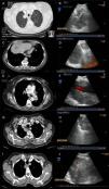
Fig. 1 shows some examples of the computed tomography (CT) and endoscopic ultrasound images obtained.

Fig. 1.

Examples of radiological and ultrasound images from four of the cases from the series. (A and B) Computed tomography (CT) and endoscopic ultrasound images of a mass located in the right upper lobe, aspirated using endobronchial ultrasound, the result of which was adenocarcinoma of pulmonary origin. (C and D) CT and endoscopic ultrasound images of a patient with a pulmonary lesion in the left lower lobe and lymphadenopathy in right mediastinal station 9. Both were aspirated using endobronchial ultrasound via the oesophagus; the result in both was colon cancer metastasis. (E and F) CT and endoscopic ultrasound images of a left hilar mass surrounding the pulmonary artery, with EBUS-guided needle aspiration diagnostic of adenocarcinoma of pulmonary origin. (G and H) CT and endoscopic ultrasound images of a right paratracheoesophageal mass possibly originating in the right upper lobe, with needle aspiration carried out via the oesophagus, resulting in a diagnosis of adenocarcinoma of pulmonary origin. (I and J) CT and endoscopic ultrasound images of a mass located in the right upper lobe infiltrating the mediastinum. EBUS-guided fine needle aspiration was performed with a result of adenocarcinoma of pulmonary origin.

(0.37MB).

No complications related with the procedures were described.

Discussion

The diagnosis of paramediastinal or central pulmonary lesions with no endobronchial lesion is a clinical problem, since conventional diagnostic techniques are not very effective for obtaining a cytohistological diagnosis, and the results depend in large part on the size of the lesions, presence or absence of the bronchus sign and the availability of new technologies.4 For lesions closer to the central airways or the digestive tract, one possible option is the use of EBUS, a technique that has been widely validated in the study of mediastinal lymphadenopathies, although there is little published evidence in these types of lesions and few series address this indication exclusively.2,3,5 In the study by Tournoy et al.,2 all the lesions were intrapulmonary and of malignant aetiology, while in the report published by Yang et al.,3 as in ours, lesions adjacent to the airways, whether intrapulmonary or mediastinal, and of benign aetiology (such as tuberculosis and mediastinal cysts) were also included. We found only one series published in Spain in the form of an abstract.5

The review of these cases confirms the value and safety of EBUS-TBNA in both the respiratory tract and the oesophagus in the diagnosis of non-endobronchial central lesions of different aetiologies. One possible limitation of this study is that we could not clearly define if a central or paratracheal lesion was of nodal origin or if it was a lung lesion that had extended beyond the bronchi or towards the mediastinum. This was the situation encountered in 17 of the 43 cases initially selected, and the reason for excluding them from the analysis, even though a diagnosis of malignancy could be made by EBUS-TBNA in all cases. This serves to further reinforce the usefulness of this technique in these types of lesions.

References
[1]
A.R. Medford, J.A. Bennett, C.M. Free, S. Agrawal.
Endobronchial ultrasound-guided transbronchial needle aspiration (EBUS-TBNA): applications in chest disease.
[2]
K.G. Tournoy, R.C. Rintoul, J.P. Meerbeeck, N.R. Carroll, M. Praet, R.C. Buttery, et al.
EBUS-TBNA for the diagnosis of central parenchymal lung lesions not visible at routine bronchoscopy.
Lung Cancer, 63 (2009), pp. 45-49
[3]
H. Yang, H. Zhao, D.H. Garfield, J. Teng, B. Han, J. Sun.
Endobronchial ultrasound-guided transbronchial needle aspiration in the diagnosis of non-lymph node thoracic lesions.
Ann Thorac Med, 1 (2013), pp. 14-21
[4]
P. Mazzone, P. Jain, A.C. Arroliga, R.A. Matthay.
Bronchoscopy and needle biopsy techniques for diagnosis and staging of lung cancer.
Clin Chest Med, 23 (2002), pp. 137-158
[5]
E. Monclou, J. Franco, P. Ordóñez, L. Gil.
Ecobroncoscopia en el diagnóstico de lesiones pulmonares.
Arch Bronconeumol, 48 (2012), pp. 153

Please cite this article as: Lourido T, et al. Diagnóstico de lesiones paratraqueobronquiales no adenopáticas mediante ecobronquial lineal. Actas Dermosifiliogr. 2013;49:337–9

Copyright © 2013. SEPAR
Archivos de Bronconeumología
Article options
Tools

Are you a health professional able to prescribe or dispense drugs?