array:24 [
  "pii" => "S1579212916300295"
  "issn" => "15792129"
  "doi" => "10.1016/j.arbr.2016.03.023"
  "estado" => "S300"
  "fechaPublicacion" => "2016-06-01"
  "aid" => "1260"
  "copyright" => "SEPAR"
  "copyrightAnyo" => "2015"
  "documento" => "simple-article"
  "crossmark" => 1
  "subdocumento" => "crp"
  "cita" => "Arch Bronconeumol. 2016;52:335-6"
  "abierto" => array:3 [
    "ES" => false
    "ES2" => false
    "LATM" => false
  ]
  "gratuito" => false
  "lecturas" => array:2 [
    "total" => 1561
    "formatos" => array:3 [
      "EPUB" => 104
      "HTML" => 1007
      "PDF" => 450
    ]
  ]
  "Traduccion" => array:1 [
    "es" => array:19 [
      "pii" => "S0300289615003749"
      "issn" => "03002896"
      "doi" => "10.1016/j.arbres.2015.09.007"
      "estado" => "S300"
      "fechaPublicacion" => "2016-06-01"
      "aid" => "1260"
      "copyright" => "SEPAR"
      "documento" => "simple-article"
      "crossmark" => 1
      "subdocumento" => "crp"
      "cita" => "Arch Bronconeumol. 2016;52:335-6"
      "abierto" => array:3 [
        "ES" => true
        "ES2" => true
        "LATM" => true
      ]
      "gratuito" => true
      "lecturas" => array:2 [
        "total" => 2217
        "formatos" => array:3 [
          "EPUB" => 94
          "HTML" => 1542
          "PDF" => 581
        ]
      ]
      "es" => array:10 [
        "idiomaDefecto" => true
        "cabecera" => "<span class="elsevierStyleTextfn">Carta al Director</span>"
        "titulo" => "Hallazgos fibrobroncosc&#243;picos en pacientes estudiados por estridor en un hospital de tercer nivel"
        "tienePdf" => "es"
        "tieneTextoCompleto" => "es"
        "paginas" => array:1 [
          0 => array:2 [
            "paginaInicial" => "335"
            "paginaFinal" => "336"
          ]
        ]
        "titulosAlternativos" => array:1 [
          "en" => array:1 [
            "titulo" => "Fiberoptic bronchoscopy findings in children with stridor in a tertiary hospital"
          ]
        ]
        "contieneTextoCompleto" => array:1 [
          "es" => true
        ]
        "contienePdf" => array:1 [
          "es" => true
        ]
        "autores" => array:1 [
          0 => array:2 [
            "autoresLista" => "Raquel Pl&#225;cido-Paias, Isabel Delgado-Pecell&#237;n, Juan Pedro Gonz&#225;lez-Valencia"
            "autores" => array:3 [
              0 => array:2 [
                "nombre" => "Raquel"
                "apellidos" => "Pl&#225;cido-Paias"
              ]
              1 => array:2 [
                "nombre" => "Isabel"
                "apellidos" => "Delgado-Pecell&#237;n"
              ]
              2 => array:2 [
                "nombre" => "Juan Pedro"
                "apellidos" => "Gonz&#225;lez-Valencia"
              ]
            ]
          ]
        ]
      ]
      "idiomaDefecto" => "es"
      "Traduccion" => array:1 [
        "en" => array:9 [
          "pii" => "S1579212916300295"
          "doi" => "10.1016/j.arbr.2016.03.023"
          "estado" => "S300"
          "subdocumento" => ""
          "abierto" => array:3 [
            "ES" => false
            "ES2" => false
            "LATM" => false
          ]
          "gratuito" => false
          "lecturas" => array:1 [
            "total" => 0
          ]
          "idiomaDefecto" => "en"
          "EPUB" => "https://multimedia.elsevier.es/PublicationsMultimediaV1/item/epub/S1579212916300295?idApp=UINPBA00003Z"
        ]
      ]
      "EPUB" => "https://multimedia.elsevier.es/PublicationsMultimediaV1/item/epub/S0300289615003749?idApp=UINPBA00003Z"
      "url" => "/03002896/0000005200000006/v1_201605260144/S0300289615003749/v1_201605260144/es/main.assets"
    ]
  ]
  "itemSiguiente" => array:19 [
    "pii" => "S1579212916300283"
    "issn" => "15792129"
    "doi" => "10.1016/j.arbr.2016.03.022"
    "estado" => "S300"
    "fechaPublicacion" => "2016-06-01"
    "aid" => "1266"
    "copyright" => "SEPAR"
    "documento" => "simple-article"
    "crossmark" => 1
    "subdocumento" => "crp"
    "cita" => "Arch Bronconeumol. 2016;52:336-7"
    "abierto" => array:3 [
      "ES" => false
      "ES2" => false
      "LATM" => false
    ]
    "gratuito" => false
    "lecturas" => array:2 [
      "total" => 1869
      "formatos" => array:3 [
        "EPUB" => 106
        "HTML" => 1248
        "PDF" => 515
      ]
    ]
    "en" => array:10 [
      "idiomaDefecto" => true
      "cabecera" => "<span class="elsevierStyleTextfn">Scientific Letter</span>"
      "titulo" => "Reactivation of <span class="elsevierStyleItalic">Mycobacterium bovis</span> Infection and Tumor Necrosis Factor Inhibitors&#46; A Mexican Case"
      "tienePdf" => "en"
      "tieneTextoCompleto" => "en"
      "paginas" => array:1 [
        0 => array:2 [
          "paginaInicial" => "336"
          "paginaFinal" => "337"
        ]
      ]
      "titulosAlternativos" => array:1 [
        "es" => array:1 [
          "titulo" => "Reactivaci&#243;n de infecci&#243;n por <span class="elsevierStyleItalic">Mycobacterium bovis</span>&#44; e inhibidores del factor de necrosis tumoral&#46; El caso de M&#233;xico"
        ]
      ]
      "contieneTextoCompleto" => array:1 [
        "en" => true
      ]
      "contienePdf" => array:1 [
        "en" => true
      ]
      "autores" => array:1 [
        0 => array:2 [
          "autoresLista" => "Alain R&#46; Rodr&#237;guez-Orozco"
          "autores" => array:1 [
            0 => array:2 [
              "nombre" => "Alain R&#46;"
              "apellidos" => "Rodr&#237;guez-Orozco"
            ]
          ]
        ]
      ]
    ]
    "idiomaDefecto" => "en"
    "Traduccion" => array:1 [
      "es" => array:9 [
        "pii" => "S0300289615003889"
        "doi" => "10.1016/j.arbres.2015.09.013"
        "estado" => "S300"
        "subdocumento" => ""
        "abierto" => array:3 [
          "ES" => true
          "ES2" => true
          "LATM" => true
        ]
        "gratuito" => true
        "lecturas" => array:1 [
          "total" => 0
        ]
        "idiomaDefecto" => "es"
        "EPUB" => "https://multimedia.elsevier.es/PublicationsMultimediaV1/item/epub/S0300289615003889?idApp=UINPBA00003Z"
      ]
    ]
    "EPUB" => "https://multimedia.elsevier.es/PublicationsMultimediaV1/item/epub/S1579212916300283?idApp=UINPBA00003Z"
    "url" => "/15792129/0000005200000006/v1_201605260126/S1579212916300283/v1_201605260126/en/main.assets"
  ]
  "itemAnterior" => array:19 [
    "pii" => "S157921291500422X"
    "issn" => "15792129"
    "doi" => "10.1016/j.arbr.2015.11.010"
    "estado" => "S300"
    "fechaPublicacion" => "2016-06-01"
    "aid" => "1263"
    "copyright" => "SEPAR"
    "documento" => "simple-article"
    "crossmark" => 1
    "subdocumento" => "crp"
    "cita" => "Arch Bronconeumol. 2016;52:334-5"
    "abierto" => array:3 [
      "ES" => false
      "ES2" => false
      "LATM" => false
    ]
    "gratuito" => false
    "lecturas" => array:2 [
      "total" => 2136
      "formatos" => array:3 [
        "EPUB" => 117
        "HTML" => 1454
        "PDF" => 565
      ]
    ]
    "en" => array:11 [
      "idiomaDefecto" => true
      "cabecera" => "<span class="elsevierStyleTextfn">Scientific Letter</span>"
      "titulo" => "Counterintuitive Pulmonary Nodules in Rheumatoid Arthritis"
      "tienePdf" => "en"
      "tieneTextoCompleto" => "en"
      "paginas" => array:1 [
        0 => array:2 [
          "paginaInicial" => "334"
          "paginaFinal" => "335"
        ]
      ]
      "titulosAlternativos" => array:1 [
        "es" => array:1 [
          "titulo" => "N&#243;dulos pulmonares contraintuitivos en artritis reumatoide"
        ]
      ]
      "contieneTextoCompleto" => array:1 [
        "en" => true
      ]
      "contienePdf" => array:1 [
        "en" => true
      ]
      "resumenGrafico" => array:2 [
        "original" => 0
        "multimedia" => array:7 [
          "identificador" => "fig0005"
          "etiqueta" => "Fig&#46; 1"
          "tipo" => "MULTIMEDIAFIGURA"
          "mostrarFloat" => true
          "mostrarDisplay" => false
          "figura" => array:1 [
            0 => array:4 [
              "imagen" => "gr1.jpeg"
              "Alto" => 1037
              "Ancho" => 1600
              "Tamanyo" => 216285
            ]
          ]
          "descripcion" => array:1 [
            "en" => "<p id="spar0005" class="elsevierStyleSimplePara elsevierViewall">CT of chest &#40;A&#41;&#44; initial CT demonstrating left upper lobe nodule with cavitation &#40;B&#41;&#44; nine month follow-up CT with resolution of left upper lobe nodule&#44; &#40;C&#41; initial CT demonstrating peripheral nodule in the lingual and &#40;D&#41; nine month follow-up CT with resolution of peripheral nodule in the lingual&#46;</p>"
          ]
        ]
      ]
      "autores" => array:1 [
        0 => array:2 [
          "autoresLista" => "Felix Daniel Zamora, Eitan Podgaetz, H&#46; Erhan Dincer"
          "autores" => array:3 [
            0 => array:2 [
              "nombre" => "Felix Daniel"
              "apellidos" => "Zamora"
            ]
            1 => array:2 [
              "nombre" => "Eitan"
              "apellidos" => "Podgaetz"
            ]
            2 => array:2 [
              "nombre" => "H&#46; Erhan"
              "apellidos" => "Dincer"
            ]
          ]
        ]
      ]
    ]
    "idiomaDefecto" => "en"
    "Traduccion" => array:1 [
      "es" => array:9 [
        "pii" => "S0300289615003853"
        "doi" => "10.1016/j.arbres.2015.09.010"
        "estado" => "S300"
        "subdocumento" => ""
        "abierto" => array:3 [
          "ES" => true
          "ES2" => true
          "LATM" => true
        ]
        "gratuito" => true
        "lecturas" => array:1 [
          "total" => 0
        ]
        "idiomaDefecto" => "es"
        "EPUB" => "https://multimedia.elsevier.es/PublicationsMultimediaV1/item/epub/S0300289615003853?idApp=UINPBA00003Z"
      ]
    ]
    "EPUB" => "https://multimedia.elsevier.es/PublicationsMultimediaV1/item/epub/S157921291500422X?idApp=UINPBA00003Z"
    "url" => "/15792129/0000005200000006/v1_201605260126/S157921291500422X/v1_201605260126/en/main.assets"
  ]
  "en" => array:14 [
    "idiomaDefecto" => true
    "cabecera" => "<span class="elsevierStyleTextfn">Scientific Letter</span>"
    "titulo" => "Fiberoptic Bronchoscopy Findings in Children With Stridor in a Tertiary Hospital"
    "tieneTextoCompleto" => true
    "saludo" => "To the Editor&#44;"
    "paginas" => array:1 [
      0 => array:2 [
        "paginaInicial" => "335"
        "paginaFinal" => "336"
      ]
    ]
    "autores" => array:1 [
      0 => array:4 [
        "autoresLista" => "Raquel Pl&#225;cido-Paias, Isabel Delgado-Pecell&#237;n, Juan Pedro Gonz&#225;lez-Valencia"
        "autores" => array:3 [
          0 => array:4 [
            "nombre" => "Raquel"
            "apellidos" => "Pl&#225;cido-Paias"
            "email" => array:1 [
              0 => "raquelpaias&#64;gmail&#46;com"
            ]
            "referencia" => array:2 [
              0 => array:2 [
                "etiqueta" => "<span class="elsevierStyleSup">a</span>"
                "identificador" => "aff0005"
              ]
              1 => array:2 [
                "etiqueta" => "<span class="elsevierStyleSup">&#42;</span>"
                "identificador" => "cor0005"
              ]
            ]
          ]
          1 => array:3 [
            "nombre" => "Isabel"
            "apellidos" => "Delgado-Pecell&#237;n"
            "referencia" => array:1 [
              0 => array:2 [
                "etiqueta" => "<span class="elsevierStyleSup">b</span>"
                "identificador" => "aff0010"
              ]
            ]
          ]
          2 => array:3 [
            "nombre" => "Juan Pedro"
            "apellidos" => "Gonz&#225;lez-Valencia"
            "referencia" => array:1 [
              0 => array:2 [
                "etiqueta" => "<span class="elsevierStyleSup">b</span>"
                "identificador" => "aff0010"
              ]
            ]
          ]
        ]
        "afiliaciones" => array:2 [
          0 => array:3 [
            "entidad" => "Servicio de Pediatr&#237;a&#44; Hospital de M&#233;rida&#44; M&#233;rida&#44; Badajoz&#44; Spain"
            "etiqueta" => "a"
            "identificador" => "aff0005"
          ]
          1 => array:3 [
            "entidad" => "Unidad de Neumolog&#237;a Pedi&#225;trica&#44; Hospitales Universitarios Virgen del Roc&#237;o&#44; Sevilla&#44; Spain"
            "etiqueta" => "b"
            "identificador" => "aff0010"
          ]
        ]
        "correspondencia" => array:1 [
          0 => array:3 [
            "identificador" => "cor0005"
            "etiqueta" => "&#8270;"
            "correspondencia" => "Corresponding author&#46;"
          ]
        ]
      ]
    ]
    "titulosAlternativos" => array:1 [
      "es" => array:1 [
        "titulo" => "Hallazgos fibrobroncosc&#243;picos en pacientes estudiados por estridor en un hospital de tercer nivel"
      ]
    ]
    "textoCompleto" => "<span class="elsevierStyleSections"><p id="par0005" class="elsevierStylePara elsevierViewall">Stridor is a musical sound generated by the rapid&#44; turbulent passage of air through a narrowed section of the airways&#46;<a class="elsevierStyleCrossRefs" href="#bib0030"><span class="elsevierStyleSup">1&#8211;3</span></a> Patients with stridor should be evaluated with a complete clinical history and physical examination&#46;<a class="elsevierStyleCrossRef" href="#bib0035"><span class="elsevierStyleSup">2</span></a> The characteristics of stridor indicate the level of the lesion&#46;<a class="elsevierStyleCrossRefs" href="#bib0030"><span class="elsevierStyleSup">1&#44;2</span></a> When it occurs on inspiration&#44; the lesion is generally supraglottic&#44; when it is expiratory&#44; it is located in the intrathoracic airway&#44;<a class="elsevierStyleCrossRefs" href="#bib0030"><span class="elsevierStyleSup">1&#44;2</span></a> and when stridor is biphasic&#44; the stenosis is located in the trachea&#46;<a class="elsevierStyleCrossRef" href="#bib0035"><span class="elsevierStyleSup">2</span></a></p><p id="par0010" class="elsevierStylePara elsevierViewall">We performed a descriptive study of children with stridor undergoing fiberoptic bronchoscopy &#40;FB&#41; from January 2009 to December 2013&#44; analyzing sex&#44; age&#44; underlying disease&#44; symptoms&#44; type of stridor&#44; fiberoptic bronchoscopy findings&#44; treatments and FB yield&#46; We used an Olympus<span class="elsevierStyleSup">&#174;</span> fiberoptic bronchoscopy &#40;2&#46;8&#44; 3&#46;6 and 4&#46;8<span class="elsevierStyleHsp" style=""></span>mm&#41;&#46;</p><p id="par0015" class="elsevierStylePara elsevierViewall">We performed 593 FBs&#44; 138 &#40;23&#37;&#41; for stridor&#59; 63&#37; in infants younger than 1 year&#44; 14&#37; in children between 1 and 2 years&#44; 17&#37; between 3 and 7 years and 6&#37; in patients older than 8 years&#59; 58&#37; were male&#46; The most predominant form of stridor was inspiratory &#40;78&#37;&#41;&#46; Diagnostic yield was 95&#37;&#44; i&#46;e&#46;&#44; 130 patients obtained diagnostic or therapeutic benefit&#44; while 18 of those patients required a repeat FB &#40;12&#37;&#41;&#46; Most &#40;81&#37;&#41; had underlying disease &#40;<a class="elsevierStyleCrossRef" href="#tbl0005">Table 1</a>&#41;&#46; Fiberoptic bronchoscopy findings are summarized in <a class="elsevierStyleCrossRef" href="#tbl0005">Table 1</a>&#46; In 91&#37; of cases we took a &#8220;wait and see&#8221; approach&#59; 12 patients &#40;9&#37;&#41; needed surgical treatment &#40;6 required tracheoplasty&#44; 2 aortopexy&#44; 2 tracheostomy&#44; 1 uvulectomy&#44; and 1 laser ablation&#41; and 2 patients with subglottic hemangioma received propanolol&#44; with good response&#46; A total of 8&#37; of the patients had mild&#44; self-limiting desaturations&#46;</p><elsevierMultimedia ident="tbl0005"></elsevierMultimedia><p id="par0020" class="elsevierStylePara elsevierViewall">Stridor is a symptom which requires an etiological diagnosis<a class="elsevierStyleCrossRefs" href="#bib0030"><span class="elsevierStyleSup">1&#8211;3</span></a> based on clinical history&#44; physical examination&#44; and direct evaluation of the airway<a class="elsevierStyleCrossRef" href="#bib0030"><span class="elsevierStyleSup">1</span></a> with fiberoptic bronchoscopy&#46;<a class="elsevierStyleCrossRefs" href="#bib0030"><span class="elsevierStyleSup">1&#8211;4</span></a> This is generally performed under spontaneous breathing&#44; allowing an anatomical and functional examination of the airways&#46;<a class="elsevierStyleCrossRefs" href="#bib0035"><span class="elsevierStyleSup">2&#44;3</span></a> Respiratory pediatricians&#44; ENT specialists&#44; pediatric surgeons and&#47;or anesthetists must work closely together to maximize the benefit of this procedure and ensure it is correctly performed&#46; In our hospital&#44; FB is performed in collaboration with anesthetists&#44; who generally apply sevoflurane&#46; In our opinion&#44; the airway must be examined in its entirety&#46;</p><p id="par0025" class="elsevierStylePara elsevierViewall">Like other authors&#44; our case series showed that laryngomalacia was the most common finding in infants with inspiratory stridor&#44;<a class="elsevierStyleCrossRefs" href="#bib0030"><span class="elsevierStyleSup">1&#8211;3</span></a> and this condition generally resolved with a &#8220;wait and see&#8221; approach&#44; since laryngomalacia is a result of immature laryngeal structures&#46;<a class="elsevierStyleCrossRefs" href="#bib0035"><span class="elsevierStyleSup">2&#44;3</span></a> The second most common disorder encountered was subglottic stenosis &#40;15&#37;&#41;&#46; This percentage was high&#44; but in our opinion there was some element of bias&#44; as our hospital is the respiratory referral center for the south of Spain&#46; Stridor can be caused by congenital or acquired diseases&#46;<a class="elsevierStyleCrossRef" href="#bib0035"><span class="elsevierStyleSup">2</span></a> The most common type is biphasic stridor associated with varying degrees of clinical airway obstruction&#46;<a class="elsevierStyleCrossRef" href="#bib0035"><span class="elsevierStyleSup">2</span></a> In our group of patients&#44; all&#44; except 1&#44; developed symptoms after intubation&#46; Subglottic stenoses with Cotton grades III and IV are treated by surgery&#44; as was the case in 9&#37; of our patients&#46; The most common technique used was tracheoplasty with rib cartilage graft &#40;5&#37;&#41;&#46;</p><p id="par0030" class="elsevierStylePara elsevierViewall">In our opinion&#44; FB is the technique of choice in the diagnosis of airway disorders&#44;<a class="elsevierStyleCrossRefs" href="#bib0045"><span class="elsevierStyleSup">4&#44;5</span></a> as it is safer and less aggressive than rigid bronchoscopy&#46; We recommend that FB be performed in any child who presents with stridor&#44; since this symptom is generally an indication of a banal disorder&#44; but it can mask a life-threatening disease&#46;<a class="elsevierStyleCrossRefs" href="#bib0030"><span class="elsevierStyleSup">1&#44;3&#44;5</span></a></p></span>"
    "pdfFichero" => "main.pdf"
    "tienePdf" => true
    "NotaPie" => array:1 [
      0 => array:2 [
        "etiqueta" => "&#9734;"
        "nota" => "<p class="elsevierStyleNotepara" id="npar0005">Please cite this article as&#58; Pl&#225;cido-Paias R&#44; Delgado-Pecell&#237;n I&#44; Gonz&#225;lez-Valencia JP&#46; Hallazgos fibrobroncosc&#243;picos en pacientes estudiados por estridor en un hospital de tercer nivel&#46; Arch Bronconeumol&#46; 2016&#59;52&#58;335&#8211;336&#46;</p>"
      ]
    ]
    "multimedia" => array:1 [
      0 => array:8 [
        "identificador" => "tbl0005"
        "etiqueta" => "Table 1"
        "tipo" => "MULTIMEDIATABLA"
        "mostrarFloat" => true
        "mostrarDisplay" => false
        "detalles" => array:1 [
          0 => array:3 [
            "identificador" => "at1"
            "detalle" => "Table "
            "rol" => "short"
          ]
        ]
        "tabla" => array:2 [
          "leyenda" => "<p id="spar0010" class="elsevierStyleSimplePara elsevierViewall">ALTE&#44; apparent life-threatening event&#46;</p>"
          "tablatextoimagen" => array:1 [
            0 => array:2 [
              "tabla" => array:1 [
                0 => """
                  <table border="0" frame="\n
                  \t\t\t\t\tvoid\n
                  \t\t\t\t" class=""><thead title="thead"><tr title="table-row"><th class="td" title="table-head  " align="left" valign="top" scope="col" style="border-bottom: 2px solid black">&#40;A&#41; Fiberoptic Bronchoscopy Findings&nbsp;\t\t\t\t\t\t\n
                  \t\t\t\t</th><th class="td" title="table-head  " align="left" valign="top" scope="col" style="border-bottom: 2px solid black">Percentage&nbsp;\t\t\t\t\t\t\n
                  \t\t\t\t</th><th class="td" title="table-head  " align="left" valign="top" scope="col" style="border-bottom: 2px solid black">&#40;B&#41; Underlying Disease of Patients Undergoing Fiberoptic Bronchoscopy&nbsp;\t\t\t\t\t\t\n
                  \t\t\t\t</th><th class="td" title="table-head  " align="left" valign="top" scope="col" style="border-bottom: 2px solid black">Percentage&nbsp;\t\t\t\t\t\t\n
                  \t\t\t\t</th></tr></thead><tbody title="tbody"><tr title="table-row"><td class="td-with-role" title="table-entry ; entry_with_role_rowhead " align="left" valign="top">Laryngomalacia&nbsp;\t\t\t\t\t\t\n
                  \t\t\t\t</td><td class="td" title="table-entry  " align="char" valign="top">46&nbsp;\t\t\t\t\t\t\n
                  \t\t\t\t</td><td class="td" title="table-entry  " align="left" valign="top">Laryngitis and recurrent wheezing &#40;<span class="elsevierStyleItalic">n</span><span class="elsevierStyleHsp" style=""></span>&#61;<span class="elsevierStyleHsp" style=""></span>50&#41;&nbsp;\t\t\t\t\t\t\n
                  \t\t\t\t</td><td class="td" title="table-entry  " align="char" valign="top">36&nbsp;\t\t\t\t\t\t\n
                  \t\t\t\t</td></tr><tr title="table-row"><td class="td-with-role" title="table-entry ; entry_with_role_rowhead " align="left" valign="top">Subglottic stenosis&nbsp;\t\t\t\t\t\t\n
                  \t\t\t\t</td><td class="td" title="table-entry  " align="char" valign="top">20&nbsp;\t\t\t\t\t\t\n
                  \t\t\t\t</td><td class="td" title="table-entry  " align="left" valign="top">Gastroesophageal reflux &#40;<span class="elsevierStyleItalic">n</span><span class="elsevierStyleHsp" style=""></span>&#61;<span class="elsevierStyleHsp" style=""></span>36&#41;&nbsp;\t\t\t\t\t\t\n
                  \t\t\t\t</td><td class="td" title="table-entry  " align="char" valign="top">26&nbsp;\t\t\t\t\t\t\n
                  \t\t\t\t</td></tr><tr title="table-row"><td class="td-with-role" title="table-entry ; entry_with_role_rowhead " align="left" valign="top">Non-specific inflammation&nbsp;\t\t\t\t\t\t\n
                  \t\t\t\t</td><td class="td" title="table-entry  " align="char" valign="top">17&nbsp;\t\t\t\t\t\t\n
                  \t\t\t\t</td><td class="td" title="table-entry  " align="left" valign="top">ALTE&#47;choking&#47;apneas &#40;<span class="elsevierStyleItalic">n</span><span class="elsevierStyleHsp" style=""></span>&#61;<span class="elsevierStyleHsp" style=""></span>11&#41;&nbsp;\t\t\t\t\t\t\n
                  \t\t\t\t</td><td class="td" title="table-entry  " align="char" valign="top">8&nbsp;\t\t\t\t\t\t\n
                  \t\t\t\t</td></tr><tr title="table-row"><td class="td-with-role" title="table-entry ; entry_with_role_rowhead " align="left" valign="top">Extrinsic tracheal&#47;bronchial compression&nbsp;\t\t\t\t\t\t\n
                  \t\t\t\t</td><td class="td" title="table-entry  " align="char" valign="top">10&nbsp;\t\t\t\t\t\t\n
                  \t\t\t\t</td><td class="td" title="table-entry  " align="left" valign="top">Congenital malformations &#40;<span class="elsevierStyleItalic">n</span><span class="elsevierStyleHsp" style=""></span>&#61;<span class="elsevierStyleHsp" style=""></span>11&#41;&nbsp;\t\t\t\t\t\t\n
                  \t\t\t\t</td><td class="td" title="table-entry  " align="char" valign="top">8&nbsp;\t\t\t\t\t\t\n
                  \t\t\t\t</td></tr><tr title="table-row"><td class="td-with-role" title="table-entry ; entry_with_role_rowhead " align="left" valign="top">Tracheomalacia&nbsp;\t\t\t\t\t\t\n
                  \t\t\t\t</td><td class="td" title="table-entry  " align="char" valign="top">10&nbsp;\t\t\t\t\t\t\n
                  \t\t\t\t</td><td class="td" title="table-entry  " align="left" valign="top">- Tracheomalacia &#40;4&#41;&nbsp;\t\t\t\t\t\t\n
                  \t\t\t\t</td><td class="td" title="table-entry  " align="" valign="top">&nbsp;\t\t\t\t\t\t\n
                  \t\t\t\t</td></tr><tr title="table-row"><td class="td-with-role" title="table-entry ; entry_with_role_rowhead " align="left" valign="top">Arytenoid paralysis&nbsp;\t\t\t\t\t\t\n
                  \t\t\t\t</td><td class="td" title="table-entry  " align="char" valign="top">5&nbsp;\t\t\t\t\t\t\n
                  \t\t\t\t</td><td class="td" title="table-entry  " align="left" valign="top">- Tracheal stenosis&nbsp;\t\t\t\t\t\t\n
                  \t\t\t\t</td><td class="td" title="table-entry  " align="" valign="top">&nbsp;\t\t\t\t\t\t\n
                  \t\t\t\t</td></tr><tr title="table-row"><td class="td-with-role" title="table-entry ; entry_with_role_rowhead " align="left" valign="top">Vocal cord paralysis&nbsp;\t\t\t\t\t\t\n
                  \t\t\t\t</td><td class="td" title="table-entry  " align="char" valign="top">3&nbsp;\t\t\t\t\t\t\n
                  \t\t\t\t</td><td class="td" title="table-entry  " align="left" valign="top">- Vascular ring due to right-sided aortic arch with aberrant left subclavian artery associated with Kommerell diverticulum &#40;1&#41;&nbsp;\t\t\t\t\t\t\n
                  \t\t\t\t</td><td class="td" title="table-entry  " align="" valign="top">&nbsp;\t\t\t\t\t\t\n
                  \t\t\t\t</td></tr><tr title="table-row"><td class="td-with-role" title="table-entry ; entry_with_role_rowhead " align="left" valign="top">Granulation tissue&nbsp;\t\t\t\t\t\t\n
                  \t\t\t\t</td><td class="td" title="table-entry  " align="char" valign="top">3&nbsp;\t\t\t\t\t\t\n
                  \t\t\t\t</td><td class="td" title="table-entry  " align="left" valign="top">- Tracheal bronchus&nbsp;\t\t\t\t\t\t\n
                  \t\t\t\t</td><td class="td" title="table-entry  " align="" valign="top">&nbsp;\t\t\t\t\t\t\n
                  \t\t\t\t</td></tr><tr title="table-row"><td class="td-with-role" title="table-entry ; entry_with_role_rowhead " align="left" valign="top">Epiglottis malformation&nbsp;\t\t\t\t\t\t\n
                  \t\t\t\t</td><td class="td" title="table-entry  " align="char" valign="top">2&nbsp;\t\t\t\t\t\t\n
                  \t\t\t\t</td><td class="td" title="table-entry  " align="left" valign="top">- Hypoplasia of right lung with pulmonary sequestration &#40;3&#41;&nbsp;\t\t\t\t\t\t\n
                  \t\t\t\t</td><td class="td" title="table-entry  " align="" valign="top">&nbsp;\t\t\t\t\t\t\n
                  \t\t\t\t</td></tr><tr title="table-row"><td class="td-with-role" title="table-entry ; entry_with_role_rowhead " align="left" valign="top">Pharynogmalacia&nbsp;\t\t\t\t\t\t\n
                  \t\t\t\t</td><td class="td" title="table-entry  " align="char" valign="top">2&nbsp;\t\t\t\t\t\t\n
                  \t\t\t\t</td><td class="td" title="table-entry  " align="" valign="top">&nbsp;\t\t\t\t\t\t\n
                  \t\t\t\t</td><td class="td" title="table-entry  " align="" valign="top">&nbsp;\t\t\t\t\t\t\n
                  \t\t\t\t</td></tr><tr title="table-row"><td class="td-with-role" title="table-entry ; entry_with_role_rowhead " align="left" valign="top">Mucocele&nbsp;\t\t\t\t\t\t\n
                  \t\t\t\t</td><td class="td" title="table-entry  " align="char" valign="top">1&nbsp;\t\t\t\t\t\t\n
                  \t\t\t\t</td><td class="td" title="table-entry  " align="left" valign="top">Prematurity &#40;<span class="elsevierStyleItalic">n</span><span class="elsevierStyleHsp" style=""></span>&#61;<span class="elsevierStyleHsp" style=""></span>9&#41;&nbsp;\t\t\t\t\t\t\n
                  \t\t\t\t</td><td class="td" title="table-entry  " align="char" valign="top">6&#46;5&nbsp;\t\t\t\t\t\t\n
                  \t\t\t\t</td></tr><tr title="table-row"><td class="td-with-role" title="table-entry ; entry_with_role_rowhead " align="left" valign="top">Subglottic hemangioma&nbsp;\t\t\t\t\t\t\n
                  \t\t\t\t</td><td class="td" title="table-entry  " align="char" valign="top">1&nbsp;\t\t\t\t\t\t\n
                  \t\t\t\t</td><td class="td" title="table-entry  " align="left" valign="top">Epilepsy &#40;<span class="elsevierStyleItalic">n</span><span class="elsevierStyleHsp" style=""></span>&#61;<span class="elsevierStyleHsp" style=""></span>8&#41;&nbsp;\t\t\t\t\t\t\n
                  \t\t\t\t</td><td class="td" title="table-entry  " align="char" valign="top">6&nbsp;\t\t\t\t\t\t\n
                  \t\t\t\t</td></tr><tr title="table-row"><td class="td-with-role" title="table-entry ; entry_with_role_rowhead " align="left" valign="top">Bronchomalacia&nbsp;\t\t\t\t\t\t\n
                  \t\t\t\t</td><td class="td" title="table-entry  " align="char" valign="top">1&nbsp;\t\t\t\t\t\t\n
                  \t\t\t\t</td><td class="td" title="table-entry  " align="left" valign="top">Interventricular communication &#40;<span class="elsevierStyleItalic">n</span><span class="elsevierStyleHsp" style=""></span>&#61;<span class="elsevierStyleHsp" style=""></span>7&#41;&nbsp;\t\t\t\t\t\t\n
                  \t\t\t\t</td><td class="td" title="table-entry  " align="char" valign="top">5&nbsp;\t\t\t\t\t\t\n
                  \t\t\t\t</td></tr><tr title="table-row"><td class="td-with-role" title="table-entry ; entry_with_role_rowhead " align="left" valign="top">Epiglottic edema&nbsp;\t\t\t\t\t\t\n
                  \t\t\t\t</td><td class="td" title="table-entry  " align="char" valign="top">0&#46;7&nbsp;\t\t\t\t\t\t\n
                  \t\t\t\t</td><td class="td" title="table-entry  " align="left" valign="top">Hypotonia &#40;<span class="elsevierStyleItalic">n</span><span class="elsevierStyleHsp" style=""></span>&#61;<span class="elsevierStyleHsp" style=""></span>6&#41;&nbsp;\t\t\t\t\t\t\n
                  \t\t\t\t</td><td class="td" title="table-entry  " align="char" valign="top">4&#46;3&nbsp;\t\t\t\t\t\t\n
                  \t\t\t\t</td></tr><tr title="table-row"><td class="td-with-role" title="table-entry ; entry_with_role_rowhead " align="left" valign="top">Vocal cord dysfunction&nbsp;\t\t\t\t\t\t\n
                  \t\t\t\t</td><td class="td" title="table-entry  " align="char" valign="top">0&#46;7&nbsp;\t\t\t\t\t\t\n
                  \t\t\t\t</td><td class="td" title="table-entry  " align="left" valign="top">Coarctation of the aorta &#40;<span class="elsevierStyleItalic">n</span><span class="elsevierStyleHsp" style=""></span>&#61;<span class="elsevierStyleHsp" style=""></span>6&#41;&nbsp;\t\t\t\t\t\t\n
                  \t\t\t\t</td><td class="td" title="table-entry  " align="char" valign="top">4&#46;3&nbsp;\t\t\t\t\t\t\n
                  \t\t\t\t</td></tr><tr title="table-row"><td class="td-with-role" title="table-entry ; entry_with_role_rowhead " align="left" valign="top">Vocal cord synechia&nbsp;\t\t\t\t\t\t\n
                  \t\t\t\t</td><td class="td" title="table-entry  " align="char" valign="top">0&#46;7&nbsp;\t\t\t\t\t\t\n
                  \t\t\t\t</td><td class="td" title="table-entry  " align="left" valign="top">Vascular ring &#40;<span class="elsevierStyleItalic">n</span><span class="elsevierStyleHsp" style=""></span>&#61;<span class="elsevierStyleHsp" style=""></span>5&#41;&nbsp;\t\t\t\t\t\t\n
                  \t\t\t\t</td><td class="td" title="table-entry  " align="char" valign="top">3&#46;6&nbsp;\t\t\t\t\t\t\n
                  \t\t\t\t</td></tr><tr title="table-row"><td class="td-with-role" title="table-entry ; entry_with_role_rowhead " align="left" valign="top">Vocal cord nodule&nbsp;\t\t\t\t\t\t\n
                  \t\t\t\t</td><td class="td" title="table-entry  " align="char" valign="top">0&#46;7&nbsp;\t\t\t\t\t\t\n
                  \t\t\t\t</td><td class="td" title="table-entry  " align="left" valign="top">Patent ductus arteriosus &#40;<span class="elsevierStyleItalic">n</span><span class="elsevierStyleHsp" style=""></span>&#61;<span class="elsevierStyleHsp" style=""></span>5&#41;&nbsp;\t\t\t\t\t\t\n
                  \t\t\t\t</td><td class="td" title="table-entry  " align="char" valign="top">3&#46;6&nbsp;\t\t\t\t\t\t\n
                  \t\t\t\t</td></tr><tr title="table-row"><td class="td-with-role" title="table-entry ; entry_with_role_rowhead " align="left" valign="top">Arytenoid dislocation&nbsp;\t\t\t\t\t\t\n
                  \t\t\t\t</td><td class="td" title="table-entry  " align="char" valign="top">0&#46;7&nbsp;\t\t\t\t\t\t\n
                  \t\t\t\t</td><td class="td" title="table-entry  " align="left" valign="top">Tracheoesophageal fistula &#40;<span class="elsevierStyleItalic">n</span><span class="elsevierStyleHsp" style=""></span>&#61;<span class="elsevierStyleHsp" style=""></span>5&#41;&nbsp;\t\t\t\t\t\t\n
                  \t\t\t\t</td><td class="td" title="table-entry  " align="char" valign="top">3&#46;6&nbsp;\t\t\t\t\t\t\n
                  \t\t\t\t</td></tr><tr title="table-row"><td class="td-with-role" title="table-entry ; entry_with_role_rowhead " align="left" valign="top">Vallecular cyst&nbsp;\t\t\t\t\t\t\n
                  \t\t\t\t</td><td class="td" title="table-entry  " align="char" valign="top">0&#46;7&nbsp;\t\t\t\t\t\t\n
                  \t\t\t\t</td><td class="td" title="table-entry  " align="left" valign="top">Cutaneous hemangioma &#40;<span class="elsevierStyleItalic">n</span><span class="elsevierStyleHsp" style=""></span>&#61;<span class="elsevierStyleHsp" style=""></span>4&#41;&nbsp;\t\t\t\t\t\t\n
                  \t\t\t\t</td><td class="td" title="table-entry  " align="char" valign="top">2&#46;9&nbsp;\t\t\t\t\t\t\n
                  \t\t\t\t</td></tr><tr title="table-row"><td class="td-with-role" title="table-entry ; entry_with_role_rowhead " align="left" valign="top">Cyst on uvula&nbsp;\t\t\t\t\t\t\n
                  \t\t\t\t</td><td class="td" title="table-entry  " align="char" valign="top">0&#46;7&nbsp;\t\t\t\t\t\t\n
                  \t\t\t\t</td><td class="td" title="table-entry  " align="left" valign="top">Down syndrome &#40;<span class="elsevierStyleItalic">n</span><span class="elsevierStyleHsp" style=""></span>&#61;<span class="elsevierStyleHsp" style=""></span>4&#41;&nbsp;\t\t\t\t\t\t\n
                  \t\t\t\t</td><td class="td" title="table-entry  " align="char" valign="top">2&#46;9&nbsp;\t\t\t\t\t\t\n
                  \t\t\t\t</td></tr><tr title="table-row"><td class="td-with-role" title="table-entry ; entry_with_role_rowhead " align="left" valign="top">Tongue compressing epiglottis&nbsp;\t\t\t\t\t\t\n
                  \t\t\t\t</td><td class="td" title="table-entry  " align="char" valign="top">0&#46;7&nbsp;\t\t\t\t\t\t\n
                  \t\t\t\t</td><td class="td" title="table-entry  " align="" valign="top">&nbsp;\t\t\t\t\t\t\n
                  \t\t\t\t</td><td class="td" title="table-entry  " align="" valign="top">&nbsp;\t\t\t\t\t\t\n
                  \t\t\t\t</td></tr><tr title="table-row"><td class="td-with-role" title="table-entry ; entry_with_role_rowhead " align="left" valign="top">Middle lobe bronchial hypoplasia&nbsp;\t\t\t\t\t\t\n
                  \t\t\t\t</td><td class="td" title="table-entry  " align="char" valign="top">0&#46;7&nbsp;\t\t\t\t\t\t\n
                  \t\t\t\t</td><td class="td" title="table-entry  " align="" valign="top">&nbsp;\t\t\t\t\t\t\n
                  \t\t\t\t</td><td class="td" title="table-entry  " align="" valign="top">&nbsp;\t\t\t\t\t\t\n
                  \t\t\t\t</td></tr></tbody></table>
                  """
              ]
              "imagenFichero" => array:1 [
                0 => "xTab1077284.png"
              ]
            ]
          ]
        ]
        "descripcion" => array:1 [
          "en" => "<p id="spar0005" class="elsevierStyleSimplePara elsevierViewall">&#40;A&#41; Diseases Identified in the 138 Fiberoptic Bronchoscopies Performed for Stridor&#59; in 43 Patients &#40;31&#37;&#41;&#44; Multiple Findings Were Found on Fiberoptic Bronchoscopy&#46; &#40;B&#41; Underlying Diseases in the 138 Patients Undergoing Fiberoptic Bronchoscopy&#46;</p>"
        ]
      ]
    ]
    "bibliografia" => array:2 [
      "titulo" => "References"
      "seccion" => array:1 [
        0 => array:2 [
          "identificador" => "bibs0005"
          "bibliografiaReferencia" => array:5 [
            0 => array:3 [
              "identificador" => "bib0030"
              "etiqueta" => "1"
              "referencia" => array:1 [
                0 => array:2 [
                  "contribucion" => array:1 [
                    0 => array:2 [
                      "titulo" => "&#191;Es el estridor un s&#237;ntoma banal en el lactante&#63;"
                      "autores" => array:1 [
                        0 => array:2 [
                          "etal" => true
                          "autores" => array:6 [
                            0 => "A&#46; Parente Hern&#225;ndez"
                            1 => "M&#46;A&#46; Garc&#237;a-Casillas"
                            2 => "J&#46;A&#46; Matute"
                            3 => "A&#46; Ca&#241;izo"
                            4 => "A&#46; La&#237;n"
                            5 => "M&#46; Fanjul"
                          ]
                        ]
                      ]
                    ]
                  ]
                  "host" => array:1 [
                    0 => array:1 [
                      "Revista" => array:5 [
                        "tituloSerie" => "An Pediatr &#40;Barc&#41;"
                        "fecha" => "2007"
                        "volumen" => "66"
                        "paginaInicial" => "559"
                        "paginaFinal" => "565"
                      ]
                    ]
                  ]
                ]
              ]
            ]
            1 => array:3 [
              "identificador" => "bib0035"
              "etiqueta" => "2"
              "referencia" => array:1 [
                0 => array:2 [
                  "contribucion" => array:1 [
                    0 => array:2 [
                      "titulo" => "An approach to stridor in infants and children"
                      "autores" => array:1 [
                        0 => array:2 [
                          "etal" => false
                          "autores" => array:3 [
                            0 => "A&#46; Boudewyns"
                            1 => "J&#46; Claes"
                            2 => "P&#46; van de Heyning"
                          ]
                        ]
                      ]
                    ]
                  ]
                  "host" => array:1 [
                    0 => array:2 [
                      "doi" => "10.1007/s00431-009-1044-7"
                      "Revista" => array:7 [
                        "tituloSerie" => "Eur J Pediatr"
                        "fecha" => "2010"
                        "volumen" => "169"
                        "paginaInicial" => "135"
                        "paginaFinal" => "141"
                        "link" => array:1 [
                          0 => array:2 [
                            "url" => "https://www.ncbi.nlm.nih.gov/pubmed/19763619"
                            "web" => "Medline"
                          ]
                        ]
                        "itemHostRev" => array:3 [
                          "pii" => "S014067360312329X"
                          "estado" => "S300"
                          "issn" => "01406736"
                        ]
                      ]
                    ]
                  ]
                ]
              ]
            ]
            2 => array:3 [
              "identificador" => "bib0040"
              "etiqueta" => "3"
              "referencia" => array:1 [
                0 => array:2 [
                  "contribucion" => array:1 [
                    0 => array:2 [
                      "titulo" => "Flexible bronchoscopy as a valuable tool in the evaluation of infants with stridor"
                      "autores" => array:1 [
                        0 => array:2 [
                          "etal" => false
                          "autores" => array:6 [
                            0 => "E&#46; Erdem"
                            1 => "Y&#46; Gokdemir"
                            2 => "F&#46; Unal"
                            3 => "R&#46; Ersu"
                            4 => "B&#46; Karadag"
                            5 => "F&#46; Karakoc"
                          ]
                        ]
                      ]
                    ]
                  ]
                  "host" => array:1 [
                    0 => array:2 [
                      "doi" => "10.1007/s00405-012-2057-9"
                      "Revista" => array:6 [
                        "tituloSerie" => "Eur Arch Otorhinolaryngol"
                        "fecha" => "2013"
                        "volumen" => "270"
                        "paginaInicial" => "21"
                        "paginaFinal" => "25"
                        "link" => array:1 [
                          0 => array:2 [
                            "url" => "https://www.ncbi.nlm.nih.gov/pubmed/22639201"
                            "web" => "Medline"
                          ]
                        ]
                      ]
                    ]
                  ]
                ]
              ]
            ]
            3 => array:3 [
              "identificador" => "bib0045"
              "etiqueta" => "4"
              "referencia" => array:1 [
                0 => array:2 [
                  "contribucion" => array:1 [
                    0 => array:2 [
                      "titulo" => "The role of rigid and flexible bronchoscopy in children"
                      "autores" => array:1 [
                        0 => array:2 [
                          "etal" => false
                          "autores" => array:1 [
                            0 => "T&#46; Nicolai"
                          ]
                        ]
                      ]
                    ]
                  ]
                  "host" => array:1 [
                    0 => array:2 [
                      "doi" => "10.1016/j.prrv.2010.10.006"
                      "Revista" => array:6 [
                        "tituloSerie" => "Paediatr Respir Rev"
                        "fecha" => "2011"
                        "volumen" => "12"
                        "paginaInicial" => "190"
                        "paginaFinal" => "195"
                        "link" => array:1 [
                          0 => array:2 [
                            "url" => "https://www.ncbi.nlm.nih.gov/pubmed/21722848"
                            "web" => "Medline"
                          ]
                        ]
                      ]
                    ]
                  ]
                ]
              ]
            ]
            4 => array:3 [
              "identificador" => "bib0050"
              "etiqueta" => "5"
              "referencia" => array:1 [
                0 => array:2 [
                  "contribucion" => array:1 [
                    0 => array:2 [
                      "titulo" => "Cl&#237;nica&#44; diagn&#243;stico y tratamiento de las estenosis traqueales"
                      "autores" => array:1 [
                        0 => array:2 [
                          "etal" => false
                          "autores" => array:4 [
                            0 => "I&#46; Delgado Pecell&#237;n"
                            1 => "J&#46;P&#46; Gonz&#225;lez Valencia"
                            2 => "M&#46; Machuca Contreras"
                            3 => "M&#46; Pineda Mantec&#243;n"
                          ]
                        ]
                      ]
                    ]
                  ]
                  "host" => array:1 [
                    0 => array:1 [
                      "Revista" => array:5 [
                        "tituloSerie" => "An Pediatr &#40;Barc&#41;"
                        "fecha" => "2009"
                        "volumen" => "70"
                        "paginaInicial" => "443"
                        "paginaFinal" => "448"
                      ]
                    ]
                  ]
                ]
              ]
            ]
          ]
        ]
      ]
    ]
  ]
  "idiomaDefecto" => "en"
  "url" => "/15792129/0000005200000006/v1_201605260126/S1579212916300295/v1_201605260126/en/main.assets"
  "Apartado" => array:4 [
    "identificador" => "49861"
    "tipo" => "SECCION"
    "en" => array:2 [
      "titulo" => "Scientific letters"
      "idiomaDefecto" => true
    ]
    "idiomaDefecto" => "en"
  ]
  "PDF" => "https://static.elsevier.es/multimedia/15792129/0000005200000006/v1_201605260126/S1579212916300295/v1_201605260126/en/main.pdf?idApp=UINPBA00003Z&text.app=https://archbronconeumol.org/"
  "EPUB" => "https://multimedia.elsevier.es/PublicationsMultimediaV1/item/epub/S1579212916300295?idApp=UINPBA00003Z"
]
Share
Journal Information
Vol. 52. Issue 6.
Pages 335-336 (June 2016)
Vol. 52. Issue 6.
Pages 335-336 (June 2016)
Scientific Letter
Full text access
Fiberoptic Bronchoscopy Findings in Children With Stridor in a Tertiary Hospital
Hallazgos fibrobroncoscópicos en pacientes estudiados por estridor en un hospital de tercer nivel
Visits
4757
Raquel Plácido-Paiasa,
Corresponding author
raquelpaias@gmail.com

Corresponding author.
, Isabel Delgado-Pecellínb, Juan Pedro González-Valenciab
a Servicio de Pediatría, Hospital de Mérida, Mérida, Badajoz, Spain
b Unidad de Neumología Pediátrica, Hospitales Universitarios Virgen del Rocío, Sevilla, Spain
This item has received
Article information
Full Text
Bibliography
Download PDF
Statistics
Tables (1)
Table 1. (A) Diseases Identified in the 138 Fiberoptic Bronchoscopies Performed for Stridor; in 43 Patients (31%), Multiple Findings Were Found on Fiberoptic Bronchoscopy. (B) Underlying Diseases in the 138 Patients Undergoing Fiberoptic Bronchoscopy.
Full Text
To the Editor,

Stridor is a musical sound generated by the rapid, turbulent passage of air through a narrowed section of the airways.1–3 Patients with stridor should be evaluated with a complete clinical history and physical examination.2 The characteristics of stridor indicate the level of the lesion.1,2 When it occurs on inspiration, the lesion is generally supraglottic, when it is expiratory, it is located in the intrathoracic airway,1,2 and when stridor is biphasic, the stenosis is located in the trachea.2

We performed a descriptive study of children with stridor undergoing fiberoptic bronchoscopy (FB) from January 2009 to December 2013, analyzing sex, age, underlying disease, symptoms, type of stridor, fiberoptic bronchoscopy findings, treatments and FB yield. We used an Olympus® fiberoptic bronchoscopy (2.8, 3.6 and 4.8mm).

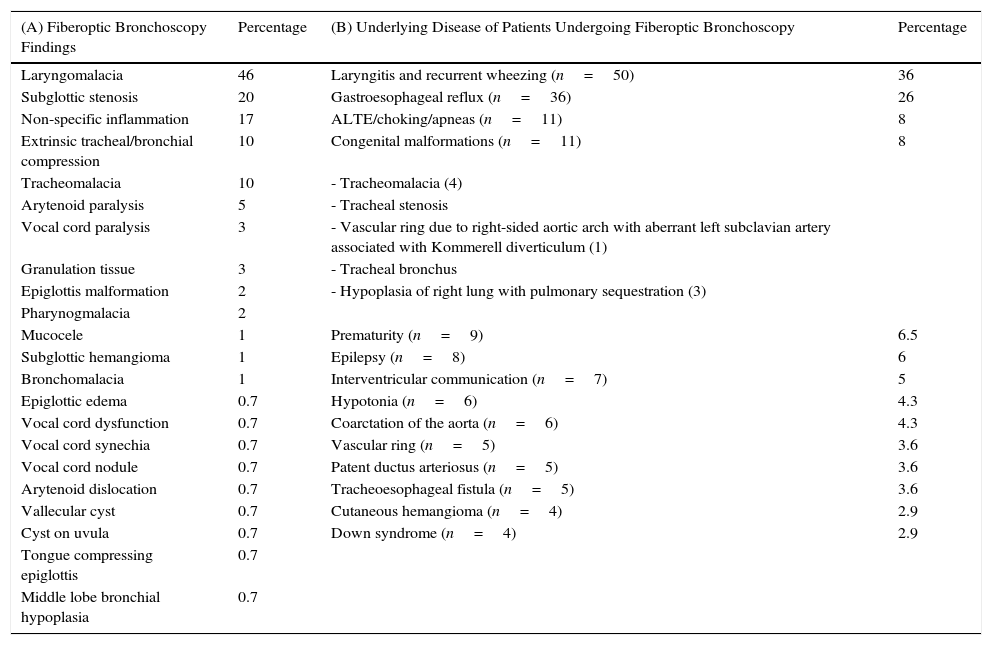
We performed 593 FBs, 138 (23%) for stridor; 63% in infants younger than 1 year, 14% in children between 1 and 2 years, 17% between 3 and 7 years and 6% in patients older than 8 years; 58% were male. The most predominant form of stridor was inspiratory (78%). Diagnostic yield was 95%, i.e., 130 patients obtained diagnostic or therapeutic benefit, while 18 of those patients required a repeat FB (12%). Most (81%) had underlying disease (Table 1). Fiberoptic bronchoscopy findings are summarized in Table 1. In 91% of cases we took a “wait and see” approach; 12 patients (9%) needed surgical treatment (6 required tracheoplasty, 2 aortopexy, 2 tracheostomy, 1 uvulectomy, and 1 laser ablation) and 2 patients with subglottic hemangioma received propanolol, with good response. A total of 8% of the patients had mild, self-limiting desaturations.

Table 1.

(A) Diseases Identified in the 138 Fiberoptic Bronchoscopies Performed for Stridor; in 43 Patients (31%), Multiple Findings Were Found on Fiberoptic Bronchoscopy. (B) Underlying Diseases in the 138 Patients Undergoing Fiberoptic Bronchoscopy.

(A) Fiberoptic Bronchoscopy Findings  Percentage  (B) Underlying Disease of Patients Undergoing Fiberoptic Bronchoscopy  Percentage 
Laryngomalacia  46  Laryngitis and recurrent wheezing (n=50)  36 
Subglottic stenosis  20  Gastroesophageal reflux (n=36)  26 
Non-specific inflammation  17  ALTE/choking/apneas (n=11) 
Extrinsic tracheal/bronchial compression  10  Congenital malformations (n=11) 
Tracheomalacia  10  - Tracheomalacia (4)   
Arytenoid paralysis  - Tracheal stenosis   
Vocal cord paralysis  - Vascular ring due to right-sided aortic arch with aberrant left subclavian artery associated with Kommerell diverticulum (1)   
Granulation tissue  - Tracheal bronchus   
Epiglottis malformation  - Hypoplasia of right lung with pulmonary sequestration (3)   
Pharynogmalacia     
Mucocele  Prematurity (n=9)  6.5 
Subglottic hemangioma  Epilepsy (n=8) 
Bronchomalacia  Interventricular communication (n=7) 
Epiglottic edema  0.7  Hypotonia (n=6)  4.3 
Vocal cord dysfunction  0.7  Coarctation of the aorta (n=6)  4.3 
Vocal cord synechia  0.7  Vascular ring (n=5)  3.6 
Vocal cord nodule  0.7  Patent ductus arteriosus (n=5)  3.6 
Arytenoid dislocation  0.7  Tracheoesophageal fistula (n=5)  3.6 
Vallecular cyst  0.7  Cutaneous hemangioma (n=4)  2.9 
Cyst on uvula  0.7  Down syndrome (n=4)  2.9 
Tongue compressing epiglottis  0.7     
Middle lobe bronchial hypoplasia  0.7     

ALTE, apparent life-threatening event.

Stridor is a symptom which requires an etiological diagnosis1–3 based on clinical history, physical examination, and direct evaluation of the airway1 with fiberoptic bronchoscopy.1–4 This is generally performed under spontaneous breathing, allowing an anatomical and functional examination of the airways.2,3 Respiratory pediatricians, ENT specialists, pediatric surgeons and/or anesthetists must work closely together to maximize the benefit of this procedure and ensure it is correctly performed. In our hospital, FB is performed in collaboration with anesthetists, who generally apply sevoflurane. In our opinion, the airway must be examined in its entirety.

Like other authors, our case series showed that laryngomalacia was the most common finding in infants with inspiratory stridor,1–3 and this condition generally resolved with a “wait and see” approach, since laryngomalacia is a result of immature laryngeal structures.2,3 The second most common disorder encountered was subglottic stenosis (15%). This percentage was high, but in our opinion there was some element of bias, as our hospital is the respiratory referral center for the south of Spain. Stridor can be caused by congenital or acquired diseases.2 The most common type is biphasic stridor associated with varying degrees of clinical airway obstruction.2 In our group of patients, all, except 1, developed symptoms after intubation. Subglottic stenoses with Cotton grades III and IV are treated by surgery, as was the case in 9% of our patients. The most common technique used was tracheoplasty with rib cartilage graft (5%).

In our opinion, FB is the technique of choice in the diagnosis of airway disorders,4,5 as it is safer and less aggressive than rigid bronchoscopy. We recommend that FB be performed in any child who presents with stridor, since this symptom is generally an indication of a banal disorder, but it can mask a life-threatening disease.1,3,5

References
[1]
A. Parente Hernández, M.A. García-Casillas, J.A. Matute, A. Cañizo, A. Laín, M. Fanjul, et al.
¿Es el estridor un síntoma banal en el lactante?.
An Pediatr (Barc), 66 (2007), pp. 559-565
[2]
A. Boudewyns, J. Claes, P. van de Heyning.
An approach to stridor in infants and children.
Eur J Pediatr, 169 (2010), pp. 135-141
[3]
E. Erdem, Y. Gokdemir, F. Unal, R. Ersu, B. Karadag, F. Karakoc.
Flexible bronchoscopy as a valuable tool in the evaluation of infants with stridor.
Eur Arch Otorhinolaryngol, 270 (2013), pp. 21-25
[4]
T. Nicolai.
The role of rigid and flexible bronchoscopy in children.
Paediatr Respir Rev, 12 (2011), pp. 190-195
[5]
I. Delgado Pecellín, J.P. González Valencia, M. Machuca Contreras, M. Pineda Mantecón.
Clínica, diagnóstico y tratamiento de las estenosis traqueales.
An Pediatr (Barc), 70 (2009), pp. 443-448

Please cite this article as: Plácido-Paias R, Delgado-Pecellín I, González-Valencia JP. Hallazgos fibrobroncoscópicos en pacientes estudiados por estridor en un hospital de tercer nivel. Arch Bronconeumol. 2016;52:335–336.

Copyright © 2015. SEPAR
Archivos de Bronconeumología
Article options
Tools

Are you a health professional able to prescribe or dispense drugs?