Lung cancer is the leading cause of cancer mortality worldwide. Cytotoxic and platinum-based chemotherapy are the standard first-line treatment for metastatic non-small cell lung cancer (NSCLC) without activating epidermal growth factor receptor (EGFR) and anaplastic lymphoma kinase (ALK) or cytoplasmic c-ros oncogene 1 translocation/re-arrangements.1 Recently, the development of immune checkpoint inhibitors (ICIs) against modulators including cytotoxic T-lymphocyte-associated protein 4 and programmed cell death protein 1 (PD-1) and its ligand (PD-L1) has created a major paradigm shift in the therapeutic management of metastatic NSCLC.1 The ICI, pembrolizumab, is a humanized monoclonal antibody against PD-1. The KEYNOTE-024 trial showed that pembrolizumab provided significantly longer progression-free survival and overall survival, with fewer adverse events, than cytotoxic and platinum-based chemotherapy.2 Compared with cytotoxic or targeted agents, however, ICIs can induce autoimmune-like toxicities known as immune-related adverse events (irAEs) by inducing the infiltration of immune cells in normal tissues3; in patients with advanced NSCLC, the common pembrolizumab-induced irAEs are thyroid dysfunction, pneumonitis, and skin reactions.3,4 Here, we describe a patient with lung adenocarcinoma with rhabdomyolysis and myositis triggered by pembrolizumab treatment, while pembrolizumab rapidly reduced lung tumor size.
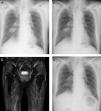
The patient is an 83-year-old non-smoking man who was diagnosed with prostate cancer at age 80 years and treated with brachytherapy for 2 years. At age 83 years, he presented with an abnormal chest X-ray (CXR) during routine follow up (Fig. 1A). Chest computed tomography (CT) revealed a 30.5mm×30.5mm mass in the right lower portion of the lung that crossed over into the adjacent middle lobe and thickened interlobular septa in the right middle and lower lobes. 18F-fluorodeoxyglucose (FDG) positron emission tomography/CT showed intense FDG uptake on both sides of the hilar and mediastinal lymph nodes. Magnetic resonance imaging (MRI) with contrast did not reveal any brain metastases. Fiberoptic bronchoscopy revealed lung adenocarcinoma. The patient was clinically staged as T2aN3M1a, stage IVA. Although no sensitizing EGFR mutations or ALK translocations were detected, the PD-L1 tumor proportion score was 95%, and the patient's Eastern Cooperative Oncology Group performance-status score was 0. Thus, we selected pembrolizumab at a dose of 200mg every 3 weeks as the first-line treatment. CXR showed a decrease in pulmonary mass size 5 days after initial pembrolizumab administration (Fig. 1B); however, the patient presented with myalgia of both proximal femurs and lower back pain 1 week after the second cycle of pembrolizumab. Two days before admission, he developed right ptosis. At admission, he was unable to walk without assistance, because of myalgia. Physical examination revealed symmetric weakness of the deltoid (medical research council [MRC] 4) and iliopsoas (MRC 4+) with hoarseness and mild dysphagia. He was administered bethanechol 25mg twice daily, silodosin 4mg twice daily, benidipine 4mg once daily, rosuvastatin 2.5mg once daily, and alfacalcidol 1μg once daily. Initial laboratory test results revealed elevated levels of creatine kinase (CK) 6417IU/L (62–287IU/L), CK-MB 176IU/L (0–25IU/L), and aldolase 74.7IU/L (2.1–6.1IU/L). The levels of thyroid-stimulating hormone and free thyroxine were within normal ranges. Test results for anti-aminoacyl tRNA synthase antibody, anti-acetylcholine receptor (AChR) antibody, and anti-muscle-specific kinase (MuSK) antibody were negative. At admission, rosuvastatin was discontinued. Short-tau inversion recovery MRI of the femur revealed diffuse increased signal in the gluteal and thigh muscles (Fig. 1C). Repetitive nerve stimulation test did not reveal gradual amplitude reduction (waning) and increment (waxing) of compound muscle action potentials. Edrophonium test results were negative. Muscle biopsy from the left musculus quadriceps femoris revealed lymphohistiocytic infiltration with muscle atrophy. The patient was then diagnosed with rhabdomyolysis with myositis, a suspected immune-related toxicity. We discontinued pembrolizumab and initiated systemic prednisone (40mg/day) soon after the muscle biopsy. Serum CK level was normalized within 3 weeks after prednisone administration. Myalgia and ptosis improved within 4 weeks after prednisone administration. CXR showed continuous decrease in pulmonary mass size 2 months after initial pembrolizumab administration (Fig. 1D). However, his performance-status score decreased from 0 to 2.
Previous trials reported that pembrolizumab-induced myositis occurred in 1–1.9% patients.2,4 The average onset of symptoms was 4.6 weeks after treatment initiation (range 1–7 weeks).5–7 Our patient presented with right ptosis in addition to myalgia and dysphagia 4 weeks after treatment initiation. Ptosis and diplopia are observed in the vast majority of myasthenia gravis (MG), whereas these symptoms are not typically observed in myositis.8 In this patient, anti-AChR antibody and anti-MuSK antibody test results were negative. Also, repetitive nerve stimulation tests did not reveal waning and waxing, and the edrophonium test result was negative. These results can make it difficult to diagnose MG. Vallet et al.5 and Haddox et al.6 reported that patients with advanced melanoma with pembrolizumab-induced myositis developed ptosis. The observations in these cases are similar to those in our case. ICIs, including pembrolizumab, can induce aberrant immune activation leading to undesired off-target inflammation and autoimmunity by blocking regulatory checkpoints9; therefore, irAE will not present with typical symptom of each disease as in our patient.
Pembrolizumab-induced rhabdomyolysis with myositis in our patient was administered systemic prednisolone. Vallet et al.5 and Haddox et al.6 used plasma exchange in addition to systemic corticosteroids. Zimmer et al. either used systemic corticosteroids or did not administer additional treatments.7 At present, there is no consensus regarding therapeutic options and treatment duration for pembrolizumab-induced myositis. Therefore, we must closely examine treatment in each case.
In several previous reports, irAEs, including skin reactions and thyroid dysfunction, were associated with a better therapy response.10–12 However, irAEs induce potentially long courses of corticosteroids and even anti-tumor necrosis factor therapy to mitigate effects.9 Furthermore, irAEs result in permanent discontinuation of treatment, long-term sequelae, and death.13 Our patient achieved good clinical response to pembrolizumab; however, pembrolizumab-induced irAE deteriorated performance-status. Therefore, it is critical to closely monitor patients treated with ICIs for early detection and appropriate management of irAE, which will not present with typical symptom of each disease as in our patient.


