We report the case of a 37-year-old man, non-intravenous drug user, who was admitted to the respiratory medicine department due to a 12-h history of rapid-onset dyspnea with no other associated symptoms.
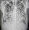
A chest X-ray was requested, showing multiple bilateral pulmonary nodules, most of which were cavitated (Fig. 1). Given the patient's history and the radiological findings, the initial diagnostic suspicion was tuberculosis, and additional tests and treatment were oriented towards an infectious process, but the patient did not improve.
The examination was completed with a computed tomography, which revealed innumerable pulmonary nodules, measuring between 1mm and 3.2cm, the vast majority of which were cavitated.
Multiple peribronchial lymphadenopathies were observed in both sides, along with a subcarinal lymph node mass, measuring 3.9cm, and a lymph node cluster in the right axilla.
The axillary mass was biopsied, and results revealed a high-grade undifferentiated sarcoma containing a high percentage of necrosis (approximately 75%).
The patient received chemotherapy with doxorubicin 25mg/m2/day, in combination with ifosfamide 3g/m2/day for 3 days. Respiratory progress was poor, and the patient died 4 weeks after admission.
Please cite this article as: Blanco MJ, Rezola F, Dueñas A. Múltiples nódulos pulmonares cavitados… ¿tuberculosis o neoplasia maligna? Arch Bronconeumol. 2017;53:691.


