
Ischemia–reperfusion (IR) lung injury has been investigated extensively on clinical and experimental models of cold ischemia. However, relatively few studies examine the detailed biochemical changes occurring during normothermic (warm) IR.
Animals and methodsSix large-white pigs underwent a lung autotransplant which entailed left pneumonectomy, ex situ cranial lobectomy, caudal lobe reimplantation and its reperfusion for 30min. Throughout the procedure, several parameters were measured in order to identify hemodynamic, gasometric and biochemical changes. Non-parametric statistical analyses were used to compare differences between periods.
ResultsAfter ischemia, a significant increase (p<.05) in lipid peroxidation metabolites, proinflammatory cytokines and chemokines (TNF-α, IL-1β and MCP-1), neutrophil activation, inducible nitric oxide synthase activity and protein kinase MAPK p38 levels were observed in lung tissue. However, constitutive nitric oxide synthase activity in lung tissue and carbon monoxide plasma levels decreased. The same held true throughout the reperfusion period, when an increase in the constitutive heme-oxygenase activity was also shown.
ConclusionsAn experimental model of normothermic lung IR injury is presented and detailed changes in hemodynamic, gasometric and biochemical parameters are shown. Both the model and the studied parameters may be clinically useful in future investigations testing new therapies to prevent normothermic IR induced lung injury.
El daño pulmonar agudo por isquemia reperfusión (IR) ha sido estudiado fundamentalmente en modelos experimentales y clínicos con IR fría. Son limitados los estudios que profundizan en las alteraciones bioquímicas durante la IR normotérmica (caliente). El objetivo del este trabajo es presentar un modelo de autotrasplante pulmonar en cerdo para el estudio de las fases más precoces del síndrome de IR normotérmica pulmonar.
Animales y métodosSeis cerdos de la raza Large-White fueron sometidos a neumonectomía izquierda, lobectomía craneal ex situ, reimplantación del lóbulo caudal y reperfusión del mismo durante 30min. Durante el procedimiento se analizaron diferentes parámetros para identificar cambios hemodinámicos, gasométricos y bioquímicos en el modelo. El estudio estadístico se realizó con pruebas no paramétricas.
ResultadosTras la isquemia, se observó en tejido pulmonar un aumento significativo (p<0,05) de metabolitos de peroxidación lipídica, de citoquinas y quemoquinas proinflamatorias (TNF-α, IL-1β y MCP-1), de actividad leucocitaria (mieloperoxidasa o MPO), de actividad óxido nítrico sintasa inducible y de la proteína quinasa MAPK p38, mientras que se observó un descenso de actividad tisular de las formas constitutivas de NOS y de monóxido de carbono sérico. Estas alteraciones se mantuvieron o acentuaron durante la reperfusión, donde se observó también una mayor actividad tisular hemo-oxigenasa constitutiva.
ConclusionesSe presenta un procedimiento experimental de IR normotérmica pulmonar describiendo en profundidad cambios hemodinámicos, gasométricos y bioquímicos. Tanto el modelo como los parámetros analizados podrían ser útiles en el estudio de nuevas terapias moduladoras del daño pulmonar agudo en situaciones clínicas de IR normotérmica.
Several clinical situations require subjecting lung tissue to more or less prolonged periods of ischemia, with the consequent risk of acute lung injury after reperfusion. Most of the studies on how to preserve the lung tissue from the effects of ischemia-reperfusion (IR) are done in experimental or clinical models of lung transplantation with cold ischemia.1 There are clinical situations, however, in which the progressive cooling of the lung is not possible, nor is it possible to perfuse it with a preservation solution before interrupting blood circulation. Among these situations are lung resections with angioplasty of the pulmonary artery2–4 and lung lobe transplants from living donors. Less frequent, almost anecdotal situations are the ex situ pulmonary resections of central tumors with reimplantation of the viable pulmonary lobe or lobes.5 In these cases, part of the lung tissue suffers a more or less prolonged period of warm (normothermic) ischemia, while reperfusion edema and the need for prolonged post-op ventilation are frequent.6
In this context, the study of lung injury due to IR and potential modulating therapies requires experimental models in which ischemia is initiated without cooling or previous lung preservation. An experimental model reproducing this situation is lung autotransplantation in animals. The objective of this present study is to present a model of lung autotransplantation in pigs in order to study the earliest phases of the IR syndrome and analyze the hemodynamic, gasometric and biochemical changes that take place during this period.
Animals and MethodsThe study was done with the approval of the Animal Experiment and Research Committee of the institution, following all times the European and Spanish guidelines for the manipulation and care of experimental animals.
In six large-white pigs, we carried out a procedure of orthotopic lung autotransplantation. Mean animal weight was 42.8kg. The intervention consisted of left pneumonectomy with cranial lobectomy ex situ, reimplantation of the caudal lobe and reperfusion for 30min. Mean procedure time was 289min (range: 232–325min) and the mean pulmonary ischemia time was 90min (range: 84–97min). At the beginning of the procedure and during the periods of ischemia and reperfusion, different parameters were analyzed to identify hemodynamic, blood gas and biochemical changes in the model.
Surgical ProcedureThe animals received no solid food for 18h before the procedure, with water available ad libitum. The premedication was done with intramuscular ketamine at 10mg/kg. During surgery, we placed a peripheral catheter, and previous 100% oxygenation was established as monitored with electrocardiogram (ECG) and pulse-oximetry. Anesthesia was induced with propofol (4mg/kg; Diprivan®, Fresenius K), fentanyl (3μg/kg; Fentanest®, Kern Pharma) and atracurium besilate (0.6mg/kg; Tracrium®, Glaxo Smith Kline) through a dorsal vein of the ear. Intubation was done with an orotracheal tube measuring 6–7mm in internal diameter. Respiratory assistance included a Dräger SA 1 ventilator. Ventilation was controlled by volume (tidal volume 8ml/kg, 12-15breaths/min, ratio between inspiration and expiration of 1:2) and it was adjusted during surgery in order to maintain in arterial blood between 35 and 40mmHg of carbonic anhydride; meanwhile, the inspired fraction of oxygen (FiO2) was maintained at 1 during the whole procedure. Surgical tracheotomy was performed and, after withdrawing the orotracheal tube, a 6mm ringed tube was introduced, which allowed easier selective intubation of the right bronchi during surgery. The anesthesia was maintained with propofol at continuous perfusion (8-10mg/kg/h), with fentanyl and atracurium in bolus, as needed. Intravenous perfusion was maintained with lactated Ringer's solution at 5-6ml/kg/h as well as a colloid substance, hydroxyethyl starch, as needed. During the intervention, the animal subjects were monitored with three-lead ECG, pulse-oximetry, capnography, invasive arterial pressure and central venous pressure, therefore the femoral vein and artery were catheterized. Through the femoral vein, we introduced a pulmonary artery catheter (thermodilution catheter 7.5F, Edwards, Irving, California, USA). In order to control diuresis, suprapubic cystostomy was carried out.
After these preliminary procedures, the animal was situated in right lateral decubitus and left thoracotomy was performed with resection of the fourth or fifth costal arch. For the pneumonectomy, we successively dissected the pulmonary artery, the pulmonary cranial vein, the pulmonary caudal vein and the left bronchus. Then, the left bronchus was cut and, with direct vision, the orotracheal tube was advanced towards the right bronchus, initiating the period of one-lung ventilation. The left pulmonary artery was occluded with a protected clamp near the fork of the main pulmonary artery and was cut distally, leaving a margin of 5-10mm for arterial anastomosis of the reimplantation. The pulmonary cranial vein was ligated near the auricle and cut. To complete the pneumonectomy, the pulmonary vein of the caudal lobe was clamped near the opening of the vein of the mediastinal lobe, cut 1 or 2mm from the clamp and sutured with continuous proline 6/0 suture. With this maneuver, we were able to keep enough length of the caudal lobe vein for venoatrial anastomosis of the reimplant. In order to prevent thrombosis of the pulmonary artery, which remained clamped during the bench surgery and the reimplant, it was heparinized with 300UI/kg in bolus at the moment of its occlusion.
This was followed by the bench surgery, and cranial lobectomy was performed. The left lung received anterograde and retrograde perfusion with University of Wisconsin solution at 10-15°C while manually ventilated (FiO2: 0.21) until a clear effluent was reached through the pulmonary artery and veins. We dissected the pedicle of the caudal lobe that was going to be reimplanted: the left pulmonary artery (after ligature and sectioning of the cranial branches), the caudal pulmonary vein (liberated from the pleural adherences until the segmental branches) and the left main bronchus (after cutting and suturing the cranial bronchus).
Finally, the caudal lobe was reimplanted by means of bronchial anastomosis with continuous suture of prolene 4/0, continuous arterial suture with prolene 5/0 and continuous venoatrial suture with prolene 6/0. Then, the ringed tube was withdrawn towards the trachea, allowing for the ventilation of the implant. Retrograde reperfusion was carried out first, unclamping the left auricle, followed by anterograde reperfusion, unclamping the pulmonary artery. The perfusion of the reimplanted lobe was maintained for 30min, after that time the animal was euthanized with terminal anesthesia and induction of cardioplegia with potassium chloride.
Parameters of the StudyWe registered: animal weight, ischemia times and total duration of the procedure and one-lung ventilation time during the intervention.
Measurement of Variables and Collection of SamplesAs a baseline study, hemodynamic and systemic arterial gasometry studies were carried out 30min after beginning of the thoracotomy, before initiating one-lung ventilation. The same hemodynamic and gasometric studies, together with lung biopsies and femoral venous vein blood extraction for the biochemical studies, were done at four other moments: pre-pneumonectomy (PreP) (before completing the pneumonectomy, with one-lung ventilation already initiated; pre-reperfusion (PreR) (before the reperfusion and ventilation of the reimplanted lobe); 10min post-reperfusion (10min after reperfusion of the reimplanted lobe); and 30min post-reperfusion (30min after reperfusion of the reimplanted lobe). Lung biopsies were taken in order to quantify the lung edema as well as to measure the biochemical parameters. The first two samples, PreP and PreR, were extracted from the cranial lobe of the left lung; the last two samples, Rep-10′ and Rep-30′, were extracted from the caudal lobe of the left lung. Each tissue sample was divided into two: one, to quantify the pulmonary edema, was frozen in polypropylene tubes at −40°C; the other was used for the biochemistry study and was immediately frozen with liquid nitrogen in cryotubes and stored at −80°C until later analysis. The femoral vein blood samples were centrifuged for 10min at 1000×g and the supernatant serum was frozen at −40°C until later analysis.
Hemodynamic StudiesThe heart rate was monitored by ECG. The arterial catheter was used to measure mean arterial pressure. The pulmonary artery catheter permitted the measurement and calculation of the following parameters: mean pulmonary artery pressure, pulmonary capillary pressure, central venous pressure, heart rate and systolic volume.
Blood Gas StudiesAt the moments mentioned, partial pressure of oxygen (PO2), carbon dioxide partial pressure (PCO2) and pH were measured in systemic arterial blood. In addition, 10min and 30min after reperfusion of the reimplanted lobe, a blood sample was taken by puncture of the pulmonary vein in order to study the gas exchange capacity of the graft, and in the sample PO2, PCO2 and pH were determined.
Biochemical Determinations in Plasma- •
Nitric oxide (NO): the serum concentration of NO was based on the Griess reaction.7
- •
Carbon monoxide (CO): to quantify the quantity of formed CO, hemoglobin was added to all the samples and the proportion of carboxyhemoglobin (CO-Hb) was determined spectrophotometrically in accordance with the methods by Omura and Sato.8
This is expressed by means of the wet/dry weight ratio. It is calculated by means of the formula wet weight-dry weight/wet weight, measuring the dry weight after incubating the samples for 24h at 60°C.
Study of the Oxidative Stress and Leukocyte Activation in the Lung TissueThe levels of lipid peroxidase (LPO) show the degree of degradation of the lipid membrane of the cells that occurs as a consequence of the oxidation. They are determined using a specific spectrophotometric kit (K-assay LPO-CC, Kamiya Biochemical Company, USA). Malondialdehyde (MDA) is a final compound of the lipid peroxidation and is a marker of cell damage. It is analyzed indirectly, quantifying the formation of thiobarbituric acids9 in the lung tissue. The myeloperoxidase (MPO) activity indicates the accumulation of polymorphonuclear neutrophils and is determined using the modified Bradley method.10
Inflammatory Mediators in the Lung Tissue: Expression of Cytokines (TNF-α, IL-1β), Nitric Oxide Synthases (Endothelial-NOSe, Neuronal-NOSn and Inducible-NOSi) and Heme Oxygenases (HO-1, HO-2)Western blot testing was done using specific antibodies: anti-TNF-α (Endogen), anti-IL-1β (Bio Genesis), anti-nitric oxide Synthase I, anti-nitric oxide Synthase II and anti-nitric oxide Synthase III (Chemicon International, Inc.), anti-heme-oxygenase I and anti-heme-oxygenase II (Chemicon International, Inc.). These are expressed as arbitrary units.
Monocyte Chemoattractant Protein-1 (MCP-1)This was measured in the lung tissue by ELISA using specific commercial kits (Biosource International).
Mitogen-Activated Protein Kinase (MAPK): MAPK p38, JNK and ERKThese were determined in lung tissue by ELISA using specific commercial kits (Oncogene).
Statistical AnalysisThe data are expressed as means and standard deviation. The statistical study was carried out using the SPSS 14.0 statistical package (SPSS Inc., Chicago, USA). The Wilcoxon test was used for paired samples to detect differences in the evolution of the variables among the different moments of the experimental procedure. The differences were considered statistically significant with a P value of <.05.
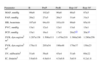
ResultsHemodynamicsThe majority of the hemodynamic constants measured remained stable throughout the procedure. However, a significant increase was registered in the pulmonary capillary pressure during the reperfusion of the reimplanted lobe (Table 1).
Hemodynamic Parameters.
| Parameter | B | PreP | PreR | Rep-10′ | Rep-30′ |
| MAP, mmHg | 99±6 | 102±5 | 90±6 | 88±5 | 87±5 |
| PAP, mmHg | 28±2 | 27±5 | 26±3 | 31±4 | 31±3 |
| HR, beats/min | 107±8 | 94±10 | 101±10 | 99±9 | 95±10 |
| PVC, mmHg | 13±1 | 12±1 | 12±1 | 13±1 | 13±1 |
| PCP, mmHg | 15±1 | 16±1 | 17±1 | 20±2a,b | 20±3a |
| SVR, dyn.seg/cm5m2 | 1.287±138 | 1.500±111 | 1.476±219 | 1.368±246 | 1.280±256 |
| PVR, dyn.seg/cm5m2 | 178±11 | 205±54 | 196±48 | 179±37 | 139±23 |
| SV, ml/beat/m2 | 51±6 | 56±8 | 45±4 | 51±6 | 69±22 |
| IC, l/min/m2 | 5.6±0.6 | 4.8±0.4 | 4.5±0.6 | 5±0.9 | 6.2±1.8 |
The data are expressed as mean±standard deviation.
B: baseline; HR: heart rate; PVR: pulmonary vascular resistance; SVR: systemic vascular resistance; SV: systolic volume; MAP: mean arterial pressure; PAP: mean pulmonary arterial pressure; PCP: pulmonary capillary pressure; PreP: pre-pneumonectomy; PreR: pre-reperfusion; PVC: central venous pressure; Rep-10′: 10min post-reperfusion; Rep-30′: 30min post-reperfusion.
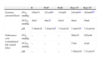
The results of the gasometric studies in systemic arterial blood and in pulmonary venous blood from the reimplanted lobe are shown in Table 2. A reduction was observed in arterial PO2 (p<.05) in the samples extracted before the pneumonectomy and 10min after reperfusion of the implanted lung. The pulmonary oxygenation capacity improved significantly after 30min of reperfusion.
Blood Gas Parameters.
| B | PreP | PreR | Rep-10′ | Rep-30′ | ||
| Systemic arterial blood | PO2, mmHg | 359±74 | 231±56a | 315±65 | 243±64a,c | 302±66b,d |
| PCO2, mmHg | 39±3 | 46±2a | 45±4 | 48±4 | 50±6 | |
| pH | 7.48±0.02 | 7.42±0.02a | 7.41±0.02 | 7.4±0.03a | 7.41±0.04 | |
| Pulmonary vein (reimplanted left caudal lobe) | PO2, mmHg | – | – | – | 260±55 | 295±46 |
| PCO2, mmHg | – | – | – | 37±6 | 42±8 | |
| pH | – | – | – | 7.56±0.05 | 7.51±0.07 | |
The data are expressed as mean±standard deviation.
B: baseline; PO2: partial pressure of oxygen; PCO2: carbon dioxide partial pressure; PreP: pre-pneumonectomy; PreR: pre-reperfusion; Rep-10′: 10min post-reperfusion; Rep-30′: 30min post-reperfusion.
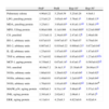
The determination of lipid peroxidation products (LPO and MDA) in the lung tissue showed a progressive increase of these in the lung tissue during the periods of ischemia and reperfusion (Table 3).
Biochemical Determinations.
| PreP | PreR | Rep-10′ | Rep-30′ | |
| Pulmonary edema | 4.96±0.22 | 5.25±0.59 | 5.25±0.28 | 4.9±0.1 |
| LPO, pmol/mg protein | 2.51±0.23 | 3.65±0.44a | 3.76±0.1a | 3.88±0.13a |
| MDA, pmol/mg protein | 3.25±0.1 | 3.95±0.03a | 4.91±0.11a,b | 5.38±0.1a,b |
| MPO, UI/mg protein | 0.06±0.006 | 0.1±0.006 | 0.19±0.005a | 0.23±0.008a |
| CO, pmol/ml | 2.513±0.11 | 2.34±0.05a | 2.47±0.12b | 2.46±0.08 |
| HO-1, arbitrary units | 0.67±0.03 | 0.647±0.07 | 0.76±0.07 | 0.79±0.04 |
| HO-2, arbitrary units | 0.667±0.2 | 0.819±0.22 | 0.908±0.21a | 0.872±0.04a |
| IL-1β, arbitrary units | 1.25±0.01 | 1.87±0.05a | 1.83±0.05a | 1.87±0.03a |
| TNF-α, arbitrary units | 0.71±0.02 | 0.89±0.03a | 0.95±0.03a,b | 1.27±0.03a,b,c |
| MCP-1, pg/mg protein | 0.339±0.2 | 0.67±0.14a | 0.43±0.1b | 0.565±0.02a,b |
| NO, nmol/ml | 40.04±7.2 | 24.4±3.3a | 25.38±6.0a | 26.09±4.5a |
| NOSe, arbitrary units | 1.66±0.01 | 1.29±0.02a | 1.41±0.04a | 1.34±0.02a |
| NOSn, arbitrary units | 1.48±0.01 | 0.97±0.04a | 1.2±0.05a,b | 1.24±0.04a |
| NOSi, arbitrary units | 1.76±0.01 | 1.81±0.05a | 1.96±0.02a,b | 2.06±0.04ab |
| MAPK p38, ng/mg protein | 4.985±0.3 | 9.51±1.6a | 5.86±0.9b | 5.57±1.01b |
| JNK, ng/mg protein | 2.381±0.3 | 2.62±0.2 | 2.08±0.1 | 1.97±0.2b |
| ERK, ng/mg protein | 4.11±0.4 | 4.23±0.3 | 4.823±0.6 | 4.02±0.4 |
The data are expressed as mean±standard deviation. Pulmonary edema is expressed by the ratio: wet/humid weight—dry weight/wet weight.
B: baseline; CO: carbon monoxide; ERK: extracellular signal-regulated kinase; HO-1, HO-2: heme-oxygenase 1, 2; IL-1β: interleukin-1β; JNK: kinase c-Jun N-terminal; LPO: lipid hydroperoxide; MCP-1: monocyte chemoattractant protein-1; MDA: malondialdehyde; MPO: myeloperoxidase; NO: nitric oxide; NOSe: endothelial nitric-oxide synthase; NOSi: inducible nitric oxide synthase; NOSn: neuronal nitric oxide synthase; MAPK p38: mitogen-activated protein kinase p38; PreP: pre-pneumonectomy; PreR: pre-reperfusion; Rep-10′: 10min post-reperfusion; Rep-30′: 30min post-reperfusion. TNF-α: tumor necrosis factor-α.
The activation of the MPO enzyme measured in the lung tissue was higher during the procedure, and significant increases were observed during the reperfusion of the implant (Table 3).
Heme-Oxygenase/Carbon Monoxide System (HO/CO)During the reperfusion, we observed an increase in the activity of the constitutive HO-2 isoform in the pulmonary tissue, while the blood levels of CO were lower than baseline in ischemia as well as in reperfusion (Table 3).
Proinflammatory CytokinesStudies with Western blot techniques showed a progressive increase of the TNF-α and IL-1β cytokines in the lung tissue during ischemia and reperfusion, as reflected in Table 3.
Monocyte Chemoattractant Protein-1The tissue concentration of MCP-1 in the lung biopsies during ischemia and reperfusion were higher than baseline levels, and this increase was notable after the period of ischemia (Table 3).
Metabolism of Nitric OxideDuring the procedure, an important decrease was detected in NO plasma levels. In the pulmonary tissue subjected to IR, we observed a decrease in the activity of the constitutive forms of the nitric oxide synthase, while we measured an increase in activity of the inducible form (Table 3).
Intracellular Signaling Pathway of the MAPKOf the different signaling routes, a significant increase in activity was observed in the MAPK p38, which presented a peak in concentration after the ischemia and maintained high levels during the reperfusion time (Table 3).
DiscussionStandard experimental models that study pulmonary IR are usually associated with the progressive cooling of the organ, perfusion with preservation solutions or extracorporeal circulation. Therefore, they are not ideal for the study of situations like the reconstruction of the pulmonary artery or lobe transplant from a living donor, in which the organ is suddenly subjected to normothermic or warm ischemia. It is necessary to develop experimental models in order to study therapies that reduce the risk of acute lung injury in these patients. The majority of the research on pulmonary warm ischemia is done in small rodents by means of vascular occlusion techniques11,12 and less frequently include surgical procedures similar to those carried out in humans. Pulmonary autotransplantation in large mammals largely meets this objective, as reported in dogs,13–17 sheep18 and pigs,19 but few of these studies thoroughly analyze the inflammatory mediators during normothermic IR.20
In this present study, we analyzed the hemodynamic, blood gas and biochemical changes during pulmonary autotransplantation procedures in pigs. Van Raemdonck et al. observed that the tolerance to warm ischemia in non-ventilated rabbit lungs was 1h.21 Yamazaki et al., in a canine model of pulmonary autotransplantation, set the limit of 120min as the maximum tolerable time of warm ischemia for the lung to be viable and for the animal to survive.22 The mean time of lung ischemia in our study, 90min, makes for a foreseeable development of a non-lethal lung injury with physiopathological manifestations that allow us to analyze the effect of potential treatments.
In experimental studies of normothermic IR, acute pulmonary injury involves increased pulmonary vascular resistance, pulmonary edema and deterioration of the gas exchange capacity. In the majority of these studies, the ischemia is maintained for 2–3h and these alterations are manifested after several hours of reperfusion.15,23,24 In our model, designed for the study of the early inflammatory response, the different hemodynamic variables were relatively stable throughout the procedure. We observed a significant increase in the pulmonary capillary pressure during the reperfusion and gasometric alterations after the start of the one-lung ventilation and 10min after initiating reperfusion. We consider that this clinical stability confers a greater reliability to the observed biochemical findings.
As for the biochemical mechanisms implicated in the IR injury of any organ, numerous studies signal the production of oxygen free radicals (OFR), the activation of polymorphonuclear leukocytes and the production of proinflammatory cytokines as important mediators in the inflammatory response.
During IR, OFR are generated, which cause cell lysis by lipid peroxidation of the free fatty acids of the membranes.25 The degree of lipid peroxidation, and indirectly the presence of OFR, can be measured by the presence of lipid peroxides (LPO) and tissue MDA. In the present model, a progressive increase of MDA and LPO is observed in the lung tissue, from the final phase of the ischemia until 30min after the reperfusion, which clearly indicates that the increase in tissue oxidative stress is a premature consequence of the IR. Similar results have been observed after normothermic ischemia, including in isolated lungs in rats26 and by using in situ occlusion of the pulmonary artery in dogs.27
In our experiment, we have observed a significant increase in tissue MPO during reperfusion, indicating recruitment and progressive activation of leucocytes in the lung tissue. Today it is known that the role of the neutrophils is important in the late phase of IR, but during the earlier phases the role of the macrophages and lymphocytes predominates.25 Eppinger et al., in a model of warm pulmonary IR in rats, demonstrated that the alterations observed during the first 30min of reperfusion were regardless of the leukocyte activation, whose effects began to become patent after 4h of reperfusion.28 Our data show that this leukocyte infiltration/activation is a phenomenon that begins a few minutes after the reperfusion of the organ.
One of the consequences of the tissue increase of OFR is the activation of the HO/CO system. HO is a microsomal enzyme that catalyzes the limiting step of the degradation of the heme group, converting it into biliverdin, CO and Fe+++. There is growing evidence of the protective role of this enzyme and its metabolites against IR injury.29 The constitutive isoform, HO-2, is expressed in baseline conditions in numerous tissues, while the inducible isoform, HO-1, also called heat shock protein 32, rises in situations of stress such as IR. On the other hand, CO presents as a potent anti-inflammatory and anti-apoptotic molecule, and its effect seems to be mediated by the activation of the mitogen-activated protein kinase (MAPK) pathway.30 Along the lines of these studies, in our study we have observed an increase of tissue HO activity during reperfusion, which indirectly implies a local increase in CO, while the decrease in CO levels in blood could be due to its peripheral consumption upon reacting with OFR and NO. Some authors have described similar results in rats during hepatic31 and pulmonary30 warm IR.
In addition to the activation of the HO/CO system, it has been reported that the OFR stimulate the synthesis and release of different cytokines and chemokines by the tissue macrophages. This activation of the macrophages has been related with the early phase of the inflammatory response to the IR.25,32 IL-1β and TNF-α are proinflammatory cytokines whose implication in the first stages of the inflammatory response to IR is well-known, but recently a crucial role in the process has been attributed to MCP-1, a chemokine that regulates the migration and activation of monocytes and macrophages, although its protective or deleterious function has still not been well defined.32,33 In our experiment, these three molecules are significantly higher in the lung tissue during ischemia, maintaining high levels after 10 and 30min of reperfusion. The progressive increase in TNF-α stands out, which would back the results of other authors which indicate this cytokine as a crucial inflammatory mediator in the initial phase as well as the late phase of IR.34 Our data could be related with a study by Krishnadasan et al., performed in mice, in which an increase in IL-1β and TNF-α is observed in the lung tissue 60min after reperfusion after a period of 90min of warm ischemia.11 Nevertheless, in the same experimental model, MCP-1 is only detected after 4h of pulmonary reperfusion,35 a discrepancy that could be due to the differences in the experimental model or to the sensitivity of the measuring technique.
The alteration of the metabolism of NO has been widely studied in different IR models, but with frequently contradicting conclusions due to the polyvalent effect of NO.25 The NO produced by the constitutive isoforms of the nitric oxide synthase enzyme, NOSe and NOSn, would be responsible for the beneficial physiological effects such as the control of the vascular tone or the aggregation of neutrophils and platelets, while NO synthesized by the inducible isoform, NOSi, would be implicated in physiopathological processes such as IR syndrome.36 We have observed in our experiment a marked decrease in blood NO and less activity of NOSe and NOSn in the lung tissue, both during ischemia as well as during reperfusion. However, there was a significant increase in the NOSi activity. To what extent these changes and their possible therapeutic modification either positively or negatively affect IR injury has still not been clarified and will have to be the object of future studies.
In order to more thoroughly analyze the biomolecular profile of the inflammatory response in this model, we have studied the activation of the MAPK. Recent studies implicate the MAPK as intracellular mediators of the inflammatory response to IR, therefore the final components of this signaling pathway are transferred to the nucleus and activate transcription factors and the expression of certain genes. The MAPK include different families of enzymes and routes of activation, and those that stand out are MAPK p38, JNK and ERK, but the specific role of each is still debated.37 The first two are activated by proinflammatory cytokines like IL-1β and TNF-α, and also OFR.38 It has also been observed that MCP-1 is capable of activating the three signaling routes.39 In addition, in vitro studies (with pulmonary endothelial cells) and in vivo ones (warm pulmonary IR in rats) show that CO has an anti-apoptotic effect during IR and that this effect is mediated by MAPK p38.30 The significant increase in MAPK p38 activity that we detect in the ischemic lung suggests the activation of this intracellular signaling pathway in response to different mediators, such as IL-1β, TNF-α, MCP-1 or CO that are also found to be high at the end of the ischemia period. Likewise, the generation of OFR, evident by the lipid peroxidation products, could be involved in this activation of the MAPK pathway. Future research should clarify to what degree this pathway amplifies and perpetuates the inflammatory response or, on the contrary, plays a protective role against IR.
In summary, we present an experimental procedure of swine lung autotransplantation as a model for studying pulmonary IR syndrome in large mammals, and consequently in humans. We have reported in detail the hemodynamic, blood gas and biochemical changes that may serve as control parameters in the study of new modulating therapies for acute lung injury due to IR. The model, in addition to presenting a great similarity with surgical procedures such as lung angioplasty or lobe transplantation from a living donor, could serve as a stepping stone to the development of lung autotransplantation techniques that, in the future, could offer real hope for surgical treatment in patients with lung cancer and low respiratory functional reserve. The association with cutting-edge procedures, such as ex vivo lung perfusion40 could widen to new extremes the field of investigation of this model.
FundingThis study has been supported by funding from the Health-Care Research Fund (Fondo de Investigación Sanitaria - FIS PI070840 and FIS PI070481) and from the Spanish Society of Pulmonology and Thoracic Surgery (SEPAR 2006/121).
Conflict of InterestThe authors declare no conflicts of interest.
Please cite this article as: Simón Adiego C, et al. Procedimiento de autotrasplante pulmonar en el cerdo como modelo experimental para el estudio del síndrome de isquemia-reperfusión. Arch Bronconeumol. 2011;47:283-9.


