array:22 [
  "pii" => "S0300289624003739"
  "issn" => "03002896"
  "doi" => "10.1016/j.arbres.2024.09.013"
  "estado" => "S200"
  "fechaPublicacion" => "2024-10-21"
  "aid" => "3662"
  "copyright" => "SEPAR"
  "copyrightAnyo" => "2024"
  "documento" => "simple-article"
  "crossmark" => 0
  "subdocumento" => "crp"
  "abierto" => array:3 [
    "ES" => false
    "ES2" => false
    "LATM" => false
  ]
  "gratuito" => false
  "lecturas" => array:1 [
    "total" => 0
  ]
  "itemSiguiente" => array:17 [
    "pii" => "S0300289624003594"
    "issn" => "03002896"
    "doi" => "10.1016/j.arbres.2024.09.001"
    "estado" => "S200"
    "fechaPublicacion" => "2024-10-21"
    "aid" => "3648"
    "copyright" => "SEPAR"
    "documento" => "article"
    "crossmark" => 0
    "subdocumento" => "rev"
    "abierto" => array:3 [
      "ES" => false
      "ES2" => false
      "LATM" => false
    ]
    "gratuito" => false
    "lecturas" => array:1 [
      "total" => 0
    ]
    "en" => array:11 [
      "idiomaDefecto" => true
      "cabecera" => "<span class="elsevierStyleTextfn">Review Article</span>"
      "titulo" => "The In-between&#58; Time to Talk About Bronchiectasis in Adolescents and Their Transition to Adult Care"
      "tienePdf" => "en"
      "tieneTextoCompleto" => "en"
      "tieneResumen" => "en"
      "contieneResumen" => array:1 [
        "en" => true
      ]
      "contieneTextoCompleto" => array:1 [
        "en" => true
      ]
      "contienePdf" => array:1 [
        "en" => true
      ]
      "resumenGrafico" => array:2 [
        "original" => 0
        "multimedia" => array:7 [
          "identificador" => "fig0005"
          "etiqueta" => "Fig&#46; 1"
          "tipo" => "MULTIMEDIAFIGURA"
          "mostrarFloat" => true
          "mostrarDisplay" => false
          "figura" => array:1 [
            0 => array:4 [
              "imagen" => "gr1.jpeg"
              "Alto" => 836
              "Ancho" => 1480
              "Tamanyo" => 147561
            ]
          ]
          "descripcion" => array:1 [
            "en" => "<p id="spar0020" class="elsevierStyleSimplePara elsevierViewall">Adolescence&#58; knowledge gaps between paediatric and adult bronchiectasis&#46; ACT&#58; airways clearance techniques&#59; LFT&#58; lung function test&#59; BAR&#58; broncho-arterial ratio&#59; PCD&#58; primary ciliary dyskinesia&#59; PID&#58; primary immunodeficiencies&#59; COPD&#58; chronic obstructive pulmonary disease&#46;</p>"
          ]
        ]
      ]
      "autores" => array:1 [
        0 => array:2 [
          "autoresLista" => "Letizia Traversi, Laura Garriga-Grimau, Antonio Moreno-Gald&#243;, Eva Polverino"
          "autores" => array:4 [
            0 => array:2 [
              "nombre" => "Letizia"
              "apellidos" => "Traversi"
            ]
            1 => array:2 [
              "nombre" => "Laura"
              "apellidos" => "Garriga-Grimau"
            ]
            2 => array:2 [
              "nombre" => "Antonio"
              "apellidos" => "Moreno-Gald&#243;"
            ]
            3 => array:2 [
              "nombre" => "Eva"
              "apellidos" => "Polverino"
            ]
          ]
        ]
      ]
    ]
    "idiomaDefecto" => "en"
    "EPUB" => "https://multimedia.elsevier.es/PublicationsMultimediaV1/item/epub/S0300289624003594?idApp=UINPBA00003Z"
    "url" => "/03002896/unassign/S0300289624003594/v1_202410210414/en/main.assets"
  ]
  "itemAnterior" => array:16 [
    "pii" => "S0300289612000968"
    "issn" => "03002896"
    "doi" => "10.1016/j.arbres.2012.02.017"
    "estado" => "S200"
    "fechaPublicacion" => "2013-05-08"
    "aid" => "621"
    "documento" => "simple-article"
    "crossmark" => 0
    "subdocumento" => "ret"
    "abierto" => array:3 [
      "ES" => false
      "ES2" => false
      "LATM" => false
    ]
    "gratuito" => false
    "lecturas" => array:2 [
      "total" => 348
      "formatos" => array:2 [
        "HTML" => 82
        "PDF" => 266
      ]
    ]
    "en" => array:8 [
      "idiomaDefecto" => true
      "titulo" => "WITHDRAWN&#58; Respiratory Muscle Assessment in Predicting Extubation Outcome in Patients With Stroke"
      "tienePdf" => "en"
      "tieneTextoCompleto" => 0
      "tieneResumen" => "en"
      "contieneResumen" => array:1 [
        "en" => true
      ]
      "contienePdf" => array:1 [
        "en" => true
      ]
      "autores" => array:1 [
        0 => array:2 [
          "autoresLista" => "Antonio A&#46;M&#46; Castro, Felipe Cortopassi, Russell Sabbag, Luis Torre-Bouscoulet, Claudia K&#252;mpel, Elias Ferreira Porto"
          "autores" => array:6 [
            0 => array:2 [
              "nombre" => "Antonio A&#46;M&#46;"
              "apellidos" => "Castro"
            ]
            1 => array:2 [
              "nombre" => "Felipe"
              "apellidos" => "Cortopassi"
            ]
            2 => array:2 [
              "nombre" => "Russell"
              "apellidos" => "Sabbag"
            ]
            3 => array:2 [
              "nombre" => "Luis"
              "apellidos" => "Torre-Bouscoulet"
            ]
            4 => array:2 [
              "nombre" => "Claudia"
              "apellidos" => "K&#252;mpel"
            ]
            5 => array:2 [
              "nombre" => "Elias Ferreira"
              "apellidos" => "Porto"
            ]
          ]
        ]
      ]
    ]
    "idiomaDefecto" => "en"
    "EPUB" => "https://multimedia.elsevier.es/PublicationsMultimediaV1/item/epub/S0300289612000968?idApp=UINPBA00003Z"
    "url" => "/03002896/unassign/S0300289612000968/v2_201305131254/en/main.assets"
  ]
  "en" => array:13 [
    "idiomaDefecto" => true
    "cabecera" => "<span class="elsevierStyleTextfn">Clinical Letter</span>"
    "titulo" => "Chest Wall CT-Fistulography for Surgical Planning in a Patient With Chronic Granulomatous Disease"
    "tieneTextoCompleto" => true
    "saludo" => "To the Director&#44;"
    "autores" => array:1 [
      0 => array:4 [
        "autoresLista" => "Abel Gonz&#225;lez-Huete, Luis Gorospe-Saras&#250;a, Gemma Mar&#237;a Mu&#241;oz-Molina"
        "autores" => array:3 [
          0 => array:4 [
            "nombre" => "Abel"
            "apellidos" => "Gonz&#225;lez-Huete"
            "email" => array:1 [
              0 => "abelgonzalezhuete@gmail.com"
            ]
            "referencia" => array:2 [
              0 => array:2 [
                "etiqueta" => "<span class="elsevierStyleSup">a</span>"
                "identificador" => "aff0005"
              ]
              1 => array:2 [
                "etiqueta" => "<span class="elsevierStyleSup">&#42;</span>"
                "identificador" => "cor0005"
              ]
            ]
          ]
          1 => array:3 [
            "nombre" => "Luis"
            "apellidos" => "Gorospe-Saras&#250;a"
            "referencia" => array:1 [
              0 => array:2 [
                "etiqueta" => "<span class="elsevierStyleSup">a</span>"
                "identificador" => "aff0005"
              ]
            ]
          ]
          2 => array:3 [
            "nombre" => "Gemma Mar&#237;a"
            "apellidos" => "Mu&#241;oz-Molina"
            "referencia" => array:1 [
              0 => array:2 [
                "etiqueta" => "<span class="elsevierStyleSup">b</span>"
                "identificador" => "aff0010"
              ]
            ]
          ]
        ]
        "afiliaciones" => array:2 [
          0 => array:3 [
            "entidad" => "Department of Radiology&#44; Hospital Universitario Ram&#243;n y Cajal&#44; Madrid&#44; Spain"
            "etiqueta" => "a"
            "identificador" => "aff0005"
          ]
          1 => array:3 [
            "entidad" => "Department of Thoracic Surgery&#44; Hospital Universitario Ram&#243;n y Cajal&#44; Madrid&#44; Spain"
            "etiqueta" => "b"
            "identificador" => "aff0010"
          ]
        ]
        "correspondencia" => array:1 [
          0 => array:3 [
            "identificador" => "cor0005"
            "etiqueta" => "&#8270;"
            "correspondencia" => "Corresponding author&#46;"
          ]
        ]
      ]
    ]
    "resumenGrafico" => array:2 [
      "original" => 0
      "multimedia" => array:7 [
        "identificador" => "fig0005"
        "etiqueta" => "Fig&#46; 1"
        "tipo" => "MULTIMEDIAFIGURA"
        "mostrarFloat" => true
        "mostrarDisplay" => false
        "figura" => array:1 [
          0 => array:4 [
            "imagen" => "gr1.jpeg"
            "Alto" => 1020
            "Ancho" => 1821
            "Tamanyo" => 245424
          ]
        ]
        "descripcion" => array:1 [
          "en" => "<p id="spar0005" class="elsevierStyleSimplePara elsevierViewall">&#40;A&#41; 3D CT reconstruction image of the patient&#39;s body surface&#46; Two cutaneous openings are clearly visible in the anterior thoracic wall &#40;arrows&#41;&#46; &#40;B&#44; C&#41; Axial contrast-enhanced thoracic CT images &#40;mediastinal window&#41; at two different levels show inflammatory changes in the anterior chest wall&#58; a collection in the right anterior pectoral muscles &#40;red arrow&#41;&#44; a small extrapleural collection adjacent to the right internal mammary vessels &#40;white arrow&#41;&#44; and several linear fistulous tracts extending toward the skin surface &#40;arrowheads&#41;&#46; &#40;D&#8211;F&#41; Thoracic CT reconstruction images &#40;D&#44; coronal maximum intensity projection &#91;MIP&#93;&#59; E&#44; sagittal MIP&#59; F&#44; sagittal 3D&#41; after the administration of diluted iodinated contrast through two skin fistulous openings on the anterior chest wall demonstrate communication between the two openings and the absence of deep soft tissue extension &#40;arrows&#41;&#46; &#40;G&#41; Post-surgical image of the anterior chest wall after the resection of the fistulous tract and the utilization of a vascularized muscle flap&#46;</p>"
        ]
      ]
    ]
    "textoCompleto" => "<span class="elsevierStyleSections"><p id="par0005" class="elsevierStylePara elsevierViewall">Chronic granulomatous disease &#40;CGD&#41; is a rare immunodeficiency caused by a defect in the nicotinamide adenine dinucleotide phosphate &#40;NADPH&#41; oxidase enzyme complex that predisposes to recurrent bacterial and fungal infections from an early age&#46;<a class="elsevierStyleCrossRefs" href="#bib0025"><span class="elsevierStyleSup">1&#44;2</span></a> Most infections are caused by bacteria &#40;<span class="elsevierStyleItalic">Staphylococcus&#44; Klebsiella&#44; Serratia</span>&#44; etc&#46;&#41;&#44; but fungal and mycobacterial infections may also occur&#46; The most affected organs are the lungs&#44; skin&#44; lymph nodes&#44; and the musculoskeletal system&#46;<a class="elsevierStyleCrossRef" href="#bib0025"><span class="elsevierStyleSup">1</span></a></p><p id="par0010" class="elsevierStylePara elsevierViewall">We present the case of a 31-year-old male with X-linked CGD&#44; on chronic antibiotic treatment&#44; with a long-standing medical history of infections &#40;pulmonary and soft-tissue&#41; and recurrent thoracic and abdominal fistulas&#44; who presented complaining of inflammation of the chest wall&#46; The patient had undergone multiple surgical interventions in the past&#44; including a right middle lobectomy for pulmonary aspergillosis and surgical debridement for soft-tissue infections&#46; On physical examination&#44; two small fistulous orifices through which fluid material was draining &#40;<a class="elsevierStyleCrossRef" href="#fig0005">Fig&#46; 1</a>A&#41; were found in the anterior chest wall&#44; with doubts about the extent in depth of the inflammatory changes&#46; A contrast-enhanced thoracic computed tomography &#40;CT&#41; confirmed the existence of inflammatory changes in the anterior chest wall &#40;<a class="elsevierStyleCrossRef" href="#fig0005">Fig&#46; 1</a>B&#44; C&#41; but ruled out the presence of pulmonary consolidations or pleural fluid collections&#46;</p><elsevierMultimedia ident="fig0005"></elsevierMultimedia><p id="par0015" class="elsevierStylePara elsevierViewall">The decision was made to perform a CT-fistulography using iso-osmolar iodinated contrast &#40;iodixanol 320<span class="elsevierStyleHsp" style=""></span>mg&#47;mL&#44; diluted to 25&#37; with saline&#41; through the two cutaneous fistulous openings&#44; due to its excellent anatomical and contrast resolution&#46; CT-fistulography revealed a subcutaneous communication between the two orifices&#44; ruled out involvement of deeper tissues &#40;<a class="elsevierStyleCrossRef" href="#fig0005">Fig&#46; 1</a>D&#8211;F&#41; and aided the thoracic and plastic surgery teams in planning the resection of the fistulous tract and the reconstruction of the anterior chest wall &#40;<a class="elsevierStyleCrossRef" href="#fig0005">Fig&#46; 1</a>G&#41;&#46;</p><p id="par0020" class="elsevierStylePara elsevierViewall">CGD is a genetically predisposed disease &#40;X-linked or autosomal recessive&#41; caused by a defect in the NADPH oxidase enzyme complex&#44; affecting phagocytosis and resulting in the inability to eliminate catalase-positive pathogens such as <span class="elsevierStyleItalic">Staphylococcus</span>&#44; <span class="elsevierStyleItalic">Mycobacteria</span>&#44; <span class="elsevierStyleItalic">Serratia</span>&#44; <span class="elsevierStyleItalic">Klebsiella</span>&#44; <span class="elsevierStyleItalic">Pseudomonas</span>&#44; and fungi&#46; Consequently&#44; recurrent infections lead to the formation of inflammatory granulomas&#46; Pulmonary involvement is the most frequent manifestation&#44; including pneumonia or lung abscesses that can extend to the chest wall&#46; It can also manifest as lymphadenopathy&#44; liver abscesses&#44; soft tissue infections&#44; osteomyelitis&#44; brain abscesses&#44; gastrointestinal infections&#44; and organomegaly&#46;<a class="elsevierStyleCrossRefs" href="#bib0025"><span class="elsevierStyleSup">1&#44;2</span></a> Occasionally&#44; thoracic surgery is necessary to treat aggressive pulmonary infections&#44; which are associated with greater complications and poorer prognosis&#46;<a class="elsevierStyleCrossRef" href="#bib0035"><span class="elsevierStyleSup">3</span></a></p><p id="par0025" class="elsevierStylePara elsevierViewall">CGD patients require prolonged antibiotic treatments and prophylactic antimicrobial therapy&#46; Hematopoietic stem cell transplantation is the curative treatment for this disease&#44; which has improved the prognosis of these patients&#46;<a class="elsevierStyleCrossRefs" href="#bib0025"><span class="elsevierStyleSup">1&#44;2</span></a></p><p id="par0030" class="elsevierStylePara elsevierViewall">CT- fistulography is a technique used for the diagnosis of abdominal wall and perianal fistulas in some patients and&#44; to our knowledge&#44; has not been used in the chest wall&#46;<a class="elsevierStyleCrossRef" href="#bib0040"><span class="elsevierStyleSup">4</span></a> In CT-fistulography&#44; acquisition of isovolumetric data sets after the instillation of contrast into the fistula delineates the tract and its components&#44; allows the acquisition of images with great anatomical detail in different spatial planes&#44; and can help surgeons plan the surgical intervention&#46;</p><p id="par0035" class="elsevierStylePara elsevierViewall">We believe that this case is interesting because CT-fistulography ruled out communication between the cutaneous-subcutaneous inflammatory process and the extrapleural fat&#44; pleura&#44; and lung parenchyma&#46; This facilitated appropriate therapeutic planning by demonstrating a communication between the two skin fistulous openings&#46; Meticulous surgical planning is essential in these patients&#44; given their higher risk of complications&#46; We have found no previous descriptions in the scientific literature of CT-fistulography of the chest wall&#46;</p><span id="sec0005" class="elsevierStyleSection elsevierViewall"><span class="elsevierStyleSectionTitle" id="sect0005">Artificial Intelligence Involvement</span><p id="par0040" class="elsevierStylePara elsevierViewall">None of the material has been partially or totally produced with the help of any artificial intelligence tool&#46;</p></span><span id="sec0010" class="elsevierStyleSection elsevierViewall"><span class="elsevierStyleSectionTitle" id="sect0010">Funding of the Study</span><p id="par0045" class="elsevierStylePara elsevierViewall">This study did not receive any specific grant from funding agencies in the public&#44; commercial&#44; or not-for-profit sectors&#46;</p></span><span id="sec0015" class="elsevierStyleSection elsevierViewall"><span class="elsevierStyleSectionTitle" id="sect0015">Conflicts of Interest</span><p id="par0050" class="elsevierStylePara elsevierViewall">The authors declare not to have any conflicts of interest that may be considered to influence directly or indirectly the content of the manuscript&#46;</p></span></span>"
    "textoCompletoSecciones" => array:1 [
      "secciones" => array:4 [
        0 => array:2 [
          "identificador" => "sec0005"
          "titulo" => "Artificial Intelligence Involvement"
        ]
        1 => array:2 [
          "identificador" => "sec0010"
          "titulo" => "Funding of the Study"
        ]
        2 => array:2 [
          "identificador" => "sec0015"
          "titulo" => "Conflicts of Interest"
        ]
        3 => array:1 [
          "titulo" => "References"
        ]
      ]
    ]
    "pdfFichero" => "main.pdf"
    "tienePdf" => true
    "multimedia" => array:1 [
      0 => array:7 [
        "identificador" => "fig0005"
        "etiqueta" => "Fig&#46; 1"
        "tipo" => "MULTIMEDIAFIGURA"
        "mostrarFloat" => true
        "mostrarDisplay" => false
        "figura" => array:1 [
          0 => array:4 [
            "imagen" => "gr1.jpeg"
            "Alto" => 1020
            "Ancho" => 1821
            "Tamanyo" => 245424
          ]
        ]
        "descripcion" => array:1 [
          "en" => "<p id="spar0005" class="elsevierStyleSimplePara elsevierViewall">&#40;A&#41; 3D CT reconstruction image of the patient&#39;s body surface&#46; Two cutaneous openings are clearly visible in the anterior thoracic wall &#40;arrows&#41;&#46; &#40;B&#44; C&#41; Axial contrast-enhanced thoracic CT images &#40;mediastinal window&#41; at two different levels show inflammatory changes in the anterior chest wall&#58; a collection in the right anterior pectoral muscles &#40;red arrow&#41;&#44; a small extrapleural collection adjacent to the right internal mammary vessels &#40;white arrow&#41;&#44; and several linear fistulous tracts extending toward the skin surface &#40;arrowheads&#41;&#46; &#40;D&#8211;F&#41; Thoracic CT reconstruction images &#40;D&#44; coronal maximum intensity projection &#91;MIP&#93;&#59; E&#44; sagittal MIP&#59; F&#44; sagittal 3D&#41; after the administration of diluted iodinated contrast through two skin fistulous openings on the anterior chest wall demonstrate communication between the two openings and the absence of deep soft tissue extension &#40;arrows&#41;&#46; &#40;G&#41; Post-surgical image of the anterior chest wall after the resection of the fistulous tract and the utilization of a vascularized muscle flap&#46;</p>"
        ]
      ]
    ]
    "bibliografia" => array:2 [
      "titulo" => "References"
      "seccion" => array:1 [
        0 => array:2 [
          "identificador" => "bibs0015"
          "bibliografiaReferencia" => array:4 [
            0 => array:3 [
              "identificador" => "bib0025"
              "etiqueta" => "1"
              "referencia" => array:1 [
                0 => array:2 [
                  "contribucion" => array:1 [
                    0 => array:2 [
                      "titulo" => "Spectrum of imaging findings of chronic granulomatous disease&#58; a single center experience"
                      "autores" => array:1 [
                        0 => array:2 [
                          "etal" => false
                          "autores" => array:5 [
                            0 => "M&#46; Lee"
                            1 => "M&#46;S&#46; Lee"
                            2 => "J&#46;S&#46; Lee"
                            3 => "S&#46;Y&#46; Ko"
                            4 => "S&#46;Y&#46; Jeong"
                          ]
                        ]
                      ]
                    ]
                  ]
                  "host" => array:1 [
                    0 => array:2 [
                      "doi" => "10.5152/dir.2017.17264"
                      "Revista" => array:6 [
                        "tituloSerie" => "Diagn Interv Radiol"
                        "fecha" => "2017"
                        "volumen" => "23"
                        "paginaInicial" => "472"
                        "paginaFinal" => "477"
                        "link" => array:1 [
                          0 => array:2 [
                            "url" => "https://www.ncbi.nlm.nih.gov/pubmed/29097349"
                            "web" => "Medline"
                          ]
                        ]
                      ]
                    ]
                  ]
                ]
              ]
            ]
            1 => array:3 [
              "identificador" => "bib0030"
              "etiqueta" => "2"
              "referencia" => array:1 [
                0 => array:2 [
                  "contribucion" => array:1 [
                    0 => array:2 [
                      "titulo" => "Chronic granulomatous disease&#58; a comprehensive review"
                      "autores" => array:1 [
                        0 => array:2 [
                          "etal" => false
                          "autores" => array:3 [
                            0 => "H&#46;-H&#46; Yu"
                            1 => "Y&#46;-H&#46; Yang"
                            2 => "B&#46;-L&#46; Chiang"
                          ]
                        ]
                      ]
                    ]
                  ]
                  "host" => array:1 [
                    0 => array:2 [
                      "doi" => "10.1007/s12016-020-08800-x"
                      "Revista" => array:6 [
                        "tituloSerie" => "Clin Rev Allergy Immunol"
                        "fecha" => "2021"
                        "volumen" => "61"
                        "paginaInicial" => "101"
                        "paginaFinal" => "113"
                        "link" => array:1 [
                          0 => array:2 [
                            "url" => "https://www.ncbi.nlm.nih.gov/pubmed/32524254"
                            "web" => "Medline"
                          ]
                        ]
                      ]
                    ]
                  ]
                ]
              ]
            ]
            2 => array:3 [
              "identificador" => "bib0035"
              "etiqueta" => "3"
              "referencia" => array:1 [
                0 => array:2 [
                  "contribucion" => array:1 [
                    0 => array:2 [
                      "titulo" => "Thoracic surgery in chronic granulomatous disease&#58; a 25-year single-institution experience"
                      "autores" => array:1 [
                        0 => array:2 [
                          "etal" => true
                          "autores" => array:6 [
                            0 => "P&#46;L&#46; Feingold"
                            1 => "H&#46;S&#46; Quadri"
                            2 => "S&#46;M&#46; Steinberg"
                            3 => "H&#46;L&#46; Malech"
                            4 => "J&#46;I&#46; Gallin"
                            5 => "C&#46;S&#46; Zerbe"
                          ]
                        ]
                      ]
                    ]
                  ]
                  "host" => array:1 [
                    0 => array:2 [
                      "doi" => "10.1007/s10875-016-0319-9"
                      "Revista" => array:6 [
                        "tituloSerie" => "J Clin Immunol"
                        "fecha" => "2016"
                        "volumen" => "36"
                        "paginaInicial" => "677"
                        "paginaFinal" => "683"
                        "link" => array:1 [
                          0 => array:2 [
                            "url" => "https://www.ncbi.nlm.nih.gov/pubmed/27497975"
                            "web" => "Medline"
                          ]
                        ]
                      ]
                    ]
                  ]
                ]
              ]
            ]
            3 => array:3 [
              "identificador" => "bib0040"
              "etiqueta" => "4"
              "referencia" => array:1 [
                0 => array:2 [
                  "contribucion" => array:1 [
                    0 => array:2 [
                      "titulo" => "Enterocutaneous fistulas&#58; a primer for radiologists with emphasis on CT and MRI"
                      "autores" => array:1 [
                        0 => array:2 [
                          "etal" => false
                          "autores" => array:2 [
                            0 => "M&#46; Tonolini"
                            1 => "P&#46; Magistrelli"
                          ]
                        ]
                      ]
                    ]
                  ]
                  "host" => array:1 [
                    0 => array:2 [
                      "doi" => "10.1007/s13244-017-0572-3"
                      "Revista" => array:6 [
                        "tituloSerie" => "Insights Imaging"
                        "fecha" => "2017"
                        "volumen" => "8"
                        "paginaInicial" => "537"
                        "paginaFinal" => "548"
                        "link" => array:1 [
                          0 => array:2 [
                            "url" => "https://www.ncbi.nlm.nih.gov/pubmed/28963700"
                            "web" => "Medline"
                          ]
                        ]
                      ]
                    ]
                  ]
                ]
              ]
            ]
          ]
        ]
      ]
    ]
  ]
  "idiomaDefecto" => "en"
  "url" => "/03002896/unassign/S0300289624003739/v1_202410210411/en/main.assets"
  "Apartado" => null
  "PDF" => "https://static.elsevier.es/multimedia/03002896/unassign/S0300289624003739/v1_202410210411/en/main.pdf?idApp=UINPBA00003Z&text.app=https://archbronconeumol.org/"
  "EPUB" => "https://multimedia.elsevier.es/PublicationsMultimediaV1/item/epub/S0300289624003739?idApp=UINPBA00003Z"
]
Share
Journal Information
Share
Share
Download PDF
More article options
Visits
408
Clinical Letter
Full text access
Available online 21 October 2024
Chest Wall CT-Fistulography for Surgical Planning in a Patient With Chronic Granulomatous Disease
Visits
408
Abel González-Huetea,
Corresponding author
abelgonzalezhuete@gmail.com

Corresponding author.
, Luis Gorospe-Sarasúaa, Gemma María Muñoz-Molinab
a Department of Radiology, Hospital Universitario Ramón y Cajal, Madrid, Spain
b Department of Thoracic Surgery, Hospital Universitario Ramón y Cajal, Madrid, Spain
This item has received
Article information
Full Text
Bibliography
Download PDF
Statistics
Figures (1)
Full Text
To the Director,

Chronic granulomatous disease (CGD) is a rare immunodeficiency caused by a defect in the nicotinamide adenine dinucleotide phosphate (NADPH) oxidase enzyme complex that predisposes to recurrent bacterial and fungal infections from an early age.1,2 Most infections are caused by bacteria (Staphylococcus, Klebsiella, Serratia, etc.), but fungal and mycobacterial infections may also occur. The most affected organs are the lungs, skin, lymph nodes, and the musculoskeletal system.1

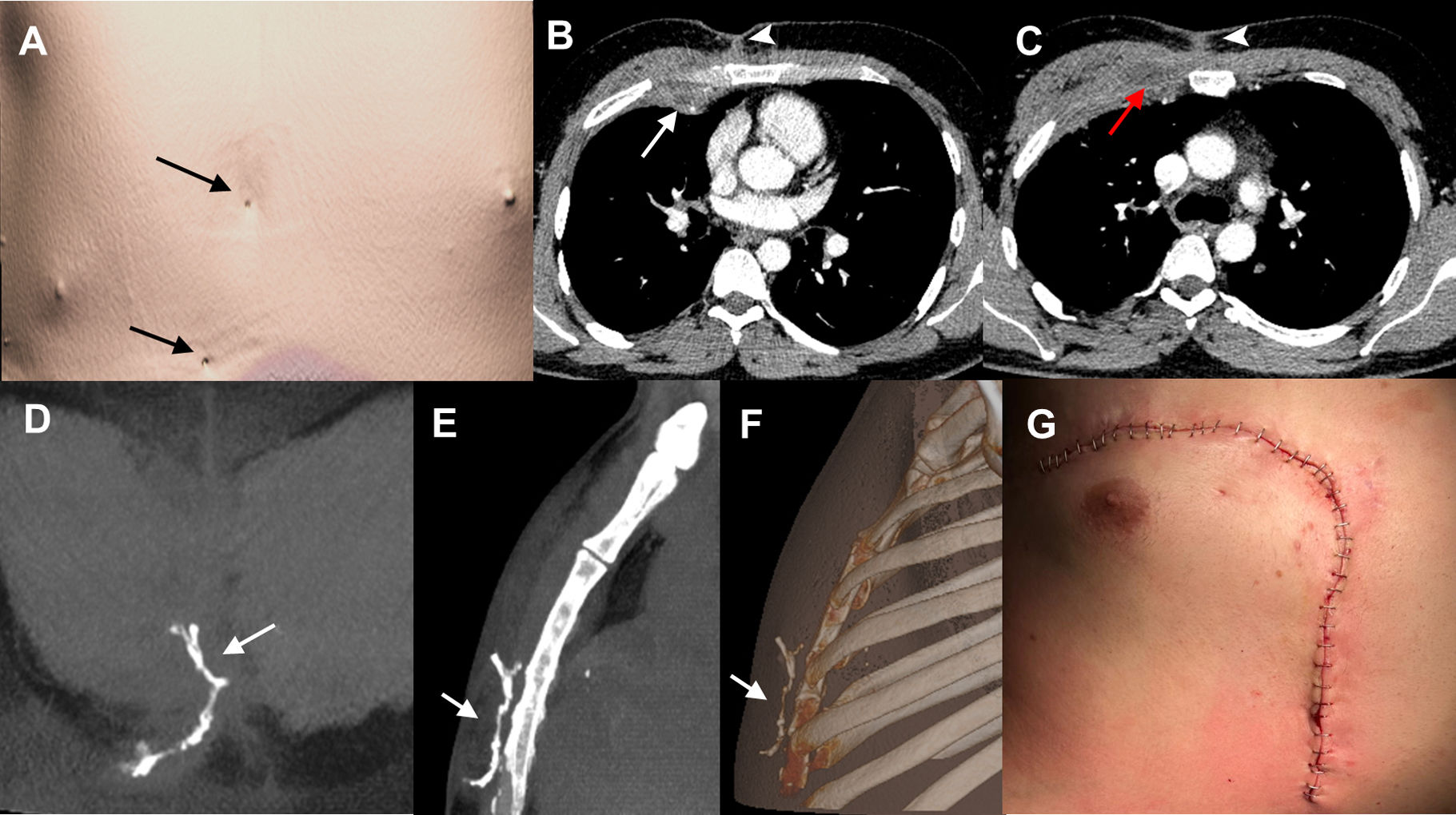
We present the case of a 31-year-old male with X-linked CGD, on chronic antibiotic treatment, with a long-standing medical history of infections (pulmonary and soft-tissue) and recurrent thoracic and abdominal fistulas, who presented complaining of inflammation of the chest wall. The patient had undergone multiple surgical interventions in the past, including a right middle lobectomy for pulmonary aspergillosis and surgical debridement for soft-tissue infections. On physical examination, two small fistulous orifices through which fluid material was draining (Fig. 1A) were found in the anterior chest wall, with doubts about the extent in depth of the inflammatory changes. A contrast-enhanced thoracic computed tomography (CT) confirmed the existence of inflammatory changes in the anterior chest wall (Fig. 1B, C) but ruled out the presence of pulmonary consolidations or pleural fluid collections.

Fig. 1.

(A) 3D CT reconstruction image of the patient's body surface. Two cutaneous openings are clearly visible in the anterior thoracic wall (arrows). (B, C) Axial contrast-enhanced thoracic CT images (mediastinal window) at two different levels show inflammatory changes in the anterior chest wall: a collection in the right anterior pectoral muscles (red arrow), a small extrapleural collection adjacent to the right internal mammary vessels (white arrow), and several linear fistulous tracts extending toward the skin surface (arrowheads). (D–F) Thoracic CT reconstruction images (D, coronal maximum intensity projection [MIP]; E, sagittal MIP; F, sagittal 3D) after the administration of diluted iodinated contrast through two skin fistulous openings on the anterior chest wall demonstrate communication between the two openings and the absence of deep soft tissue extension (arrows). (G) Post-surgical image of the anterior chest wall after the resection of the fistulous tract and the utilization of a vascularized muscle flap.

(0.23MB).

The decision was made to perform a CT-fistulography using iso-osmolar iodinated contrast (iodixanol 320mg/mL, diluted to 25% with saline) through the two cutaneous fistulous openings, due to its excellent anatomical and contrast resolution. CT-fistulography revealed a subcutaneous communication between the two orifices, ruled out involvement of deeper tissues (Fig. 1D–F) and aided the thoracic and plastic surgery teams in planning the resection of the fistulous tract and the reconstruction of the anterior chest wall (Fig. 1G).

CGD is a genetically predisposed disease (X-linked or autosomal recessive) caused by a defect in the NADPH oxidase enzyme complex, affecting phagocytosis and resulting in the inability to eliminate catalase-positive pathogens such as Staphylococcus, Mycobacteria, Serratia, Klebsiella, Pseudomonas, and fungi. Consequently, recurrent infections lead to the formation of inflammatory granulomas. Pulmonary involvement is the most frequent manifestation, including pneumonia or lung abscesses that can extend to the chest wall. It can also manifest as lymphadenopathy, liver abscesses, soft tissue infections, osteomyelitis, brain abscesses, gastrointestinal infections, and organomegaly.1,2 Occasionally, thoracic surgery is necessary to treat aggressive pulmonary infections, which are associated with greater complications and poorer prognosis.3

CGD patients require prolonged antibiotic treatments and prophylactic antimicrobial therapy. Hematopoietic stem cell transplantation is the curative treatment for this disease, which has improved the prognosis of these patients.1,2

CT- fistulography is a technique used for the diagnosis of abdominal wall and perianal fistulas in some patients and, to our knowledge, has not been used in the chest wall.4 In CT-fistulography, acquisition of isovolumetric data sets after the instillation of contrast into the fistula delineates the tract and its components, allows the acquisition of images with great anatomical detail in different spatial planes, and can help surgeons plan the surgical intervention.

We believe that this case is interesting because CT-fistulography ruled out communication between the cutaneous-subcutaneous inflammatory process and the extrapleural fat, pleura, and lung parenchyma. This facilitated appropriate therapeutic planning by demonstrating a communication between the two skin fistulous openings. Meticulous surgical planning is essential in these patients, given their higher risk of complications. We have found no previous descriptions in the scientific literature of CT-fistulography of the chest wall.

Artificial Intelligence Involvement

None of the material has been partially or totally produced with the help of any artificial intelligence tool.

Funding of the Study

This study did not receive any specific grant from funding agencies in the public, commercial, or not-for-profit sectors.

Conflicts of Interest

The authors declare not to have any conflicts of interest that may be considered to influence directly or indirectly the content of the manuscript.

References
[1]
M. Lee, M.S. Lee, J.S. Lee, S.Y. Ko, S.Y. Jeong.
Spectrum of imaging findings of chronic granulomatous disease: a single center experience.
Diagn Interv Radiol, 23 (2017), pp. 472-477
[2]
H.-H. Yu, Y.-H. Yang, B.-L. Chiang.
Chronic granulomatous disease: a comprehensive review.
Clin Rev Allergy Immunol, 61 (2021), pp. 101-113
[3]
P.L. Feingold, H.S. Quadri, S.M. Steinberg, H.L. Malech, J.I. Gallin, C.S. Zerbe, et al.
Thoracic surgery in chronic granulomatous disease: a 25-year single-institution experience.
J Clin Immunol, 36 (2016), pp. 677-683
[4]
M. Tonolini, P. Magistrelli.
Enterocutaneous fistulas: a primer for radiologists with emphasis on CT and MRI.
Insights Imaging, 8 (2017), pp. 537-548
Copyright © 2024. SEPAR
Archivos de Bronconeumología
Article options
Tools

Are you a health professional able to prescribe or dispense drugs?