Journal Information
Share
Share
Download PDF
More article options
Visits
603
Original Article
Full text access
Available online 22 October 2024
Unraveling the Molecular Mechanisms of OSA-Related Cardiovascular Event Recurrence: A Post Hoc Analysis From the ISAACC Study
Visits
603
Andrea Zapatera,b,1, Lucía Pinillaa,b,1, Esther Gracia-Lavedanb,c, Adriano Targab,c, Gerard Torresb,c, Olga Mínguezc, Lydia Pascualc, Anunciación Cortijoc, Dolores Martínezc, Ivan David Benítezb,c, Maria Coronada García-Hidalgob,c, Jordi De Batlleb,c, Jorge Abadb,d, Joaquín Duran-Cantollab,e, Amaia Urrutiaf, Olga Medianob,g, María José Masdeub,h, Estrella Ordax-Carbajoi, Juan Fernando Masab,j, Mónica De la Peñak..., Mercè Mayosb,l, Ramon Colomam, Josep María Montserratb,n, Eusebi Chinero, Alejandra Roncerop, David Sanz-Rubioq, Ferran Barbéb,c, Manuel Sánchez-de-la-Torreb,r,
Corresponding author
sanchezdelatorre@gmail.com

Corresponding author.
,
on behalf of the Spanish Sleep Network Ver más
a Group of Precision Medicine in Chronic Diseases, University Hospital Arnau de Vilanova and Santa María, IRBLleida, Lleida, Spain
b Centro de Investigación Biomédica en Red de Enfermedades Respiratorias (CIBERES), Madrid, Spain
c Translation Research in Respiratory Medicine, Hospital Universitari Arnau de Vilanova-Santa Maria, IRBLleida, Lleida, Spain
d Respiratory Department, Hospital Universitari Germans Trias I Pujol, Badalona, Barcelona, Spain
e Servicio de Investigación OSI, Hospital Universitario Araba, IIS Bioaraba, Vitoria, Álava, Spain
f Servicio Neurologia, Hospital Universitario Cruces, Bizkaia, Spain
g Respiratory Department, Hospital Universitario de Guadalajara, Guadalajara, Spain
h Respiratory and Sleep Department, Hospital Universitari Parc Taulí, Institut Investigació i Innovació Parc Taulí (I3PT), Universitat Autònoma de Barcelona, Sabadell, Spain
i Respiratory Department, Hospital Universitario de Burgos, Burgos, Spain
j Respiratory Department, Hospital San Pedro Alcántara, Cáceres, Spain
k Clinic Analysis and Respiratory Services, Hospital Universitari Son Espases, Institut de investigació sanitaria de Palma (IdisPa), Palma de Mallorca, Spain
l Sleep Unit, Department of Respiratory Medicine, Hospital de la Santa Creu i Sant Pau, Barcelona, Spain
m Respiratory Department, Hospital General Universitario de Albacete, Spain
n Respiratory Department, Hospital Clinic, Barcelona, Spain
o Respiratory Department, Hospital Universitari Sant Joan d’Alacant, Alicante, Spain
p Unidad Multidisciplinar del Sueño, Servicio de Neumología, Hospital San Pedro, Logroño, Spain
q Precision Medicine in Respiratory Diseases (PRES) Group, Unidad de Investigación Traslacional, Instituto de Investigación Sanitaria de Aragón-IISA, Hospital Universitario Miguel Servet, Zaragoza, Spain
r Group of Precision Medicine in Chronic Diseases, Hospital Nacional de Parapléjicos, IDISCAM, Department of Nursing, Physiotherapy and Occupational Therapy, Faculty of Physiotherapy and Nursing, University of Castilla-La Mancha, Toledo, Spain
Ver más
This item has received
Received 16 April 2024. Accepted 21 September 2024
Article information
Abstract
Full Text
Bibliography
Download PDF
Statistics
Figures (4)
Show moreShow less
Tables (1)
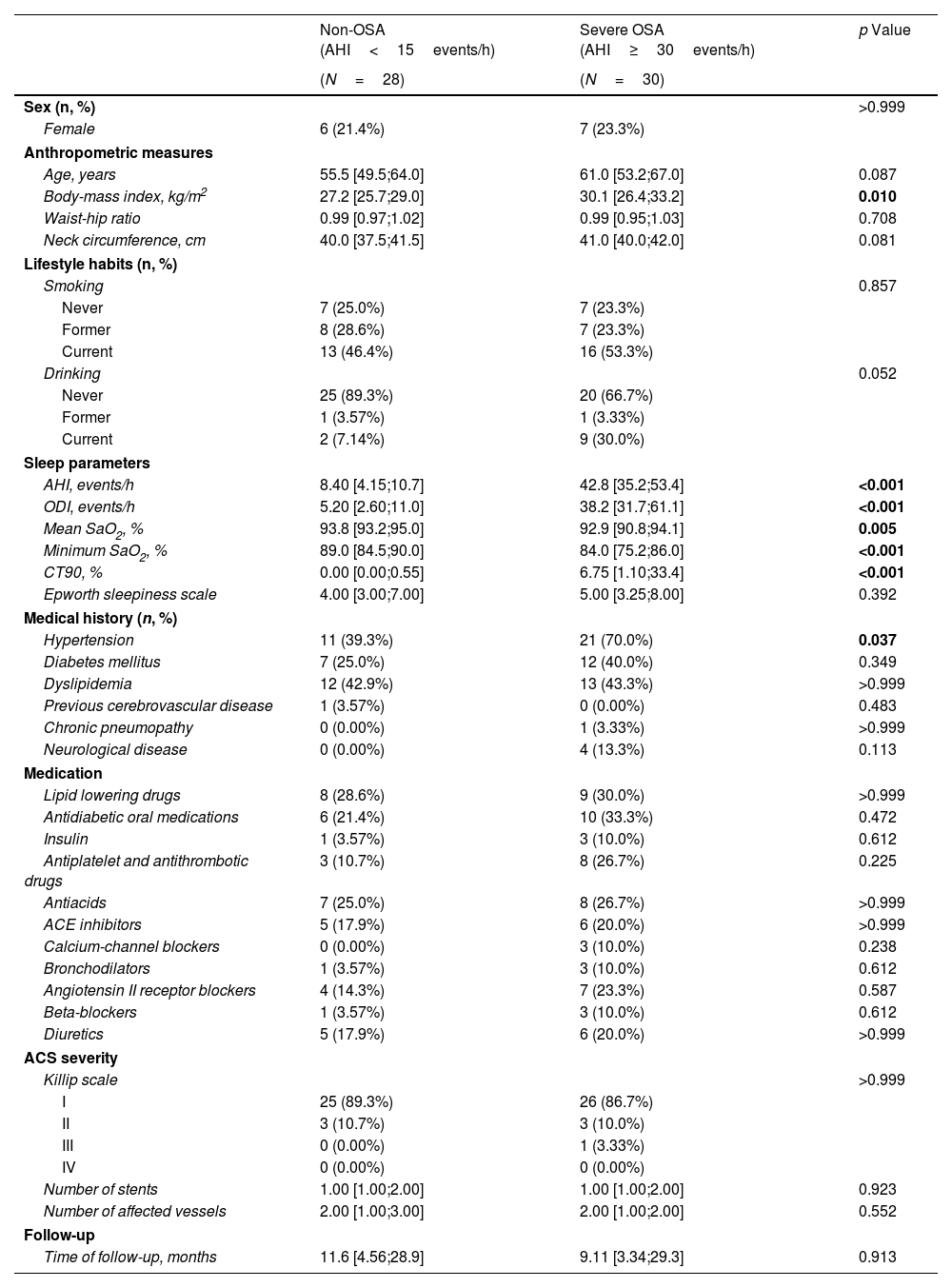
Table 1. Baseline Characteristics of ACS Patients With Recurrent CVEs According to OSA Condition (Non-OSA vs. Untreated Severe OSA).
Additional material (1)
Abstract
Rationale

Although obstructive sleep apnea (OSA) is a prevalent condition among patients with acute coronary syndrome (ACS), the impact of OSA on cardiovascular event (CVE) recurrence is not homogeneous. We previously defined a specific phenotype of first-ACS patients without previous cardiovascular disease who are at increased risk of OSA-related CVE recurrence. However, the pathobiological mechanisms whereby OSA leads to adverse cardiovascular outcomes in this singular ACS phenotype remain to be investigated.

Objective

To characterize the molecular pathways that relate OSA with CVE recurrence.

Methods

This post hoc analysis of the ISAACC study (NCT01335087) included subjects without previous cardiovascular disease who were hospitalized for a first ACS and developed a recurrent CVE during the follow-up. Patients underwent respiratory polygraphy and fasting blood extraction during hospitalization. Two study groups were established on the basis of the apnea–hypopnea index (AHI): untreated severe OSA (AHI30events/h) and non-OSA (AHI<15events/h) groups. Proteomic profiling analysis included 276 cardiovascular and inflammatory-related plasma proteins via Olink® technology.

Results

Proteomics was performed in 58 patients (77.6% male, median [p25;p75] age 58.0 [51.2;65.8] years, and median BMI 28.6 [25.8;31.2]kg/m2). Thirty patients had severe OSA, and 28 subjects were considered non-OSA controls. A total of 24 plasma proteins were differentially expressed between the groups. Among these proteins, 18 were significantly associated with OSA severity parameters derived from respiratory polygraphy. Further bioinformatic analyses of OSA-related proteins revealed their involvement in several molecular pathways, mostly related to immune function, cell signaling, and inflammatory processes.

Conclusion

A specific proteomic profile related to OSA presence and severity was identified in the plasma of ACS patients who developed recurrent CVEs. This analysis suggests the activation of key OSA-mediated molecular pathways with potential implications for cardiovascular prognosis.

Keywords:
Obstructive sleep apnea
Acute coronary syndrome
Proteomics
Biomarkers
Molecular pathways
Cardiovascular disease
Full Text
Introduction

Obstructive sleep apnea (OSA) is a chronic sleep disorder affecting 20–30% of the adult population and is considered a major public health concern.1 This condition is characterized by recurrent episodes of partial or complete upper airway collapse during sleep. Disease severity is currently gauged by the frequency of apneas and hypopneas per hour of sleep, which is termed the apnea–hypopnea index (AHI).2

OSA is associated with diminished quality of life and has been linked with metabolic, neurologic, and cardiovascular consequences.3,4 As such, it is considered an independent risk factor for cardiovascular morbidity and mortality.5 Furthermore, a high prevalence (40–60%) of OSA is present in cardiovascular disease (CVD) populations.6 There is a substantial body of evidence suggesting a role for OSA in the initiation and/or progression of several CVDs.7

Acute coronary syndrome (ACS) is often the first manifestation of CVD and has been established as a major cause of morbidity and mortality worldwide. Despite OSA being prevalent among ACS patients, its impact on cardiovascular prognosis is not homogeneous.6,8 Our group has defined a specific phenotype of ACS patients with heightened susceptibility to the deleterious consequences of OSA in the cardiovascular sphere.9 Specifically, we observed that OSA was associated with an increased risk of cardiovascular event (CVE) recurrence in patients without previous CVD who were experiencing their first ACS episode. Additionally, we recently identified a protein-based circulating signature in this specific phenotype of ACS patients with OSA, which was able to predict CVE recurrence in the long term.10 These findings constitute a potential prognostic tool that may provide a new direction for cardiovascular risk stratification and the clinical management of ACS patients with OSA. However, the specific physiopathological mechanisms promoted by OSA that lead to CVE recurrence remain unclear. Recent advances in proteomics provide a valuable opportunity to help establish the molecular underpinnings of this association.

In the present study, we aimed to evaluate the association of OSA with the circulating expression of proteomic markers in patients with ACS who further develop CVEs. We hypothesized that the deleterious pathobiological mechanisms that relate OSA to an increased risk of CVE recurrence would be translated into differential expression of specific plasma proteins. This analysis aims to shed light on the currently unexplored molecular mechanisms whereby OSA impacts cardiovascular function in this specific phenotype of patients who are more susceptible to the detrimental effects of OSA.

MethodsSleep Study and Baseline Procedures

This is an ancillary analysis of the ISAACC study, a multicenter, open-label, parallel, prospective, randomized controlled trial performed throughout 15 hospitals in Spain (NCT01335087).11 The ethics committee of each participating center approved the study (approval number in the coordinating center, University Hospital Arnau de Vilanova and Santa María de Lleida, Spain: 2010-852), and all patients provided written informed consent to participate in the study. Full details related to the trial protocol and detailed inclusion and exclusion criteria can be found elsewhere.12 Briefly, eligible patients aged18 years were admitted to the coronary care units or cardiology hospitalization wards for ACS, defined as the acute presentation of coronary disease with or without ST elevation infarction, unstable angina, or type 1 myocardial infarction.

Demographic and anthropometric data, medical history, and usual pharmacological treatment were recorded at the initial visit. Excessive daytime sleepiness was assessed through the Spanish validated version of the Epworth sleepiness scale (ESS). Patients who did not present excessive daytime sleepiness (inclusion criteria), defined as an ESS score of ≤10, underwent respiratory polygraphy (Embletta, ResMed, Bella Vista, NSW, Australia) during the first 24–72h after hospitalization, according to the national clinical practice guidelines and regulations.13 An apnea episode was defined as an interruption in airflow for ≥10s. A hypopnea episode was defined as a reduction in airflow for ≥10s associated with a decrease in arterial oxygen saturation (SaO2)4%. The OSA severity parameters derived from the sleep study included the AHI (average number of apneas plus hypopneas per hour of sleep), oxygen desaturation index (ODI; number of episodes of SaO2 decrease4%), minimum and mean SaO2, and percentage of total sleep time spent with SaO2<90% (CT90).

Study Groups and Outcomes

In the ISAACC trial, patients with an AHI<15events/h were considered controls (non-OSA), and those with an AHI15events/h (OSA) were randomly assigned (1:1) to receive either continuous positive airway pressure (CPAP) therapy plus usual care or usual care alone. Following the initial visit, patients were evaluated after 1, 3, 6, 12, 18, 24, 30, and 36 months. After this period, the assessments were performed annually. The minimum follow-up time was one year for all patients. The primary outcome in the ISAACC trial was a composite of CVEs, including cardiovascular death or nonfatal events (acute myocardial infarction, nonfatal stroke, hospital admission for heart failure, and new hospitalizations for unstable angina or transient ischemic attack). The follow-up time was defined as the time between the baseline visit and the end of the study or the occurrence of an adverse CVE, whichever occurred first.

For the current post hoc analysis, we focused the study population on the specific phenotype of ACS patients in which OSA is associated with increased cardiovascular risk, i.e., patients without previous heart disease who were admitted for their first ACS.9 Specifically, as our primary aim was to study the molecular mechanisms that relate OSA to the risk of CVE recurrence, we selected patients who presented with recurrent CVEs during the follow-up period and classified them according to OSA severity. OSA patients allocated to the CPAP arm were excluded. To maximize proteomic differences and ensure that the observed associations were related to OSA physiopathology, patients with moderate OSA (15<AHI<30events/h) were removed from the analysis. Therefore, two study groups were established: a non-OSA group (AHI<15events/h) and an untreated severe OSA group (AHI30events/h) that did not receive CPAP treatment (Fig. 1).

Fig. 1.

Flowchart of the study. Of the 1851 participants enrolled in the ISAACC trial, after applying the exclusion criteria described in the Methods section, this proteomic post hoc analysis included 58 untreated patients with a first ACS episode: 28 were included in the non-OSA group, and 30 were included in the severe OSA group. Definitions of abbreviations: ACS=acute coronary syndrome; AHI=apnea–hypopnea index; CPAP=continuous positive airway pressure; CVD=cardiovascular disease; CVE=cardiovascular event; OSA=obstructive sleep apnea.

(0.33MB).
Proteomic Analysis

Fasting venous blood samples obtained at baseline during hospital admission at each participating center were processed to obtain the plasma fraction. Details about the blood collection, processing, and storage procedures are outlined in the supplementary material. The selected plasma samples were transferred to 96-well plates and shipped to Olink® (Uppsala, Sweden). A total of 276 plasma proteins were measured via 3 predefined panels: Target 96 Cardiovascular II, Cardiovascular III, and Inflammation. Additional information can be found in the supplementary material and on the manufacturer's website (https://olink.com/resources-support/document-download-center/).

Statistical Analysis

Data are presented as the median [25th percentile–75th percentile] or n (%), and differences between groups were assessed via the Mann–Whitney U test for continuous variables or Fisher's exact test for categorical variables. Differentially expressed proteins between the study groups were evaluated via empirical Bayes methods and linear models for arrays.14 A volcano plot was generated to illustrate the results. The false discovery rate (FDR) was defined by the p value corrected for multiple testing via the Benjamini–Hochberg procedure. The analysis was adjusted for age, sex, body mass index (BMI) and the presence of diagnosed hypertension. The associations between protein levels and respiratory polygraphy parameters were explored via multiple linear regression models adjusted for confounders. All variables were previously standardized. A Voronoi diagram was used to summarize the proteins with significant differences between the study groups, which also revealed a significant dose–response relationship with one or more respiratory polygraphy parameters. Statistical analyses were conducted via R, version 3.6.15 Two-sided p values were reported, and statistical significance was set at 0.05.

Bioinformatic Analysis

The Search Tool for the Retrieval of Interacting Genes/Proteins (STRING) database, with an interaction score of 0.700 (high confidence), was used to establish protein–protein interaction networks.16 To perform the functional pathway enrichment analysis, the STRING, Reactome and Gene Ontology databases were used.17 The identified protein set associated with OSA severity was used for both in silico analyses. Additionally, the cell, tissue, and organ expression patterns of the selected proteins were evaluated via data from the Genotype-Tissue Expression (GTEx) Portal (www.gtexportal.org).

ResultsStudy Population

Our study population focused on individuals with a high OSA-related cardiovascular risk phenotype (subjects without previous cardiovascular disease who were hospitalized for a first ACS episode), who later experienced recurrent CVEs during the follow-up period. Proteomic data were generated for the 58 available patients (Table 1), which were predominantly males (77.6%), with a median [p25;p75] age of 58 [51.2;65.8] years and a median BMI of 28.6 [25.8;31.2]kg/m2. Among them, 30 patients had been diagnosed with severe OSA (AHI30events/h), whereas 28 patients were included in the non-OSA group (AHI<15events/h) (Fig. 1). As anticipated, severe OSA patients had a higher BMI, a greater proportion of males, and a greater incidence of hypertension than controls did. The severity of ACS, according to the Killip scale and the number of affected vessels and implanted stents, was similar between the groups. No significant differences were found regarding prior pharmacological treatments between the groups.

Table 1.

Baseline Characteristics of ACS Patients With Recurrent CVEs According to OSA Condition (Non-OSA vs. Untreated Severe OSA).

  Non-OSA (AHI<15events/h)  Severe OSA (AHI30events/h)  p Value 
  (N=28)  (N=30)   
Sex (n, %)      >0.999 
Female  6 (21.4%)  7 (23.3%)   
Anthropometric measures
Age, years  55.5 [49.5;64.0]  61.0 [53.2;67.0]  0.087 
Body-mass index, kg/m2  27.2 [25.7;29.0]  30.1 [26.4;33.2]  0.010 
Waist-hip ratio  0.99 [0.97;1.02]  0.99 [0.95;1.03]  0.708 
Neck circumference, cm  40.0 [37.5;41.5]  41.0 [40.0;42.0]  0.081 
Lifestyle habits (n, %)       
Smoking      0.857 
Never  7 (25.0%)  7 (23.3%)   
Former  8 (28.6%)  7 (23.3%)   
Current  13 (46.4%)  16 (53.3%)   
Drinking      0.052 
Never  25 (89.3%)  20 (66.7%)   
Former  1 (3.57%)  1 (3.33%)   
Current  2 (7.14%)  9 (30.0%)   
Sleep parameters       
AHI, events/h  8.40 [4.15;10.7]  42.8 [35.2;53.4]  <0.001 
ODI, events/h  5.20 [2.60;11.0]  38.2 [31.7;61.1]  <0.001 
Mean SaO2, %  93.8 [93.2;95.0]  92.9 [90.8;94.1]  0.005 
Minimum SaO2, %  89.0 [84.5;90.0]  84.0 [75.2;86.0]  <0.001 
CT90, %  0.00 [0.00;0.55]  6.75 [1.10;33.4]  <0.001 
Epworth sleepiness scale  4.00 [3.00;7.00]  5.00 [3.25;8.00]  0.392 
Medical history (n, %)       
Hypertension  11 (39.3%)  21 (70.0%)  0.037 
Diabetes mellitus  7 (25.0%)  12 (40.0%)  0.349 
Dyslipidemia  12 (42.9%)  13 (43.3%)  >0.999 
Previous cerebrovascular disease  1 (3.57%)  0 (0.00%)  0.483 
Chronic pneumopathy  0 (0.00%)  1 (3.33%)  >0.999 
Neurological disease  0 (0.00%)  4 (13.3%)  0.113 
Medication       
Lipid lowering drugs  8 (28.6%)  9 (30.0%)  >0.999 
Antidiabetic oral medications  6 (21.4%)  10 (33.3%)  0.472 
Insulin  1 (3.57%)  3 (10.0%)  0.612 
Antiplatelet and antithrombotic drugs  3 (10.7%)  8 (26.7%)  0.225 
Antiacids  7 (25.0%)  8 (26.7%)  >0.999 
ACE inhibitors  5 (17.9%)  6 (20.0%)  >0.999 
Calcium-channel blockers  0 (0.00%)  3 (10.0%)  0.238 
Bronchodilators  1 (3.57%)  3 (10.0%)  0.612 
Angiotensin II receptor blockers  4 (14.3%)  7 (23.3%)  0.587 
Beta-blockers  1 (3.57%)  3 (10.0%)  0.612 
Diuretics  5 (17.9%)  6 (20.0%)  >0.999 
ACS severity       
Killip scale      >0.999 
25 (89.3%)  26 (86.7%)   
II  3 (10.7%)  3 (10.0%)   
III  0 (0.00%)  1 (3.33%)   
IV  0 (0.00%)  0 (0.00%)   
Number of stents  1.00 [1.00;2.00]  1.00 [1.00;2.00]  0.923 
Number of affected vessels  2.00 [1.00;3.00]  2.00 [1.00;2.00]  0.552 
Follow-up       
Time of follow-up, months  11.6 [4.56;28.9]  9.11 [3.34;29.3]  0.913 

The data are presented as n (%) or medians [25th percentile; 75th percentile]. Significant p values (p<0.05) are presented in bold. Abbreviations: ACE=angiotensin-converting enzyme; ACS=acute coronary syndrome; AHI=apnea–hypopnea index; CT90=% of time spent with SaO2<90%; CVE=cardiovascular event; ODI=oxygen desaturation index; OSA=obstructive sleep apnea; SaO2=oxygen saturation.

Proteomic Profiling AnalysisQuality Control

All the samples passed quality control. Among the 276 tested proteins, 20 had >25% values below the LOD and were consequently excluded from all the analyses. Additionally, 10 proteins were duplicated and analyzed in two different Olink® panels. Consistently, a strong correlation between panels was observed (Fig. E1), and one of the two was deleted at random.

Differentially Expressed Proteins Between Untreated Severe OSA Patients and Non-OSA Patients

After adjustment for confounding factors, 24 plasma proteins displayed significant differential values between untreated patients with severe OSA and patients without OSA (Fig. 2A, Table E1). We subsequently explored the associations of the differentially expressed proteins with relevant respiratory polygraphy parameters commonly used to assess OSA severity, including the AHI, ODI, mean and minimum SaO2, and CT90. This analysis revealed a significant linear association between the 18 proteins and at least one OSA severity parameter (Fig. 2B). These 18 plasma proteins were considered to be related to OSA physiopathology and were thus included in further bioinformatic analyses. A total of 16 out of the 18 OSA-related proteins were upregulated in severe OSA patients, whereas the remaining 2 were downregulated (Fig. 2C and Fig. E2).

Fig. 2.

Analysis of differential protein detection according to OSA condition (non-OSA vs. untreated severe OSA) in ACS patients with recurrent CVEs. (A) Volcano plot showing the negative logarithm of the p value (y-axis) versus the log2-fold change (x-axis) for each analyzed plasma protein. The horizontal dashed line indicates the cutoff for the p value defining statistical significance (0.05). The black dots denote the significantly differentially expressed proteins between the groups. (B) Linear associations between the differentially expressed proteins (y-axis) and the OSA severity parameters derived from respiratory polygraphy (x-axis). All variables were standardized. The slopes between the differentially expressed proteins and the respiratory polygraphy parameters are represented through a color scale, with red being related to positive associations and blue to negative associations. Asterisks denote statistical significance (p<0.05). (C) Voronoi diagrams illustrating the differentially expressed proteins between the study groups, which were significantly correlated with at least one OSA severity parameter. The size of the polygons reflects the magnitude of the fold change. The upregulated and downregulated proteins in severe OSA patients are represented in gray and black, respectively. All analyses were adjusted for confounding factors (age, sex, BMI, and the presence of hypertension). Definitions of abbreviations: ACS=acute coronary syndrome; AHI=apnea–hypopnea index; CT90=time with SaO2<90%; CVE=cardiovascular event; ODI=oxygen desaturation index; OSA=obstructive sleep apnea; SaO2=oxygen saturation.

(0.29MB).
Bioinformatic Analysis of OSA-Related Proteins

An in silico analysis, including the identified OSA-related proteins, was conducted to explore the molecular pathways linking OSA to CVE recurrence. To understand the biological functions of the identified proteins, pathway enrichment analysis and protein–protein interaction network analysis were performed with STRING software. The canonical pathways enriched in the proteins associated with OSA severity are detailed in Table E2, with the top 25 significantly enriched pathways displayed in Fig. 3A (FDR cutoff of 0.05). The enriched pathways with the strongest associations included signal transduction and cellular signaling pathways, regulation of apoptosis, and immune system-related processes. Several additional pathways related to the cellular response to chemical and organic stimuli and the inflammatory response were also identified. An interaction network representing protein–protein relationships is shown in Fig. E3, which revealed few interactions between the identified proteins and no central edges.

Fig. 3.

Bioinformatic analyses including the differentially expressed proteins between the study groups (non-OSA vs. untreated severe OSA), which were significantly associated with OSA severity parameters derived from respiratory polygraphy. (A) Gene set pathway enrichment analysis performed via STRING. The graph shows the FDR values (x-axis) of the top twenty-five identified biological processes (y-axis). The size of each bubble is proportional to the number of proteins included within each set. (B) Tissue and organ expression enrichment analysis via genotype-tissue expression (GTEx). Hierarchical clustering shows each identified tissue and/or organ on the bottom and each included protein on the right. Definition of abbreviations: FDR=false discovery rate; OSA=obstructive sleep apnea.

(0.43MB).

To further elucidate the biological role of the identified proteins, a functional enrichment analysis was performed via Reactome software, revealing 46 significantly enriched pathways associated with OSA severity (Table E3), involving different signaling pathways. Moreover, the Gene Ontology software revealed 8 significant pathways, which were related mainly to the immune system and hematopoiesis processes. Finally, considering the tissue/organ enrichment analysis via GTEx, the selected proteins were generally expressed in the lung and in other tissues and organs in a minor fraction (Fig. 3B).

Discussion

Here, we present the results of the first proteomic investigation conducted on the specific phenotype of ACS patients in which OSA has been identified as a contributing factor to the development of recurrent CVEs. Our goal was to illuminate the potential OSA-related drivers of CVD progression, which may mediate the recurrence of CVEs. In this study, we identified a specific plasma proteomic profile associated with OSA and its severity in the context of patients without previous cardiovascular disease who were hospitalized for a first ACS and developed a recurrent CVE during the follow-up period. This analysis suggests the activation of key OSA-mediated molecular pathways with potential implications for cardiovascular prognosis in patients with ACS.

Previous research conducted by our group demonstrated that OSA was associated with an increased risk of CVEs among patients admitted to the hospital for a first ACS episode without prior CVD.9 Additionally, in a post hoc analysis of the referred study, we identified a specific blood-based proteomic signature that was capable of predicting CVE recurrence in patients with OSA.10 Nevertheless, the mechanisms through which OSA specifically induces drivers of cardiovascular damage in this patient phenotype have not been explored. The scope of the current study is to shed light on this matter by exploring protein-based markers and molecular pathways associated with OSA, which could contribute to increased cardiovascular risk in this patient profile. The blood-based profile that we identified here was highly correlated not only with the AHI, which is the primary disease-defining metric for OSA, but also with other respiratory polygraphy parameters commonly used to assess OSA severity, including the ODI, minimum and mean SaO2, and CT90. A further bioinformatic analysis was performed to identify and characterize the main OSA-relevant pathways potentially related to CVE recurrence. Pathway enrichment analyses of the OSA-related proteins revealed their involvement in distinct processes and signaling pathways, including apoptotic, immune system, and stress-response processes. Furthermore, the tissue and organ enrichment in silico analysis revealed that the identified plasma proteins were expressed in several organs and tissues, with a remarkable concentration in the lung. Importantly, biomarkers associated with clinically relevant outcomes, such as the cardiovascular prognostic implications studied here, hold promise as potential targets for therapeutic interventions and tools for cardiovascular risk prognostication.

Prior investigations indicate that OSA may play a role in the initiation and/or progression of CVDs.7 The mechanisms contributing to these adverse outcomes are thought to be related to oxidative stress, systemic inflammation, sympathetic activation, hypercoagulability, endothelial dysfunction, and metabolic dysregulation.18 Notably, intermittent hypoxia, one of the main hallmarks of OSA, has been identified as a crucial factor for the impaired endothelial function consistently observed in OSA patients, which is mediated through the generation of reactive oxygen species (ROS) and the release of inflammatory molecules.19 Markers of oxidative stress are closely linked to the severity of OSA and potentially to its cardiovascular consequences.20 Both animal models and human studies have reported a pronounced inflammatory process in OSA,21 which is correlated with an increase in the expression of inflammatory markers, such as HIF-1α, NF-kB and IL6.22 Elevated levels of NF-kB have been documented in OSA patients, suggesting a potential role as an inflammatory and cardiovascular mediator of OSA consequences.23 Intermittent hypoxia increases the production of both free oxygen radicals and inflammatory cytokines while promoting vascular alterations dependent on nitric oxide production, collectively leading to endothelial dysfunction and thereby contributing to the development of CVD.24 The role of intermittent hypoxia in endothelial dysfunction has been previously demonstrated in both in vitro and in vivo hypoxic models, as well as in patients with OSA.25

The findings regarding the biological role of the proteins identified in this study suggest their potential involvement in the recurrence of CVEs. Specific proteins (TEK, IDUA, hOSCAR, PSGL-1, FABP2, PSP-D and TNF-β) have previously been recognized for their roles in OSA pathogenesis and their relationship with hypoxia, vascular inflammation, and other conditions, such as cognitive impairment.26,27 Specifically, TEK, PSGL-1, FABP2, PSP-D and TNF-β are linked to the activation of intermediate mechanisms and may serve as reliable indicators for specific populations at increased cardiovascular risk in the context of OSA.28 Additionally, our prior research revealed lower plasma levels of STK4 among OSA patients. STK4, a key component of the Hippo pathway, is intricately involved in cardiovascular remodeling.29 In addition, DECR1 and MMP10 are implicated in the progression of atherosclerosis, with DECR1 increasing the inflammatory response mediated by hypoxia and MMP10 stimulating inflammation, development, and complications of atheroma plaques.30 DECR1 plays a relevant role in cardiac apoptosis and has been proposed as a prognostic marker for heart failure.31 MMP-10 is thought to play a vital role in pulmonary vascular remodeling associated with hypoxia and pulmonary arterial hypertension.32 MMP-10, STK4, and Flt3L are also associated with inflammation, apoptosis, and cell proliferation through the Akt pathway.33 Flt3L enhances early hematopoietic cell proliferation through the activation of FLT3.34 Notably, TEK has also been related to hematopoiesis after hypoxia, further indicating an association between hematopoiesis and OSA in the context of CVD.35 Flt3L, hOSCAR and PSGL-1 play roles in immunity by stimulating dendritic cells, which promote inflammation, oxidative stress, and cytokine production, thereby increasing cardiovascular risk and atherosclerosis.36 Several of the proteins identified here, including TNFRSF13B and MERTK, are postulated to play a role in the destabilization of atherosclerotic plaques.37 Additionally, other identified proteins, such as TNFRSF13B, PSGL-1 and TRAIL-R2, are key components of the NF-kB pathway, which is implicated in sleep regulation and CVD progression.38 We also detected dysregulation of the plasma expression of cytokines such as TNF-β or interleukins such as IL1RL2, which are involved in the activation of inflammatory pathways that contribute to the development of cardiovascular consequences.39 Finally, VEGF-D, BOC and CTSL1 have been shown to play a role in cardiac remodeling and intermediate mechanisms related to the manifestation of different types of CVD.40 Overall, the current results provide a framework for potential targeted interventions and strategies against the development of further OSA-related CVD in patients with ACS.

Strengths and Limitations

The strengths of this study include its multicentric and prospective design with a relevant follow-up period. Standardized methods were uniformly applied for data collection across all participating centers. All sleep studies were performed with the same respiratory polygraphy model and were scored by certified technicians following international guidelines. Major adverse cardiovascular outcomes specified in the study protocol were documented and adjudicated by a blinded external committee. Additionally, this study involves the use of a sensitive, specific, and robust proteomic method and strict quality control of the proteomic data.

This study has several limitations that should be noted. First, the observational nature of the study precludes conclusions about the causative role of OSA in promoting cardiovascular damage in this specific ACS phenotype. To mitigate this limitation and ensure that the observed associations were related to OSA physiopathology, patients with moderate OSA (15<AHI<30events/h) were removed from the analysis to maximize proteomic differences. Additionally, we further evaluated the correlation of the differentially expressed proteins with commonly assessed OSA severity parameters derived from sleep studies. Only those proteins that were significantly linearly associated with one or more respiratory polygraphy variables were included in the subsequent bioinformatic analyses. Nevertheless, cause–effect relationships remain unproven. Second, our findings should not be extrapolated to different populations other than the specific phenotype of patients without preexisting cardiovascular conditions who were admitted to the hospital for their first ACS. Additionally, for ethical reasons, patients with excessive daytime sleepiness were excluded from the ISAACC trial, limiting the applicability of the results to this profile of OSA patients. Notably, the cohort was largely male, leaving females underrepresented. In addition, data on race and ethnicity were not collected as part of this study. Given that the trial was conducted in Spain, nonwhite ethnicities may be underrepresented. Validation of this work in further external cohorts involving different clinical settings with more diverse demographic and clinical characteristics and larger populations is warranted, although it falls beyond the scope of the current initial investigation. Third, owing to the exploratory nature of the study and the relatively low prevalence of recurrent CVEs within our cohort, a nonprobability sampling method was applied for sample selection, in which all available participants were considered for the proteomic analysis, and the study groups were not matched. To counteract this limitation, all analyses in this study were adjusted for relevant confounding factors.

Conclusions

This study reveals a circulating protein profile that differs between untreated severe OSA patients and non-OSA patients within the specific phenotype of individuals without previous cardiovascular disease who were admitted for a first ACS episode and subsequently developed recurrent CVEs during the follow-up period. Remarkably, three-quarters of these proteins exhibited a significant dose–response linear association with OSA severity parameters derived from the sleep study. Further bioinformatic analyses of OSA severity-related proteins revealed specific enriched pathways that could establish a link between OSA physiopathology and the poor cardiovascular prognosis observed in patients with this phenotype. These findings collectively emphasize protein-based markers and molecular pathways that provide insight into the potential physiopathological mechanisms by which OSA could contribute to the elevated cardiovascular risk observed in this patient profile. Future larger-scale studies are warranted to confirm these initial findings.

Funding

Instituto de Salud Carlos III (ISCIII) (PI10/02763, PI10/02745, PI18/00449, PI21/00337), co-funded by the European Union, IRBLleida-Fundació Dr. Pifarré, CERCA Programme/Generalitat de Catalunya, SEPAR, ResMed Ltd. (Australia), Esteve-Teijin (Spain), Oxigen Salud (Spain), Associació Lleidatana de Respiratori (ALLER) and Sociedad Española de Sueño (SES). AZ held a predoctoral fellowship “Ajuts 2021 de Promoció de la Recerca en Salut-9ª edició” from IRBLleida/Diputació de Lleida. JdB acknowledges receiving financial support from ISCIII (Miguel Servet 2019: CP19/00108), co-funded by the European Social Fund (ESF), “Investing in your future”. MS has received financial support from a “Ramón y Cajal” grant (RYC2019-027831-I) from the “Ministerio de Ciencia e Innovación – Agencia Estatal de Investigación” co-funded by the European Social Fund (ESF)/“Investing in your future” FB is supported by the ICREA Academia program from the Generalitat de Catalunya.

Authors’ Contributions

AZ, LPi, EG, AT, IB, FB and MSdT contributed to the study concept and design; GT, OM, LPa, AC, DM, JA, JDC, AU, OMe, MJM, EOC, JFM, MP, MM, RC, JMM, EC and FB contributed to the data acquisition; AZ, LPi, EG, AT, IB, MCGH, JB, AR, DSR, FB and MSdT contributed to the data analysis and interpretation; and all authors contributed to the drafting of the manuscript, critically revised the manuscript for important intellectual content and approved the final version. MSdT is the guarantor of the paper.

Conflicts of Interest

FB received a research grant from ResMed (an Australian company that develops products related to sleep apnea), the Health Research Fund, the Spanish Ministry of Health, the Spanish Respiratory Society, the Catalonian Cardiology Society, Esteve-Teijin (Spain), Oxigen Salud (Spain), and ALLER to develop the ISAACC trial. ResMed partly funded the ISAACC study but did not participate or decision in study development or the writing of the present manuscript. All other authors declare no competing interests.

Acknowledgements

We are grateful to the individuals who participated in this trial and their respective families, the clinical and research teams of the participating sleep and cardiology departments, and the Spanish Sleep Network for their work on the ISAACC study.

Appendix B
Supplementary Data

The followings are the supplementary data to this article:

References
[1]
R. Heinzer, S. Vat, P. Marques-Vidal, H. Marti-Soler, D. Andries, N. Tobback, et al.
Prevalence of sleep-disordered breathing in the general population: the HypnoLaus study.
Lancet Respir Med, 3 (2015), pp. 310-318
[2]
O. Mediano, N. González Mangado, J.M. Montserrat, M.L. Alonso-Álvarez, I. Almendros, A. Alonso-Fernández, et al.
Documento internacional de consenso sobre apnea obstructiva del sueño.
Arch Bronconeumol, 41 (2021), pp. 7-9
[3]
H. Nakashima, M. Kurobe, K. Minami, S. Furudono, Y. Uchida, K. Amenomori, et al.
Effects of moderate-to-severe obstructive sleep apnea on the clinical manifestations of plaque vulnerability and the progression of coronary atherosclerosis in patients with acute coronary syndrome.
Eur Heart J Acute Cardiovasc Care, 4 (2015), pp. 75-84
[4]
O. Mediano, I. Cano-Pumarega, M. Sánchez-de-la-Torre, M.L. Alonso-Álvarez, M.F. Troncoso, F. García-Río, et al.
Upcoming scenarios for the comprehensive management of obstructive sleep apnea: an overview of the Spanish sleep network.
Arch Bronconeumol, 56 (2020), pp. 35-41
[5]
J.M. Marin, S.J. Carrizo, E. Vicente, A.G.N. Agusti.
Long-term cardiovascular outcomes in men with obstructive sleep apnoea–hypopnoea with or without treatment with continuous positive airway pressure: an observational study.
Lancet, 365 (2005), pp. 1046-1053
[6]
T.D. Bradley, J.S. Floras.
Obstructive sleep apnoea and its cardiovascular consequences.
[7]
M. Sánchez-de-la-Torre, F. Campos-Rodriguez, F. Barbé.
Obstructive sleep apnoea and cardiovascular disease.
Lancet Respir Med, 1 (2013), pp. 61-72
[8]
M. Florés, J. de Batlle, A. Sánchez-De-La-Torre, M. Sánchez-De-La-Torre, A. Aldomá, F. Worner, et al.
Central sleep apnoea is related to the severity and short-term prognosis of acute coronary syndrome.
PLOS ONE, 11 (2016), pp. e0167031
[9]
A. Zapater, M. Sánchez-de-la-Torre, I.D. Benítez, A. Targa, S. Bertran, G. Torres, et al.
The effect of sleep apnea on cardiovascular events in different acute coronary syndrome phenotypes.
Am J Respir Crit Care Med, 202 (2020), pp. 1698-1706
[10]
A. Zapater, E. Gracia-Lavedan, G. Torres, O. Mínguez, L. Pascual, A. Cortijo, et al.
Proteomic profiling for prediction of recurrent cardiovascular event in patients with acute coronary syndrome and obstructive sleep apnea: a post-hoc analysis from the ISAACC study.
Biomed Pharmacother, 158 (2022), pp. 114125
[11]
M. Sanchez-de-la-Torre, A. Sanchez-de-la-Torre, S. Bertran, J. Abad, J. Duran Cantolla, V. Cabriada, et al.
Effect of obstructive sleep apnoea and its treatment with continuous positive airway pressure on the prevalence of cardiovascular events in patients with acute coronary syndrome (ISAACC study): a randomised controlled trial.
Lancet Respir Med, 8 (2019), pp. 359-367
[12]
C. Esquinas, M. Sánchez-De-La Torre, A. Aldomá, M. Florés, M. Martínez, A. Barcelõ, et al.
Rationale and methodology of the impact of continuous positive airway pressure on patients with ACS and nonsleepy OSA: the ISAACC trial.
Clin Cardiol, 36 (2013), pp. 495-501
[13]
Consenso Nacional sobre el Síndrome de Apneas–Hipopneas del Sueño (SAHS).
Resumen.
Arch Bronconeumol, 41 (2005), pp. 7-9
[14]
M.E. Ritchie, B. Phipson, D. Wu, Y. Hu, C.W. Law, W. Shi, et al.
Limma powers differential expression analyses for RNA-sequencing and microarray studies.
Nucleic Acids Res, 43 (2015), pp. e47
[15]
R Core Team (2021). R: a language and environment for statistical computing. R Foundation for Statistical Computing. 2021.
[16]
D. Szklarczyk, A.L. Gable, K.C. Nastou, D. Lyon, R. Kirsch, S. Pyysalo, et al.
The STRING database in 2021: customizable protein-protein networks, and functional characterization of user-uploaded gene/measurement sets.
Nucleic Acids Res, 49 (2021), pp. D605-D612
[17]
B. Jassal, L. Matthews, G. Viteri, C. Gong, P. Lorente, A. Fabregat, et al.
The reactome pathway knowledgebase.
Nucleic Acids Res, 48 (2020), pp. D498-D503
[18]
P. Lévy, M. Kohler, W.T. McNicholas, et al.
Obstructive sleep apnoea syndrome.
Nat Rev Dis Prim, 1 (2015), pp. 15015
[19]
M. Quintero, M.D.C. Gonzalez-Martin, V. Vega-Agapito, C. Gonzalez, A. Obeso, R. Farré, et al.
The effects of intermittent hypoxia on redox status, NF-κB activation, and plasma lipid levels are dependent on the lowest oxygen saturation.
Free Radic Biol Med, 65 (2013), pp. 1143-1154
[20]
P. Fiedorczuk, A. Stróżyński, E. Olszewska.
Is the oxidative stress in obstructive sleep apnea associated with cardiovascular complications? Systematic review.
J Clin Med, 9 (2020), pp. 1-25
[21]
A. Maniaci, G. Iannella, S. Cocuzza, C. Vicini, G. Magliulo, S. Ferlito, et al.
Oxidative stress and inflammation biomarker expression in obstructive sleep apnea patients.
J Clin Med, 10 (2021), pp. 1-19
[22]
W.T. McNicholas.
Obstructive sleep apnea and inflammation.
Prog Cardiovasc Dis, 51 (2009), pp. 392-399
[23]
D. Lu, A. Abulimiti, T. Wu, A. Abudureyim, N. Li.
Pulmonary surfactant-associated proteins and inflammatory factors in obstructive sleep apnea.
Sleep Breath, 22 (2018), pp. 99-107
[24]
J.F. Garvey, C.T. Taylor, W.T. McNicholas.
Cardiovascular disease in obstructive sleep apnoea syndrome: the role of intermittent hypoxia and inflammation.
Eur Respir J, 33 (2009), pp. 1195-1205
[25]
G. Orrù, M. Storari, A. Scano, V. Piras, R. Taibi, D. Viscuso.
Obstructive sleep apnea, oxidative stress, inflammation and endothelial dysfunction – an overview of predictive laboratory biomarkers.
Eur Rev Med Pharmacol Sci, 24 (2020), pp. 6939-6948
[26]
A.D. McGown, H. Makker, C. Elwell, P.G. al Rawi, A. Valipour, S.G. Spiro.
Measurement of changes in cytochrome oxidase redox state during obstructive sleep apnea using near-infrared spectroscopy.
Sleep, 26 (2003), pp. 710-716
[27]
C. Lal, G. Hardiman, S. Kumbhare, C. Strange.
Proteomic biomarkers of cognitive impairment in obstructive sleep apnea syndrome.
Sleep Breath, 23 (2019), pp. 251-257
[28]
D. Lu, N. Li, X. Yao, L. Zhou.
Potential inflammatory markers in obstructive sleep apnea–hypopnea syndrome.
Bosn J Basic Med Sci, 17 (2017), pp. 47
[29]
D. Gozal, A. Khalyfa, Z. Qiao, D.L. Smith, M.F. Philby, D. Koren, et al.
Angiopoietin 2 and soluble tie-2 receptor plasma levels in children with obstructive sleep apnea and obesity.
Obesity (Silver Spring), 25 (2017), pp. 1083
[30]
A. Purroy, C. Roncal, J. Orbe, O. Meilhac, M. Belzunce, G. Zalba, et al.
Matrix metalloproteinase-10 deficiency delays atherosclerosis progression and plaque calcification.
Atherosclerosis, 278 (2018), pp. 124-134
[31]
L. Dai, Y. Xie, W. Zhang, X. Zhong, M. Wang, H. Jiang, et al.
Weighted gene co-expression network analysis identifies ANGPTL4 as a key regulator in diabetic cardiomyopathy via FAK/SIRT3/ROS pathway in cardiomyocyte.
Front Endocrinol (Lausanne), 12 (2021), pp. 1
[32]
C. Wang, F. Wang, Q. Cao, Z. Li, L. Huang, S. Chen.
The effect of Mecp2 on heart failure.
Cell Physiol Biochem, 47 (2018), pp. 2380-2387
[33]
G.d. Verde, M. Mochizuki, V. Lorenz, J. Roux, L. Xu, L. Ramin-Wright, et al.
Fms-like tyrosine kinase 3 is a regulator of the cardiac side population in mice.
Life Sci Alliance, 5 (2022), pp. e202101112
[34]
P.L. Chi, C.C. Cheng, C.C. Hung, M.T. Wang, H.Y. Liu, M.W. Ke, et al.
MMP-10 from M1 macrophages promotes pulmonary vascular remodeling and pulmonary arterial hypertension.
Int J Biol Sci, 18 (2022), pp. 331
[35]
L. Matilla, C. Roncal, J. Ibarrola, V. Arrieta, A. Garciá-Penã, A. Fernández-Celis, et al.
A Role for MMP-10 (matrix metalloproteinase-10) in calcific aortic valve stenosis.
Arterioscler Thromb Vasc Biol, 40 (2020), pp. 1370-1382
[36]
Z. Ye, L. Zhong, S. Zhu, Y. Wang, J. Zheng, S. Wang, et al.
The P-selectin and PSGL-1 axis accelerates atherosclerosis via activation of dendritic cells by the TLR4 signaling pathway.
Cell Death Dis, 10 (2019), pp. 507
[37]
M.I. Dregoesc, A.B. Ţigu, S. Bekkering, C.D.C.C. van der Heijden, S.D. Bolboac¿, L.A.B. Joosten, et al.
Relation between plasma proteomics analysis and major adverse cardiovascular events in patients with stable coronary artery disease.
Front Cardiovasc Med, 9 (2022), pp. 731325
[38]
E. Thorp, D. Cui, D.M. Schrijvers, G. Kuriakose, I. Tabas.
Mertk receptor mutation reduces efferocytosis efficiency and promotes apoptotic cell accumulation and plaque necrosis in atherosclerotic lesions of apoe−/− mice.
Arterioscler Thromb Vasc Biol, 28 (2008), pp. 1421-1428
[39]
K. Kakareko, A. Rydzewska-Rosołowska, E. Zbroch, T. Hryszko.
TRAIL and cardiovascular disease-a risk factor or risk marker: a systematic review.
J Clin Med, 10 (2021), pp. 1-17
[40]
N. Cauwenberghs, F. Sabovčik, A. Magnus, F. Haddad, T. Kuznetsova.
Proteomic profiling for detection of early-stage heart failure in the community.
ESC Heart Fail, 8 (2021), pp. 2928

Co-first authors.

Copyright © 2024. SEPAR
Archivos de Bronconeumología
Article options
Tools

Are you a health professional able to prescribe or dispense drugs?