


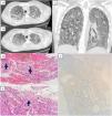
A 26-year-old female patient with a medical history of acute myeloid leukemia (on clinical remission after consolidation treatment) and bipolar disorder was admitted from the emergency department due to fever, dyspnea and severe hypoxemia (PaO2 53mmHg room air). Initial management included initiation of empiric Piperacillin–Tazobactam and high-flow-nasal-cannula oxygen support. Chest computed tomography demonstrated diffuse ground glass opacities with a crazy paving pattern, indicative of pulmonary alveolar proteinosis (PAP) (Fig. 1 A, B and C). A bronchoscopy with a bronchoalveolar lavage initially yielded no identifiable pathogens but it had a milky appearance and was positive for Periodic Acid-Schiff staining. Transbronchial lung biopsies were performed, confirming PAP but also revealing cells with eosinophilic inclusions (Fig. 1D). Immunochemistry confirmed cytomegalovirus (CMV) infection in these cells (Fig. 1F) and DNA PCR levels for CMV were over 10,000,000UI/mL. Despite prompt treatment with ganciclovir and three segmental lung lavages using 3L of saline solution each, the patient's condition deteriorated rapidly, leading to her unfortunate passing away. PAP is characterized by the accumulation of surfactant in the alveoli and in rare cases may be associated to hematological or infectious diseases.1 There have been reports of CMV-associated infections, which are more common in immunocompromised patients.2,3 This case underscores the potential association of PAP with CMV infection in immunocompromised patients.

(A–C) Chest computed tomography (sagittal – A and B-l and coronal – C – views) displaying crazy paving pattern in PAP; D: Pulmonary parenchyma with scarce inflammatory infiltrate with scattered cells displaying eosinophilic nuclear inclusions (arrows). (E) An eosinophilic cotton-like finding is consistent with alveolar proteinosis (arrow F): The cells with inclusions tested positive for immunochemistical staining for CMV.
The authors state that they have no conflict of interests.


