Journal Information
Visits
680
Vol. 60. Issue 3.
Pages 180 (March 2024)
Clinical Image
Full text access
Lung Ultrasound in ARDS: B-lines Pattern and Shred Sign
Visits
680
Juan Francisco Muñoz Moreno
Corresponding author
juanfran9juanfran9@gmail.com

Corresponding author.
, Ester Rubio Prieto, María Ángela Magro Martín
Hospital General Universitario de Ciudad Real, Ciudad Real, Spain
This item has received
Article information
Full Text
Bibliography
Download PDF
Statistics
Figures (2)
Additional material (1)
Full Text

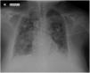
A 56-year-old woman was admitted to the Intensive Care Unit due to urological septic shock and hypoxemic respiratory failure. The chest X-ray revealed bilateral pulmonary infiltrates with an alveolar pattern, and the patient was diagnosed with adult respiratory distress syndrome (Fig. 1).

Fig. 1.

Bilateral and symmetrical ground-glass opacities. Signs of adult respiratory distress syndrome.

(0.07MB).

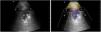
Pleuropulmonary ultrasound shows the two most frequent patterns of this respiratory pathology: 1st. “Comet tail” images or B Pattern, with unstructured pleural line, due to thickening of the interlobular septa due to inflammation (Video left). 2nd. Shred sign, characteristic of non-translobar subpleural consolidations/collections, with the shape of an inverted triangle with irregular and poorly defined edges (Video right and Fig. 2).1

Fig. 2.

Lung ultrasound showing shred sign (non-translobar subpleural collections, with the shape of an inverted triangle). C: ribs; M: intercostal muscles; S: acoustic shadowing; TCS: subcutaneous cellular tissue.

(0.05MB).

ARDS lung lesions are characterized by B pattern (at least three mixed hyperechoic shadows perpendicular to the pleural line). B-Lines are not uniform and appear patchy throughout both lungs, finding normal lung tissues (denoted by the A-line) between B patterns. This distribution is known as non-uniform interstitial syndrome; existing focal changes that include mild diffuse lesions and other regions with severe damage, with shred sign and translobar consolidations, more frequent in less ventilated posterior areas around 30%–50%.2

Authors’ Contributions

The three authors, as a work team, have contributed in a similar way both in the diagnosis and treatment of the patient, and in the preparation and writing of the manuscript.

Funding

No funding to complete the manuscript.

Conflict of Interests

The authors state that they have no conflict of interests.

Appendix B
Supplementary data

The following are the supplementary data to this article:

(1.3MB)

Lung ultrasound. Left: pattern B ultrasound (Comet tail), with unstructured pleural line, due to thickening of the interlobular septa due to inflammation. Right: shred sign, characteristic of non-translobar subpleural consolidations/collections.

References
[1]
D. Lichtenstein.
Lung ultrasound in the critically ill.
Curr Opin Crit Care, 20 (2014), pp. 315-322
[2]
C. Lock, C.M. Nix.
Use of point-of-care lung ultrasonography in the critical care setting as an aid to identifying the correct diagnosis in an acutely desaturating patient with COVID-19-related acute respiratory distress syndrome.
BMJ Case Rep, 14 (2021), pp. e240891
Copyright © 2023. SEPAR
Archivos de Bronconeumología
Article options
Tools

Are you a health professional able to prescribe or dispense drugs?