Journal Information
Share
Share
Download PDF
More article options
Visits
958
Original Article
Full text access
Available online 12 December 2024
New Classifications of COPD Severity Based on Impulse Oscillometry: A SAIO Grade Base on ECOPD Cohort in China
Visits
958
Lifei Lua,1, Qi Wana,1, Gaoying Tanga,1, Fan Wua, Zhishan Denga, Jieqi Penga,b, Cuiqiong Daia, Kunning Zhoua, Xiaohui Wua, Shuqing Yua, Yongqing Huangc, Changli Yangd, Shengtang Chend, Pixin Rana,b,
Corresponding author
pxran@gzhmu.edu.cn

Corresponding authors.
, Yumin Zhoua,b,
Corresponding author
zhouyumin410@126.com

Corresponding authors.
a Department of Pulmonary and Critical Care Medicine, State Key Laboratory of Respiratory Disease, National Clinical Research Center for Respiratory Disease, National Center for Respiratory Medicine, Guangzhou Institute of Respiratory Health, The First Affiliated Hospital of Guangzhou Medical University, Guangzhou, China
b Guangzhou National Laboratory, Guangzhou, China
c Lianping County People's Hospital, Heyuan, China
d Wengyuan County People's Hospital, Shaoguan, China
This item has received
Received 13 August 2024. Accepted 10 October 2024
Article information
Abstract
Full Text
Bibliography
Download PDF
Statistics
Figures (5)
Show moreShow less
Tables (1)
Table 1. Demographics.
Additional material (1)
Abstract
Background

Recently, the severities of chronic obstructive pulmonary disease (COPD) can also be assessed by impulse oscillometry (IOS). This study aimed to explore a new classification of severity of COPD based on IOS and associations with acute exacerbations (AE) in patients with COPD.

Methods

The data of our study were based on the baseline and 2-year follow-up data of a prospective cohort in China. COPD was defined as post-bronchodilator FEV1/FVC <0.70. A new severity classification (staging of airflow obstruction by IOS, SAIO) was evaluated based on IOS parameters (R5, R5–R20, and X5 z-scores). We quantified using the weighted Bangdiwala B for agreement of severities of COPD between IOS parameters and FEV1%pred. The differences among SAIO stages were performed in symptom scores and imaging using analysis of covariance, and in the AE using Poisson regression.

Results

Overall, 833 patients with COPD were included in this study. The weighted Bangdiwala B of R5, R5–R20, X5 z-scores, and FEV1%pred for evaluating agreement of the severities of COPD was 0.68, 0.70 and 0.83, respectively. The SAIO classifications system identified a greater number of patients with stage III–IV. SAIO provided significant discrimination between the stage I and stage III, IV for symptom scores, emphysema, and air trapping. SAIO provided significant discrimination between the stage I and other stages for AE.

Conclusions

The SAIO classifications provide discrimination between the stage I and stage III, IV for symptom scores, emphysema, air trapping, and AE, similar to the GOLD classifications.

Trial registration

Chinese Clinical Trial Registry, ChiCTR1900024643. Registered on 19 July, 2019.

Keywords:
Impulse oscillometry
Severity of chronic obstructive pulmonary disease
Lung function decline
Acute exacerbations
Graphical abstract
Full Text
Introduction

Chronic obstructive pulmonary disease (COPD) ranks as the third leading cause of death globally, following ischemic heart disease and stroke.1 Characteristics of COPD are persistent airflow limitation and corresponding respiratory symptoms such as dyspnea, cough, and sputum production. COPD is usually diagnosed at an advanced stage, resulting in a heavy disease burden with high rates of mortality and disability. With the aging population increasing in high-income countries and rising smoking rates in developing nations, the number of deaths due to COPD is projected to exceed 5.4 million by 2060.2 A national cross-sectional study in China reported that approximately 100 million individuals have COPD, and prevalence was higher in men than in women (11.9% vs 5.4%).3 COPD in women has distinct characteristics expressed differently compared to men in terms of respiratory symptoms and disease outcomes. Sex differences in COPD management and therapy also warrant attention.4–6 The Global Initiative for Chronic Obstructive Lung Disease (GOLD) guidelines recommend diagnosing COPD with a post-bronchodilator forced expiratory volume in 1s (FEV1)/forced vital capacity (FVC) ratio <0.70 and using FEV1% predicted to assess the severity of the obstruction.1 FEV1% predicted was associated with increased symptoms and long-term mortality.7,8 Recently, some studies have proposed new classifications of severity of the obstruction, including FVC-adjusted FEV1% predicted, the FEV1/FVC ratio, and FEV1z-scores.7,9–11 However, regardless of the obstruction standard employed, these measures are all based on spirometry tests. Spirometry tests require patients to exert maximal effort in exhalation, and certain clinical patients with severe dyspnea, may not complete spirometry tests. Consequently, clinicians faced significant challenges in assessing the severity of COPD to guide pharmacological treatment. Therefore, there is an urge to find supplementary methods to indirectly evaluate the severity of COPD.

Impulse oscillometry (IOS) is a technique to measure airway resistance during calm breathing but not require patient effort. IOS plays a crucial role in assessing small airway dysfunction, expiratory flow limitation, monitoring disease recovery, and disease progression.12–16 Recently, Liang et al. proposed new obstruction severity classifications using IOS z-scores, establishing cutoff values for grading the severity of COPD using IOS parameters[resistance at 5Hz (R5), difference between resistance at 5Hz and 20Hz (R5–R20), reactance at 5Hz (X5) z-scores]: stage I, II, III, IV (staging of airflow obstruction by IOS, SAIO).17 However, it remained unclear whether SAIO grades were correlated with respiratory symptoms, image abnormalities, and prognosis and had similar results to the GOLD classifications.

The objective of our study was to identify a supplementary method to assess COPD severity and guide treatment of patients with COPD. We hypothesized that SAIO grades may correlate with respiratory symptoms, image abnormalities, and prognosis similarly to GOLD classifications. To test this hypothesis, we conducted a prospective cohort study in China to investigate the associations between SAIO grades and symptom scores, imaging abnormalities, acute exacerbations (AE) and lung function decline at a 2-year follow-up.

MethodSettings and Participants

We analyzed data from a prospective population-based cohort study in Guangdong Province, China. The details of the cohort design have been published previously.18 Briefly, individuals aged 40–80 years were enrolled in the study between July 2019 and August 2021, including approximately 2000 individuals with never, current, or former cigarette smoking history with or without COPD (post-bronchodilator FEV1/FVC <0.7). Individuals were excluded if they met any of the following criteria at baseline: (1) age <40 years or >80 years; (2) incomplete IOS tests; (3) respiratory infection or exacerbations within four weeks prior to screening; (4) heart attack (myocardial infarction or malignant arrhythmia) within the past three months. The previous cohort design report contained more details. Our study only included patients with COPD (post-bronchodilator FEV1/FVC <0.7). At baseline, participants filled up questionnaires including the modified Medical Research Council dyspnea scale (mMRC) and COPD assessment test (CAT) scores.19 Information regarding medical history and risk factors was collected including family history of respiratory diseases, occupational exposure, and biomass exposure.

To avoid airway constriction after spirometry testing that could affect airway resistance measurements, pre-bronchodilator IOS (CareFusion, Hochberg, Germany) testing was performed according to European Respiratory Society (ERS) 2003 guidelines prior to spirometry testing.20 IOS parameters include resistance at 5Hz (R5), resistance at 20Hz (R20), the difference between R5 and R20 (R5–R20), reactance at 5Hz (X5), reactance area (AX), and resonant frequency (Fres). The z-scores of IOS parameters were calculated using the predicted value formulas of IOS in healthy Chinese subjects.21

Subsequently, spirometry (CareFusion, Yorba Linda, CA, USA) tests were included for analysis if they met the American Thoracic Society (ATS)/ERS acceptability and repeatability criteria, with post-bronchodilator spirometry being performed after inhalations of 400μg albuterol.22 These participants completed questionnaires, pre-bronchodilator IOS tests, and pre- and post-bronchodilator spirometry tests at each visit. COPD was defined as post-bronchodilator FEV1/FVC <0.70.

This study adhered to the ethical guidelines outlined in the Declaration of Helsinki. The research protocol received approval from the Ethics Committee of the First Affiliated Hospital of Guangzhou Medical University (Approval No. 2018-53) prior to initiation. Written informed consent was obtained from all participants prior to their enrollment in the study.

Imaging

Quantitative assessment of CT images was performed using the 3D Slicer 4.11 software within the Chest Imaging Platform. Emphysema was defined as the percentage of voxels with attenuation values less than −950 Hounsfield units at maximal inspiration (LAA−950), while air trapping was quantified as the percentage of voxels with attenuation values less than −856 Hounsfield units at end-expiration (LAA−856).23,24

Definitions and Outcomes

A new severity classification (staging of airflow obstruction by IOS, SAIO) from the Liang et al. study17 was evaluated based on IOS parameters (R5, R5–R20, X5 z-scores). As we explored the effect of the new classification in patients with COPD, we adjusted the COPD severities definitions based on SAIO stages as follows:

  • 1.

    R5 z-scores. Stage I: z-scores ≤1.645; stage II: 1.645 z-scores ≤2.5; stage III: 2.5 z-scores ≤4; stage IV: R5 z-scores >4.

  • 2.

    R5–R20 z-scores. Stage I: z-scores ≤1.645; stage II: 1.645 z-scores ≤3; stage III: 3 z-scores ≤5; stage IV: R5–R20 z-scores >5.

  • 3.

    X5 z-scores. Stage I: z-scores ≥−1.645; stage II: −1.645 z-scores ≤−4.5; stage III: −4.5 z-scores ≤−8.5; stage IV: X5 z-scores >−8.5.

Meanwhile, the severity of obstruction was graded (GOLD stages) using spirometry parameters FEV1% predicted value based on European Coal and Steel Community 1993: stage I, FEV1% predicted ≥80%; stage II, 50% ≤FEV1% predicted <80%; stage III, 30% ≤FEV1% predicted <50%; stage IV, FEV1% predicted <30%.1

We defined AE as the onset or worsening of at least two of the following symptoms: cough, sputum production, purulent sputum, wheezing, and dyspnea lasting for at least 48h after excluding self-reported left and right heart dysfunction, pulmonary embolism, pneumothorax, pleural effusion, arrhythmia, and other diseases.18 Moderate–severe AE were defined as those resulting in outpatient or emergency department visits, even hospitalization, or the need for COPD medication.25,26

Statistical Analyses

Firstly, we assessed concordance between the GOLD stages and SAIO stages using Bangdiwala plots, and quantified using the weighted Bangdiwala B for agreement between multiple classes, which adjusts for the frequency of each severity class. Subsequently, we compared the difference among different SAIO stages groups in mMRC score, CAT score, %LAA−950, and %LAA−856 using analysis of covariance at baseline.

We then constructed a Poisson regression analysis to assess the associations between SAIO grade and AE. The model included covariates: age, sex, smoking status, pack-years, body mass index (BMI), family history of respiratory diseases, occupational exposure, and biomass exposure. To account for differences in individual follow-up time, the natural logarithm (ln) of follow-up time was adjusted as an offset variable. A linear mixed-effects model was applied to explore the associations between decline in FEV1 and the SAIO grades. The model included covariates: age, sex, smoking status, pack-years, body mass index (BMI), family history of respiratory diseases, occupational exposure, biomass exposure, and post-bronchodilator FEV1. All analyses were conducted using IBM SPSS 27.0 and R statistical package v4.3.3, with a two-sided alpha of 0.05 considered statistically significant.

Results

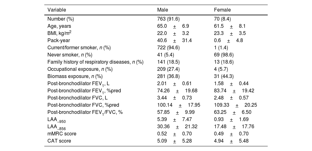
Patients with COPD who had acceptable IOS data were enrolled in our study, of 833 patients. Table 1 presents the demographic characteristics of patients with COPD stratified by sex. Of the patients, 91.6% were male and 8.4% were female. Among males, 94.6% were current or former smokers, while 44.3% of females had a history of biomass exposure. The mean±standard deviation (SD) FEV1 of male patients was 2.01±0.61L. Table S1 illustrates the categorization of airflow obstruction using SAIO and GOLD stages. The proportion of grade I–IV assessed by R5 z-score was 42.3%, 11.6%, 19.8%, 26.3%; assessed by R5–R20 was 38.7%, 15.7%, 16.8%, 28.8%; assessed by X5 was 48.7%, 25.5%, 14.2%, 11.6%; assessed by FEV1%pred was 44.1%, 43.5%, 10.6%, 1.9%. Most subjects who changed categorization shifted from a less severe FEV1% predicted category to a more severe R5 z-score or R5–R20 z-score (Fig. 1).

Table 1.

Demographics.

Variable  Male  Female 
Number (%)  763 (91.6)  70 (8.4) 
Age, years  65.0+6.9  61.5+8.1 
BMI, kg/m2  22.0+3.2  23.3+3.5 
Pack-year  40.6+31.4  0.6+4.8 
Current/former smoker, n (%)  722 (94.6)  1 (1.4) 
Never smoker, n (%)  41 (5.4)  69 (98.6) 
Family history of respiratory diseases, n (%)  141 (18.5)  13 (18.6) 
Occupational exposure, n (%)  209 (27.4)  4 (5.7) 
Biomass exposure, n (%)  281 (36.8)  31 (44.3) 
Post-bronchodilator FEV1, L  2.01+0.61  1.58+0.44 
Post-bronchodilator FEV1, %pred  74.26+19.68  83.74+19.42 
Post-bronchodilator FVC, L  3.44±0.73  2.48±0.57 
Post-bronchodilator FVC, %pred  100.14+17.95  109.33+20.25 
Post-bronchodilator FEV1/FVC, %  57.85+9.99  63.25+6.50 
LAA−950  5.39+7.47  0.93+1.69 
LAA−856  30.36+21.32  17.48+17.76 
mMRC score  0.52+0.70  0.49+0.70 
CAT score  5.09+5.28  4.94+5.48 

BMI: body mass index; FEV1: forced expiratory volume in 1s; FVC: forced vital capacity; LAA−950: low-attenuation area of the lung with attenuation values below −950 Hounsfield units; LAA−856: low-attenuation area of the lung with attenuation values below −856 Hounsfield units; mMRC, modified British medical research council score; CAT, COPD assessment test.

Fig. 1.

Airflow obstruction categorization for SAIO and GOLD stages. R5: resistance at 5Hz; R5–R20: difference from R5 to R20; X5: reactance at 5Hz; FEV1: forced expiratory volume in 1s; FVC: forced vital capacity.

(0.17MB).
Concordance Between GOLD and SAIO Classes

Fig. 2 illustrates the Bangdiwala plots depicting agreement between GOLD and R5 z-scores, R5–R20 z-scores, and X5 z-scores. The agreement, measured by the weighted Bangdiwala B, between GOLD stages and R5 z-scores, R5–R20 z-scores, and X5 z-scores was 0.68, 0.70, and 0.83, respectively.

Fig. 2.

Bangdiwala agreement charts comparing classification of severity of airflow obstruction using GOLD and SAIO severity schema. R5: resistance at 5Hz; R5–R20: difference from R5 to R20; X5: reactance at 5Hz; GOLD: Global Initiative for Chronic Obstructive Lung Disease.

(0.21MB).
Symptom score, Emphysema, Air trapping Among SAIO Stages and GOLD Stages

Fig. 3 displays the difference of symptom score, emphysema, air trapping across SAIO stages. After adjusting for age, sex, smoking status, pack-years, BMI, family history of respiratory diseases, occupational exposure, and biomass exposure, the analysis of covariance indicated that SAIO stages III and IV were associated with higher mMRC and CAT scores, as well as more severe emphysema and air trapping, compared to stage I. However, no significant differences were observed between SAIO stage I and II in symptom scores, emphysema, and air trapping. Meanwhile, the GOLD stages demonstrated significant differentiation in terms of stages I–IV in symptom scores, emphysema, and air trapping (Fig. S1).

Fig. 3.

The difference among SAIO stages in symptom score, emphysema, and air trapping. Data are mean±SE. R5: resistance at 5Hz; R5–R20: difference from R5 to R20; X5: reactance at 5Hz; LAA−950: low-attenuation area of the lung with attenuation values below −950 Hounsfield units; LAA−856: low-attenuation area of the lung with attenuation values below −856 Hounsfield units; mMRC: modified British medical research council score; CAT: COPD assessment test. ***P<0.001; **P<0.05.

(0.21MB).
AE

The SAIO stages exhibited similar discriminatory power to GOLD stages in predicting AE at 2-year follow-up between stage I and stage II–IV. After adjusting for age, sex, smoking status, pack-years, BMI, family history of respiratory diseases, occupational exposure, biomass exposure and follow-up time. Compared with SAIO stage I, the incidence rate ratio (IRR) for the presence of SAIO stages assessed by R5 z-scores II through IV airflow obstruction were 1.33 (95% CI 1.03–1.71), 1.30 (95% CI 1.05–1.60), and 1.91 (95% CI 1.60–2.28). The IRR for the presence of SAIO stages assessed by R5–R20 z-scores II through IV airflow obstruction were 1.38 (95% CI 1.09–1.74), 1.38 (95% CI 1.10–1.74), and 2.02 (95% CI 1.68–2.43); The IRR for the presence of SAIO stages assessed by X5 z-scores II through IV airflow obstruction were 1.04 (95% CI 0.86–1.26), 1.79 (95% CI 1.48–2.18), and 1.38 (95% CI 1.10–1.73). Similarly, compared with GOLD stage I, IRR for the presence of GOLD stages II through IV airflow obstruction were 1.61 (95% CI 1.37–1.90), 1.47 (95% CI 1.14–1.90), and 2.08 (95% CI 1.29–3.37) (Fig. 4). We additionally analyzed the differences in moderate-to-severe AE among the SAIO stages and GOLD stages. Our results showed that SAIO stages III–IV had higher risk of moderate-to-severe AE than stage I, but no differences in moderate-to-severe AE were observed between SAIO stages I and II (Fig. S2). Then subgroup analysis stratified by sex was conducted, our results showed that significant differences in AE or moderate-to-severe AE between SAIO stages III–IV and stages I was observed in male individuals but hardly any in female individuals (Figs. S3–S6).

Fig. 4.

The difference among SAIO stages in acute exacerbations in overall individuals. Data are mean±SE. AE: acute exacerbations; R5: resistance at 5Hz; R5–R20: difference from R5 to R20; X5: reactance at 5Hz; GOLD: Global Initiative for Chronic Obstructive Lung Disease; IRR: the incidence rate ratio.

(0.3MB).
Lung Function Decline

After adjusting for age, sex, smoking status, pack-years, BMI, family history of respiratory diseases, occupational exposure, biomass exposure and post-bronchodilator FEV1, a linear mixed-effects model indicated no significant differences in the decline in FEV1 among SAIO grade I–IV. However, our results showed that GOLD grade I had a faster decline in FEV1 than grade IV (Fig. S7).

Discussion

Our study presents a novel severity classification based on IOS parameters (SAIO stages) and investigates their associations with symptoms, imaging abnormalities, AE and lung function decline. Our study has reported several significant findings. Initially, SAIO stages evaluated by X5 z-scores demonstrated a stronger agreement with GOLD stages in severity classifications compared to other parameters. Secondly, the SAIO grades identify a higher proportion of patients in stages III–IV compared to the GOLD grades. Finally, SAIO grade I and III–IV exhibited similar discrimination in symptom scores, emphysema, air trapping symptoms, and AE compared to GOLD grades.

The SAIO stages were defined based on severity classifications of COPD using FEV1z-scores.17 However, the clinical significance of the SAIO stages in COPD remains unclear. In this study, we utilized FEV1%pred instead of FEV1z-score to evaluate COPD severity. Several factors were considered: (1) the ATS/ERS and GOLD guidelines recommend using FEV1%pred for severity classifications. (2) Although the FEV1z-score can eliminate biases related to age, sex, height, and race,27 in terms of mortality, a study by Hegewald et al. indicated that FEV1%pred was more accurate in predicting the risk of 5-year mortality than FEV1z-score.7 Therefore, we focused on the agreement between the GOLD stages as assessed by FEV1%pred and SAIO stages. We found that the SAIO stages assessed by X5 z-scores demonstrated better agreement with GOLD stages in severity classifications compared to other parameters. This result indicated that the reactance value was more appropriate than the resistance values for reflecting GOLD stages using FEV1%pred. We further analyzed the distributions of SAIO and GOLD stages. Compared to the GOLD classifications, the SAIO classifications, as defined by R5 and R5–R20, divided the GOLD stage II into SAIO III–IV. We believed that this phenomenon was attributable to the different physiological mechanisms of each indicator. Firstly, Rrs (R5 and R5–R20) primarily reflects changes in airway diameter, particularly the degree of small airway stenosis.28 Small airway disease represented an early lesion in the progression to COPD.29 Conversely, FEV1%pred primarily reflected the obstruction of large airway, and may not be sensitive to small airway lesions.30,31 Thus, small airway disease may occur before abnormalities in FEV1 become evident. This result also explained why the SAIO classifications defined by R5 and R5–R20 divided GOLD stage II into SAIO III–IV.

The strength of our study lies in exploring the distribution of the new SAIO classifications in COPD and analyzing the differences in physiological indicators and AE among SAIO stages. Clinically, some patients cannot cooperate with spirometry due to the exertion of maximal effort in exhalation, which makes it challenging for clinicians to assess disease severity in these cases. Our study aimed to provide an indirect, effortless tool for evaluating the severity of COPD patients who cannot cooperate with lung function tests. We further analyzed the discriminatory ability of SAIO grades in assessing severity through symptom scores and imaging. Our study showed that SAOI stage III–IV had higher mMRC and CAT scores, more severe emphysema, and air trapping than those with stage I. Similar findings were also observed in GOLD grades. Meanwhile, GOLD stage II showed higher mMRC and CAT scores, more severe emphysema, and air trapping compared to GOLD stage I. This finding was consistent with that reported by Bhatt et al., who found that mMRC score and emphysema worsen with increasing GOLD grade severity in the Pittsburgh Cohort.9 However, no significant differences were observed in symptom scores, emphysema, or air trapping between SAIO grade I and II. This is not surprising. Patients with GOLD grade I and II have already exhibited imaging abnormalities (emphysema, air trapping), whereas SAIO grade I and II only exhibit early lesions (such as SAD), and these patients may not present with specific imaging abnormalities or obvious respiratory symptoms. Of course, our results showed that SAIO classifications may be more effectively used to assist in the evaluation of patients with GOLD III to IV who cannot cooperate with lung function. Due to severe dyspnea typically occurred in these patients. It was difficult for the patients to complete spirometry. IOS only requires calm breathing and has a similar distinction of symptoms, imaging, and AE as spirometry. Therefore, IOS can be used to assist in evaluating the severity of patients with GOLD III–IV to guide treatment.

The goal of COPD treatment is to alleviate current symptoms and reduce the risk of future AE.1 We analyzed the differences in the number of AE between GOLD and SAIO classifications based on 2-year follow-up data. We found that GOLD grades demonstrated results similar to SAIO grades in predicting AE. Patients with grade II–IV experienced a higher number of AE over the following two years compared to those with grade I. This result indicated that both the GOLD and SAIO grades were effective in predicting the risk of AE, suggesting their potential as alternative tools in clinical practice. Identifying high-risk patients with grades II–IV can assist clinicians in developing personalized treatment plans that emphasize aggressive intervention and management to mitigate the occurrence of AE. Interestingly, it has been observed that patients classified in stage III of the X5 parameter presented a higher number of exacerbations than those in stage IV. We considered possible explanations, such as limited sample size or patient heterogeneity across these stages. Additionally, it would be beneficial to reflect on the need for future studies with larger samples to verify this trend and gain a better understanding of the behavior of the X5 parameter in COPD. Finally, in lung function decline, we only found GOLD grade I had a faster decline in FEV1 than grade IV. The result was consistent with previous studies that have reported faster lung function decline in patients with GOLD I compared to patients with advanced COPD.32,33 However, no differences were found in the FEV1 decline among SAIO grades. We also guessed that the distribution gap of SAIO and GOLD grades could play an important role, especially as some patients with GOLD grade II were classified as SAIO grade I, III–IV. Lung function decline was measured on the basis of FEV1, and there was a so-called “horse race effect” that patients with advanced COPD had slower lung function decline than mild to moderate COPD because of a lower baseline FEV1. Inconsistent GOLD classifications exist in all SAIO grades, which also affects disease prognosis to some extent. Long-term follow-up is needed in the future to explain this potential mechanism.

There were some limitations to our research. First of all, due to a small number of patients with GOLD grade III–IV in our study, in the more advanced stages, where variability in exacerbations could influence the results. Future large cohort studies will be necessary to further elucidate the relationship between SAIO classifications and the prognosis of COPD. Secondly, our study only demonstrated that SAIO classifications and GOLD classifications had similar effects in predicting AE, but we found that SAIO classifications do not offer advantages in distinguishing certain physiological reactions than GOLD classifications in patients with grade I and II. Therefore, new physiological indicators are needed to explore the advantages of SAIO grades. Thirdly, all-cause mortality is an important indicator for assessing disease prognosis.2 Since our cohort was followed up for only 2 years and there have been few fatalities, we could not compare differences in all-cause mortality between SAIO and GOLD classifications. Furthermore, the SAIO classifications were based on the IOS parameter z-scores, which were affected by race,28 and lacked validation of results in other countries.34,35 Therefore, the generalizability of our study's findings was limited. Moreover, ΔX5, the difference between inspiratory and expiratory reactance at 5Hz, was an indicator of assessing expiratory flow limitation in COPD. ΔX5 showed significantly correlated with FEV1, and reflected the severity of COPD to some extent.36,37 Therefore, exploring whether ΔX5 can be used as a new classification of COPD is worth considering. However, we currently lack a cutoff value for ΔX5 to effectively assess the severity classifications.

Conclusion

The new severity classifications, SAIO grades, provide discrimination between the stage I and stage III, IV for symptom score, emphysema, air trapping, and AE similar to the GOLD classifications. The SAIO classifications method offer an alternative assessment for patients unable to complete spirometry, thereby assisting clinicians in gaining a comprehensive understanding of a patient's disease status, leading to more accurate diagnosis and treatment decisions. With further research and clinical validation, the SAIO classifications are expected to become a crucial tool in the management of COPD.

CRediT Authorship Contribution Statement

P.X.R., Y.M.Z., and L.F.L. designed the project and planned the statistical analysis. L.F.L. drafted and revised the paper. L.F.L., Q.W., G.Y.T., F.W., Z.S.D., J.Q.P., C.Q.D., K.N.Z., X.H.W., S.Q.Y., Y.Q.H., C.L.Y., S.T.C., P.X.R., and Y.M.Z. collected and monitored the data collection. All authors approved the final draft of the manuscript for publication. L.F.L. take responsibility for the integrity of the data and the accuracy of the data analysis. L.F.L. is the study guarantors.

Ethics Approval and Consent to Participate

This study adhered to the ethical guidelines outlined in the Declaration of Helsinki. The research protocol received approval from the Ethics Committee of the First Affiliated Hospital of Guangzhou Medical University (Approval No. 2018-53) prior to initiation. Written informed consent was obtained from all participants prior to their enrollment in the study.

Consent for Publication

Not applicable.

Funding

This work was supported by Foundation of Guangzhou National Laboratory (SRPG22-018 and SRPG22-016), the National Natural Science Foundation of China (81970045, 81970038, 82270043), the Clinical and Epidemiological Research Project of State Key Laboratory of Respiratory Disease (SKLRD-L-202402), and Major Clinical Research Project of Guangzhou Medical University's Scientific Research Capability Improvement Plan (GMUCR2024-01012) the Guangzhou Science and Technology Plans (No. 202201020372), the grant of State Key Laboratory of Respiratory Disease (SKLRD-Z-202315), and Research Capability Strengthening and Fundamental Improvement Project of Guangzhou Medical University (2024SRP080).

Acknowledgements

We thank all the participants who participated in the study. We would like to express our appreciation to Xiang Wen, Shan Xiao, Peiyu Huang, Bijia Lin, Shaodan Wei, Xiaopeng Ling, Heshen Tian, Zihui Wang (State Key Laboratory of Respiratory Disease, National Clinical Research Center for Respiratory Disease, National Center for Respiratory Medicine, Guangzhou Institute of Respiratory Health, The First Affiliated Hospital of Guangzhou Medical University), Jianhui Huang, and Xiangwen Luo (Lianping County People's Hospital) for their effort in collecting and verifying the data.

Appendix B
Supplementary Data

The following are the supplementary data to this article:

References
[1]
A. Agustí, B.R. Celli, G.J. Criner, D. Halpin, A. Anzueto, P. Barnes, et al.
Global Initiative for Chronic Obstructive Lung Disease 2023 Report: GOLD Executive Summary.
[2]
A.I. Papaioannou, G. Hillas, S. Loukides, T. Vassilakopoulos.
Mortality prevention as the centre of COPD management.
[3]
C. Wang, J. Xu, L. Yang, Y. Xu, X. Zhang, C. Bai, et al.
Prevalence and risk factors of chronic obstructive pulmonary disease in China (the China Pulmonary Health [CPH] study): a national cross-sectional study.
Lancet, 391 (2018), pp. 1706-1717
[4]
T.A. Perez, E.G. Castillo, J. Ancochea, M.T. Pastor Sanz, P. Almagro, P. Martínez-Camblor, et al.
Sex differences between women and men with COPD: a new analysis of the 3CIA study.
Respir Med, 171 (2020), pp. 106105
[5]
P. Rogliani, F. Cavalli, B.L. Ritondo, M. Cazzola, L. Calzetta.
Sex differences in adult asthma and COPD therapy: a systematic review.
Respir Res, 23 (2022), pp. 222
[6]
M.G. Matera, J. Ora, L. Calzetta, P. Rogliani, M. Cazzola.
Sex differences in COPD management.
Expert Rev Clin Pharmacol, 14 (2021), pp. 323-332
[7]
M.J. Hegewald, D.S. Collingridge, T.W. DeCato, R.L. Jensen, A.H. Morris.
Airflow obstruction categorization methods and mortality.
Ann Am Thorac Soc, 15 (2018), pp. 920-925
[8]
Y. Zhou, M.R. Ampon, M.J. Abramson, A.L. James, G.P. Maguire, R. Wood-Baker, et al.
Respiratory symptoms, disease burden, and quality of life in Australian adults according to GOLD spirometry grades: data from the BOLD Australia Study.
Int J Chron Obstruct Pulmon Dis, 18 (2023), pp. 2839-2847
[9]
S.P. Bhatt, A. Nakhmani, S. Fortis, M.J. Strand, E.K. Silverman, F.C. Sciurba, et al.
FEV(1)/FVC severity stages for chronic obstructive pulmonary disease.
Am J Respir Crit Care Med, 208 (2023), pp. 676-684
[10]
P.M.A. Calverley.
A STAR is born: a new approach to assessing chronic obstructive pulmonary disease severity.
Am J Respir Crit Care Med, 208 (2023), pp. 647-648
[11]
H. Backman, L. Vanfleteren, D.M. Mannino, M. Ekström.
Severity of airflow obstruction based on FEV(1)/FVC vs FEV(1)% of predicted in the general US population.
Am J Respir Crit Care Med, 210 (2024), pp. 1308-1316
[12]
L. Lu, J. Peng, F. Wu, H. Yang, Y. Zheng, Z. Deng, et al.
Clinical characteristics of airway impairment assessed by impulse oscillometry in patients with chronic obstructive pulmonary disease: findings from the ECOPD study in China.
BMC Pulm Med, 23 (2023), pp. 52
[13]
L. Lu, J. Peng, N. Zhao, F. Wu, H. Tian, H. Yang, et al.
Discordant spirometry and impulse oscillometry assessments in the diagnosis of small airway dysfunction.
Front Physiol, 13 (2022), pp. 892448
[14]
M.K. Johnson, M. Birch, R. Carter, J. Kinsella, R.D. Stevenson.
Measurement of physiological recovery from exacerbation of chronic obstructive pulmonary disease using within-breath forced oscillometry.
Thorax, 62 (2007), pp. 299-306
[15]
R.L. Dellacà, N. Duffy, P.P. Pompilio, A. Aliverti, N.G. Koulouris, A. Pedotti, et al.
Expiratory flow limitation detected by forced oscillation and negative expiratory pressure.
Eur Respir J, 29 (2007), pp. 363-374
[16]
S.C. Zimmermann, J. Huvanandana, C.D. Nguyen, A. Bertolin, J.C. Watts, A. Gobbi, et al.
Day-to-day variability of forced oscillatory mechanics for early detection of acute exacerbations in COPD.
[17]
X. Liang, J. Zheng, Y. Gao, Z. Zhang, W. Han, J. Du, et al.
Clinical application of oscillometry in respiratory diseases: an impulse oscillometry registry.
[18]
F. Wu, Y. Zhou, J. Peng, Z. Deng, X. Wen, Z. Wang, et al.
Rationale and design of the early chronic obstructive pulmonary disease (ECOPD) study in Guangdong, China: a prospective observational cohort study.
J Thorac Dis, 13 (2021), pp. 6924-6935
[19]
P.W. Jones, G. Harding, P. Berry, I. Wiklund, W.H. Chen, N. Kline Leidy.
Development and first validation of the COPD Assessment Test.
Eur Respir J, 34 (2009), pp. 648-654
[20]
E. Oostveen, D. MacLeod, H. Lorino, R. Farre, Z. Hantos, K. Desager, et al.
The forced oscillation technique in clinical practice: methodology, recommendations and future developments.
Eur Respir J, 22 (2003), pp. 1026-1041
[21]
X.-L. Liang, Y. Gao, W.-J. Guan, J. Du, L. Chen, W. Han, et al.
Reference values of respiratory impedance with impulse oscillometry in healthy Chinese adults.
J Thorac Dis, 13 (2021), pp. 3680
[22]
M.R. Miller, J. Hankinson, V. Brusasco, F. Burgos, R. Casaburi, A. Coates, et al.
Standardisation of spirometry.
Eur Respir J, 26 (2005), pp. 319-338
[23]
D.P. Capaldi, N. Zha, F. Guo, D. Pike, D.G. McCormack, M. Kirby, et al.
Pulmonary imaging biomarkers of gas trapping and emphysema in COPD: 3He MR imaging and CT parametric response maps.
Radiology, 279 (2016), pp. 597-608
[24]
A. Busacker, J.D. Newell Jr., T. Keefe, E.A. Hoffman, J.C. Granroth, M. Castro, et al.
A multivariate analysis of risk factors for the air-trapping asthmatic phenotype as measured by quantitative CT analysis.
Chest, 135 (2009), pp. 48-56
[25]
V. Kim, S.D. Aaron.
What is a COPD exacerbation? Current definitions, pitfalls, challenges and opportunities for improvement.
[26]
N.R. Anthonisen, J. Manfreda, C.P. Warren, E.S. Hershfield, G.K. Harding, N.A. Nelson.
Antibiotic therapy in exacerbations of chronic obstructive pulmonary disease.
Ann Intern Med, 106 (1987), pp. 196-204
[27]
P.H. Quanjer, J.J. Pretto, D.J. Brazzale, P.W. Boros.
Grading the severity of airways obstruction: new wine in new bottles.
Eur Respir J, 43 (2014), pp. 505-512
[28]
G.G. King, J. Bates, K.I. Berger, P. Calverley, P.L. de Melo, R.L. Dellacà, et al.
Technical standards for respiratory oscillometry.
[29]
A. Higham, A.M. Quinn, J.E.D. Cançado, D. Singh.
The pathology of small airways disease in COPD: historical aspects and future directions.
Respir Res, 20 (2019), pp. 49
[30]
W. McNulty, O.S. Usmani.
Techniques of assessing small airways dysfunction.
Eur Clin Respir J, 1. (2014),
[31]
N. Lazarinis, E. Fouka, A. Linden, A. Bossios.
Small airways disease in chronic obstructive pulmonary disease.
Expert Rev Respir Med, 18 (2024), pp. 539-552
[32]
C. Tantucci, D. Modina.
Lung function decline in COPD.
Int J Chron Obstruct Pulmon Dis, 7 (2012), pp. 95-99
[33]
S.P. Bhatt, X. Soler, X. Wang, S. Murray, A.R. Anzueto, T.H. Beaty, et al.
Association between functional small airway disease and FEV1 decline in chronic obstructive pulmonary disease.
Am J Respir Crit Care Med, 194 (2016), pp. 178-184
[34]
L. Gochicoa-Rangel, D. Martínez-Briseño, S. Guerrero-Zúñiga, J. Contreras-Morales, D. Arias-Jiménez, R. Del-Río-Hidalgo, et al.
Reference equations using segmented regressions for impulse oscillometry in healthy subjects aged 2.7–90 years.
[35]
K.I. Berger, M. Wohlleber, R.M. Goldring, J. Reibman, M.R. Farfel, S.M. Friedman, et al.
Respiratory impedance measured using impulse oscillometry in a healthy urban population.
[36]
D. Junhasavasdikul, I. Telias, D.L. Grieco, L. Chen, C.M. Gutierrez, T. Piraino, et al.
Expiratory flow limitation during mechanical ventilation.
[37]
B.B. Aarli, P.M. Calverley, R.L. Jensen, R. Dellacà, T.M. Eagan, P.S. Bakke, et al.
The association of tidal EFL with exercise performance, exacerbations, and death in COPD.
Int J Chron Obstruct Pulmon Dis, 12 (2017), pp. 2179-2188

These authors contributed equally to this work.

Archivos de Bronconeumología
Article options
Tools

Are you a health professional able to prescribe or dispense drugs?