Journal Information
Vol. 57. Issue 7.
Pages 496 (July 2021)
Share
Share
Download PDF
More article options
Vol. 57. Issue 7.
Pages 496 (July 2021)
Clinical image
Full text access
Giant Pulmonary Sequestration Presented as a Paravertebral Mass
Secuestro pulmonar gigante que se presenta como masa paravertebral
Visits
1914
Veysel Ayyildiza, Yener Aydinb,c, Hayri Ogulc,d,
Corresponding author
drhogul@gmail.com

Corresponding author.
a Department of Radiology, Medical Faculty, Suleyman Demirel University, Isparta, Turkey
b Department of Thoracic Surgery, Medical Faculty, Ataturk University, Erzurum, Turkey
c Anesthesiology, Clinical Research Office, Ataturk University, Erzurum, Turkey
d Department of Radiology, Medical Faculty, Ataturk University, Erzurum, Turkey
This item has received
Article information
Full Text
Bibliography
Download PDF
Statistics
Figures (1)
Full Text

A 18-year-old female presented for evaluation of recurrent cough and hemoptysis. A giant pulmonary sequestration mimicking a paravertebral mass was detected in computed tomography (Fig. 1A–D).

Fig. 1.

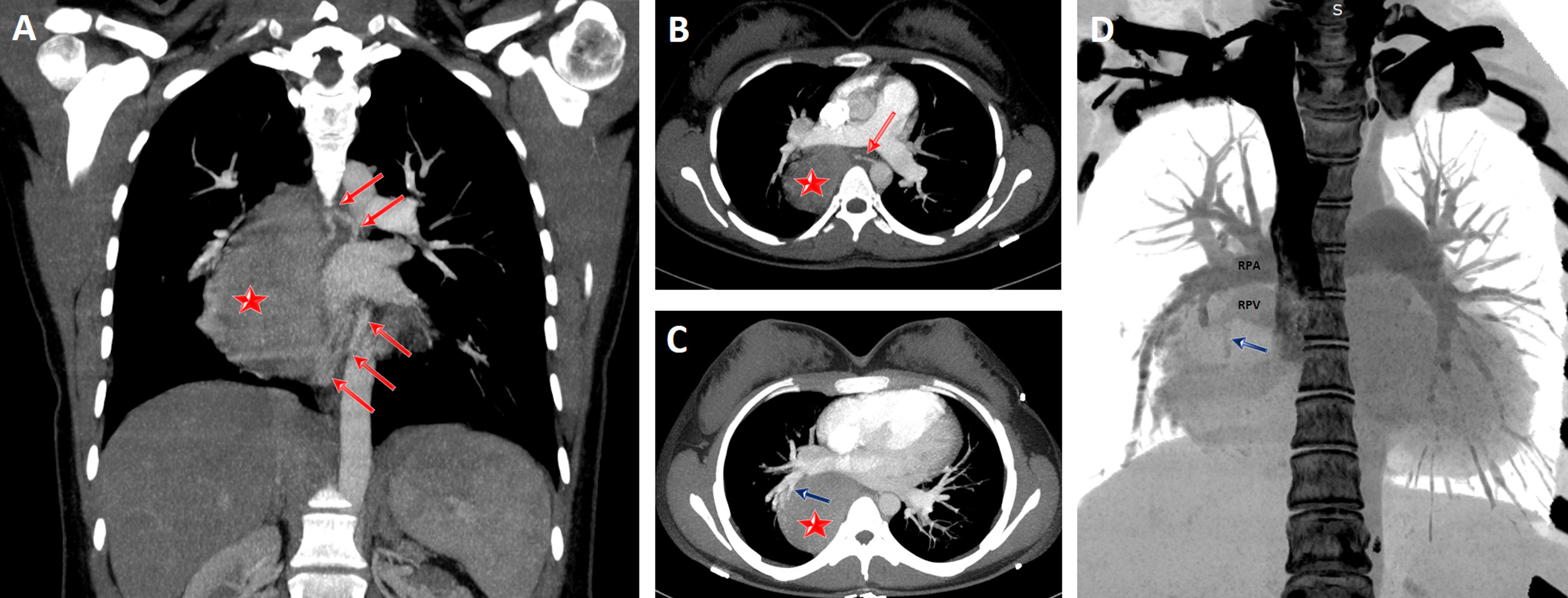
Coronal (A) and axial (B) CT scans show a giant pulmonary sequestration (star) feeding by systemic arteries (red arrows). Maximum intensity projection (MIP) axial CT scan (C) also reveals that the pulmonary sequestration was drained by the pulmonary vein (blue arrow) of the right lower lobe. Coronal volume rendering negative MIP image shows clearly the drainage vein (blue arrow). RPA, right main pulmonary artery; RPV, right main pulmonary vein.

(0.4MB).

Pulmonary sequestration is a rare congenital lung anomaly. It is defined by nonfunctional lung tissue originating from the normal bronchopulmonary system and bleeding from the aberrant systemic artery. Pulmonary sequestration is classified as extralobar and intralobar. About three quarters of the cases are localized in the left lower lobe. In rare cases, it can mimic a benign or malign thoracic lesions.1,2 Detecting pulmonary sequestration before surgery is very important to prevent serious intraoperative complications.

References
[1]
Y. Wei, F. Li.
Pulmonary sequestration: a retrospective analysis of 2625 cases in China.
Eur J Cardiothorac Surg, 40 (2011), pp. e39-e42
[2]
Y. Aydin, H. Ogul, B. Altuntas, A. Eroglu.
Pulmonary sequestration mimicking a hydatid cyst.
Eur J Cardiothorac Surg, 48 (2015), pp. 514
Copyright © 2021. SEPAR
Archivos de Bronconeumología
Article options
Tools

Are you a health professional able to prescribe or dispense drugs?