An 82-year-old man, former smoker, total laryngectomy over 30 years previously, with a permanent tracheostomy, monitored for severe COPD, stable with home oxygen and aerosol therapy.
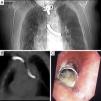
In a recent check-up, he complained of repeated exacerbations in the last few months, with greater need of aerosol therapy, cough, and some bloody sputum. A chest radiograph (Fig. 1A) and a chest computed tomography (CT) (Fig. 1B) were requested, revealing1 a foreign body of metallic density in the distal part of the trachea and tracheal carina, entering the left main bronchus, approximately 6.5cm in length, consistent with migration of a tracheotomy cannula to the bronchial tree. In view of this finding, a fiberoptic bronchoscopy was performed under general anesthesia via the tracheostomy (Fig. 1C). The metal cannula was seen appearing out2 of the tracheal carina, reducing the lumen of the entrance to the right main bronchus by 50%, and entering the left main bronchus. It was extracted with the assistance of an ENT specialist.
Despite the potential severity of the episode, the patient tolerated the foreign body for 2–3 months (he could not remember any time when the event may have taken place). He only began to show clinical symptoms when the lumen of the migrated cannula began to become obstructed.
Please cite this article as: Perez Torres C, Dominguez Perez LD, Rial Morilla F. Cuerpo extraño traqueobronquial inusual. Arch Bronconeumol. 2017;53:159.


