To the Editor: Bronchial carcinoid tumors represent 1% to 2% of all lung cancers.1 Their nomenclature has undergone constant revision over recent decades. According to the criteria drawn up by the World Health Organization in 1982 they were classified into two groups, typical and atypical. In the classification devised by Dresler et al2 in 1997, the type of tumor previously classified as a typical carcinoid in the earlier criteria was redefined as a type 1 neuroendocrine carcinoma. This denomination reflects the fact that this is not a benign process, but rather a carcinoma of low-grade malignancy in the neuroendocrine pulmonary tumor scale. In a study carried out by Pareja et al,3 over two thirds of resected carcinoid tumors were type 1.
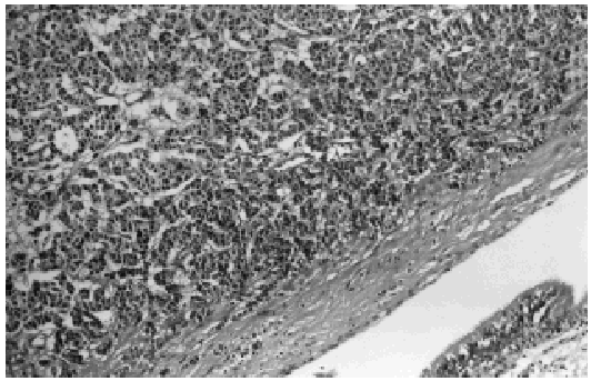
We present the case of an otherwise healthy 27-year-old woman diagnosed with a bronchial carcinoid tumor (pT1N1M0) in whom a bilobectomy (lower and middle lobes) was performed. Histology revealed a tumor composed of medium-sized polygonal cells, with slightly granular cytoplasm and round uniform nuclei, which were only slightly hyperchromatic. Mitosis was scant. These cells were organized such that the tumor presented a pattern of organoid growth (Figure). Immunohistochemistry revealed a very strong expression of chromogranin. The margin of the bronchial resection was free of neoplasia.
Figure. Normal bronchial epithelium (lower right-hand corner), with proliferation of neoplastic tissue in the bronchial airway.
The patient remained disease-free for 7 years, at which time a broadening of the mediastinum was observed in a routine x-ray. Further exploration of the area using computed tomography (CT) revealed right paratracheal adenopathy that had not been present in earlier studies (a lymph node 2.5 in diameter). Positron emission tomography showed intense uptake in the right paratracheal region in an area that coincided with the lesion visible in the CT scan. Fiberoptic bronchoscopy detected a tumorous growth of 0.5 cm in the bronchial stump. Bronchial biopsy revealed a carcinoid tumor, which tested positive for chromogranin in the immunohistochemical panel performed.
When the diagnosis of recurrent carcinoid tumor had been confirmed, a second operation was performed to complete the pneumonectomy, to perform a hilar and mediastinal lymphadenectomy, and to remove the affected right paratracheal node, which measured 5 cm in diameter. The pathological diagnosis was recurrent bronchial carcinoid tumor with metastasis to a paratracheal node. Other nodes and the margin of resection were not affected by neoplasia (pT1N2M0). At no time did the patient present the general symptoms of carcinoid syndrome.
According to a multicenter study conducted in Spain by the Spanish Society of Pulmonology and Thoracic Surgery (the EMETNE-SEPAR study), mediastinal node involvement is found in 4.15% of type I neuroendocrine carcinomas.4 In that study, 2 out of 261 patients developed local recurrence. Overall survival rates at 5 and 10 years were over 93%. Filosso et al5 found significant differences in the prognosis of patients with nodal involvement and/or recurrence. However, correct evaluation of the effect of recurrence and mediastinal involvement on prognosis is particularly difficult in typical carcinoids given the limited number of cases.
With respect to treatment, differences of opinion can be found in the literature regarding the value of conservative, parenchymal-sparing surgery. While some authors defend the hypothesis that the long-term survival of patients undergoing conservative resections is unaffected by nodal involvement,6 others believe that nodal spread of the disease represents a contraindication for bronchoplasty.1
In the case under discussion, two aggressive mechanisms uncommon in typical carcinoids were present: local recurrence and mediastinal nodal involvement. It is therefore difficult to establish the prognostic implications with exactitude. However, this case does highlight the importance of performing oncologically curative surgery and a routine lymphadenectomy that will make it possible to stage these patients correctly.


