To the Editor: When video-assisted thoracoscopy (VATS) is used to treat spontaneous pneumothorax (SP), the goal is to eliminate the lesions that caused the air leak and to perform pleurodesis to minimize recurrence.1 In our facility, iodine in hydroalcoholic solution has been used to treat primary SP since 1993. The only important postoperative complication has been the recurrence of pneumothorax (in 6.2% of cases).2 We report the first case in our experience of hemithoracic retraction after VATS and instillation of a chemical agent--in this case a hydroalcoholic solution of povidone iodine.
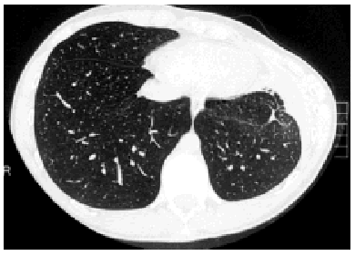
A 17-year-old male with no known relevant medical history diagnosed with a third recurrence of primary SP was admitted for definitive treatment by VATS, which was carried out with three trocars. The pleural cavity and the surface of the lung were carefully inspected and no resectable lesion causing the air leak was found. Pleurodesis was performed with 60 mg of a hydroalcoholic solution of povidone iodine instilled until a uniform distribution was obtained in the pleural cavity and across the surface of the lung. The patient had an uneventful postoperative recovery, did not present air leaks, and was discharged 5 days following the procedure. He was referred to respiratory physiotherapy because a small left pleural effusion was observed on the x-ray. The patient was well at early follow-up visits; he was also well by clinical standards at the 6-month visit and had apparently normal thoracic x-rays. However, he complained of a deformity in the operated hemithorax. Physical examination revealed a left-sided retraction and marked left-right asymmetry. A computed axial tomography scan (Figure) and magnetic resonance of the chest were obtained, and spirometry was performed. Both the computed tomography scan and the magnetic resonance image revealed posttreatment alterations in the pleura without pleural thickening, a small left loculated pleural effusion, a small scar-like infiltrate near the operated region, and a marked retraction of the left hemithorax. No muscular or osseous alterations of the thoracic wall that could have caused the retraction were found. Forced vital capacity was 4.19 (86%) and forced expired volume in 1 second was 3.79 (92%). Complete biochemical analyses with immunological studies were requested and were normal. Based on clinical course and complementary test findings the diagnosis was asymmetry of the thoracic wall due to surgery.
Figure. The thoracic computed tomography scan reveals retraction of the right hemithorax.
VATS was added to the therapeutic armamentarium for SP in 1990 and is currently the most common procedure used.3 Chemical pleurodesis has been used to treat SP since 1906 and is one of the options for treatment.4 Talc is the most commonly used substance.5 We use a hydroalcoholic solution of povidone iodine in patients diagnosed with primary SP who require surgery. In our 9-year experience with this technique3 we have observed no severe complications, having recorded persistent air leaks lasting more than 5 days (12.3%), self-limited fever (6.1%), operative wound infection (2.4%), and recurrence during recovery (6.1%). In the case we present, a young patient diagnosed with recurrent primary SP and without relevant medical history developed left thoracic retraction after undergoing chemical pleurodesis with povidone iodine by VATS. Localized pleural thickening, due to excessive accumulation of the chemical agent, alteration of its composition, or an idiosyncratic reaction to povidone iodine, would have been the most likely cause of the lesion. The composition and volume of the povidone iodine used in the hemithorax were the usual ones, although the postoperative pleural effusion could have contained an excess amount of povidone iodine, which if accumulated can cause localized pleural thickening. To rule out an allergic reaction of the pleura to the chemical agent, immunological tests were requested and were normal. Imaging techniques did not reveal pleural thickening with sufficient severity to explain retraction of the thoracic cavity. Chemical pleurodesis with povidone iodine solution is used in our facility in patients with primary SP to create pleural adhesions that are looser than those obtained with talc, in order to avoid a granulomatous reaction which, if thick enough, could hinder future resective surgery in that lung. The use of povidone iodine solution also circumvents the side effects due to talc application,6 which we use in older patients with secondary SP. Was the pleurodesis so intense it caused the thoracic cavity to retract? The degree of observed pleural thickening does not support that hypothesis. Furthermore, the tests ruled out a systemic process or an allergy to povidone iodine. This complication has not been reported after any pleurodesis technique to date, even after talc pleurodesis, and the case remains unexplained.


