array:24 [
  "pii" => "S1579212919302393"
  "issn" => "15792129"
  "doi" => "10.1016/j.arbr.2018.12.027"
  "estado" => "S300"
  "fechaPublicacion" => "2019-10-01"
  "aid" => "2057"
  "copyright" => "SEPAR"
  "copyrightAnyo" => "2018"
  "documento" => "article"
  "crossmark" => 1
  "subdocumento" => "sco"
  "cita" => "Arch Bronconeumol. 2019;55:532"
  "abierto" => array:3 [
    "ES" => false
    "ES2" => false
    "LATM" => false
  ]
  "gratuito" => false
  "lecturas" => array:1 [
    "total" => 0
  ]
  "Traduccion" => array:1 [
    "es" => array:19 [
      "pii" => "S0300289618304708"
      "issn" => "03002896"
      "doi" => "10.1016/j.arbres.2018.12.006"
      "estado" => "S300"
      "fechaPublicacion" => "2019-10-01"
      "aid" => "2057"
      "copyright" => "SEPAR"
      "documento" => "article"
      "crossmark" => 1
      "subdocumento" => "sco"
      "cita" => "Arch Bronconeumol. 2019;55:532"
      "abierto" => array:3 [
        "ES" => true
        "ES2" => true
        "LATM" => true
      ]
      "gratuito" => true
      "lecturas" => array:2 [
        "total" => 135
        "formatos" => array:2 [
          "HTML" => 91
          "PDF" => 44
        ]
      ]
      "es" => array:11 [
        "idiomaDefecto" => true
        "cabecera" => "<span class="elsevierStyleTextfn">Imagen cl&#237;nica</span>"
        "titulo" => "Amiloidosis traqueobronquial como hallazgo incidental en un paciente con neoplasia pulmonar"
        "tienePdf" => "es"
        "tieneTextoCompleto" => "es"
        "paginas" => array:1 [
          0 => array:1 [
            "paginaInicial" => "532"
          ]
        ]
        "titulosAlternativos" => array:1 [
          "en" => array:1 [
            "titulo" => "Tracheobronchial Amyloidosis as an Incidental Finding in a Patient With Lung Cancer"
          ]
        ]
        "contieneTextoCompleto" => array:1 [
          "es" => true
        ]
        "contienePdf" => array:1 [
          "es" => true
        ]
        "resumenGrafico" => array:2 [
          "original" => 0
          "multimedia" => array:7 [
            "identificador" => "fig0005"
            "etiqueta" => "Figura 1"
            "tipo" => "MULTIMEDIAFIGURA"
            "mostrarFloat" => true
            "mostrarDisplay" => false
            "figura" => array:1 [
              0 => array:4 [
                "imagen" => "gr1.jpeg"
                "Alto" => 1062
                "Ancho" => 917
                "Tamanyo" => 167998
              ]
            ]
            "descripcion" => array:1 [
              "es" => "<p id="spar0005" class="elsevierStyleSimplePara elsevierViewall">A y B&#58; tomograf&#237;a axial computarizada y PET&#46; C y D&#58; placas eritematosas sobreelevadas e hipervascularizadas en tercio medio derecho traqueal y carina principal con extensi&#243;n a lo largo de la pared medial del bronquio principal derecho&#46; E y F&#58; presencia de dep&#243;sitos acid&#243;filos en la submucosa correspondientes a sustancia amiloide tras tinci&#243;n espec&#237;fica con rojo congo y tioflavina T&#46;</p>"
            ]
          ]
        ]
        "autores" => array:1 [
          0 => array:2 [
            "autoresLista" => "Blanca de Vega S&#225;nchez, Carlos Disdier Vicente, Gerardo Mart&#237;nez Garc&#237;a"
            "autores" => array:3 [
              0 => array:2 [
                "nombre" => "Blanca"
                "apellidos" => "de Vega S&#225;nchez"
              ]
              1 => array:2 [
                "nombre" => "Carlos"
                "apellidos" => "Disdier Vicente"
              ]
              2 => array:2 [
                "nombre" => "Gerardo"
                "apellidos" => "Mart&#237;nez Garc&#237;a"
              ]
            ]
          ]
        ]
      ]
      "idiomaDefecto" => "es"
      "Traduccion" => array:1 [
        "en" => array:9 [
          "pii" => "S1579212919302393"
          "doi" => "10.1016/j.arbr.2018.12.027"
          "estado" => "S300"
          "subdocumento" => ""
          "abierto" => array:3 [
            "ES" => false
            "ES2" => false
            "LATM" => false
          ]
          "gratuito" => false
          "lecturas" => array:1 [
            "total" => 0
          ]
          "idiomaDefecto" => "en"
          "EPUB" => "https://multimedia.elsevier.es/PublicationsMultimediaV1/item/epub/S1579212919302393?idApp=UINPBA00003Z"
        ]
      ]
      "EPUB" => "https://multimedia.elsevier.es/PublicationsMultimediaV1/item/epub/S0300289618304708?idApp=UINPBA00003Z"
      "url" => "/03002896/0000005500000010/v1_201910021029/S0300289618304708/v1_201910021029/es/main.assets"
    ]
  ]
  "itemSiguiente" => array:19 [
    "pii" => "S157921291930240X"
    "issn" => "15792129"
    "doi" => "10.1016/j.arbr.2018.12.028"
    "estado" => "S300"
    "fechaPublicacion" => "2019-10-01"
    "aid" => "2070"
    "copyright" => "SEPAR"
    "documento" => "article"
    "crossmark" => 1
    "subdocumento" => "sco"
    "cita" => "Arch Bronconeumol. 2019;55:533"
    "abierto" => array:3 [
      "ES" => false
      "ES2" => false
      "LATM" => false
    ]
    "gratuito" => false
    "lecturas" => array:1 [
      "total" => 0
    ]
    "en" => array:11 [
      "idiomaDefecto" => true
      "cabecera" => "<span class="elsevierStyleTextfn">Clinical Image</span>"
      "titulo" => "Tracheal Stenosis Due to IgG4-Related Fibrosing Mediastinitis"
      "tienePdf" => "en"
      "tieneTextoCompleto" => "en"
      "paginas" => array:1 [
        0 => array:1 [
          "paginaInicial" => "533"
        ]
      ]
      "titulosAlternativos" => array:1 [
        "es" => array:1 [
          "titulo" => "Estenosis traqueal por mediastinitis fibrosante relacionada con enfermedad por IgG4"
        ]
      ]
      "contieneTextoCompleto" => array:1 [
        "en" => true
      ]
      "contienePdf" => array:1 [
        "en" => true
      ]
      "resumenGrafico" => array:2 [
        "original" => 0
        "multimedia" => array:7 [
          "identificador" => "fig0005"
          "etiqueta" => "Fig&#46; 1"
          "tipo" => "MULTIMEDIAFIGURA"
          "mostrarFloat" => true
          "mostrarDisplay" => false
          "figura" => array:1 [
            0 => array:4 [
              "imagen" => "gr1.jpeg"
              "Alto" => 673
              "Ancho" => 903
              "Tamanyo" => 111414
            ]
          ]
          "descripcion" => array:1 [
            "en" => "<p id="spar0005" class="elsevierStyleSimplePara elsevierViewall">&#40;A&#41; Bronchoscopy and CT image of tracheal stenosis&#46; &#40;B&#41; Outcome after treatment with corticosteroids and stenting&#44; after 4 months&#46; &#40;C&#41; Argon plasma coagulation&#44; after biopsy&#46; &#40;D&#41; Placement of Dumon&#174; stent&#46;</p>"
          ]
        ]
      ]
      "autores" => array:1 [
        0 => array:2 [
          "autoresLista" => "Laura Arroyo Pareja, Ana Trivi&#241;o, Carolina Espa&#241;a"
          "autores" => array:3 [
            0 => array:2 [
              "nombre" => "Laura"
              "apellidos" => "Arroyo Pareja"
            ]
            1 => array:2 [
              "nombre" => "Ana"
              "apellidos" => "Trivi&#241;o"
            ]
            2 => array:2 [
              "nombre" => "Carolina"
              "apellidos" => "Espa&#241;a"
            ]
          ]
        ]
      ]
    ]
    "idiomaDefecto" => "en"
    "Traduccion" => array:1 [
      "es" => array:9 [
        "pii" => "S0300289619300146"
        "doi" => "10.1016/j.arbres.2018.12.014"
        "estado" => "S300"
        "subdocumento" => ""
        "abierto" => array:3 [
          "ES" => true
          "ES2" => true
          "LATM" => true
        ]
        "gratuito" => true
        "lecturas" => array:1 [
          "total" => 0
        ]
        "idiomaDefecto" => "es"
        "EPUB" => "https://multimedia.elsevier.es/PublicationsMultimediaV1/item/epub/S0300289619300146?idApp=UINPBA00003Z"
      ]
    ]
    "EPUB" => "https://multimedia.elsevier.es/PublicationsMultimediaV1/item/epub/S157921291930240X?idApp=UINPBA00003Z"
    "url" => "/15792129/0000005500000010/v1_201910100934/S157921291930240X/v1_201910100934/en/main.assets"
  ]
  "itemAnterior" => array:19 [
    "pii" => "S1579212919302484"
    "issn" => "15792129"
    "doi" => "10.1016/j.arbr.2019.08.001"
    "estado" => "S300"
    "fechaPublicacion" => "2019-10-01"
    "aid" => "2134"
    "copyright" => "SEPAR"
    "documento" => "article"
    "crossmark" => 1
    "subdocumento" => "fla"
    "cita" => "Arch Bronconeumol. 2019;55:526-31"
    "abierto" => array:3 [
      "ES" => false
      "ES2" => false
      "LATM" => false
    ]
    "gratuito" => false
    "lecturas" => array:2 [
      "total" => 7
      "formatos" => array:2 [
        "HTML" => 3
        "PDF" => 4
      ]
    ]
    "en" => array:13 [
      "idiomaDefecto" => true
      "cabecera" => "<span class="elsevierStyleTextfn">Original Article</span>"
      "titulo" => "Lung Cancer Screening&#58; Use of Low-Dose Computed Tomography"
      "tienePdf" => "en"
      "tieneTextoCompleto" => "en"
      "tieneResumen" => array:2 [
        0 => "en"
        1 => "es"
      ]
      "paginas" => array:1 [
        0 => array:2 [
          "paginaInicial" => "526"
          "paginaFinal" => "531"
        ]
      ]
      "titulosAlternativos" => array:1 [
        "es" => array:1 [
          "titulo" => "Diagn&#243;stico precoz del c&#225;ncer de pulm&#243;n&#58; utilidad de la tomograf&#237;a computarizada de baja dosis de radiaci&#243;n"
        ]
      ]
      "contieneResumen" => array:2 [
        "en" => true
        "es" => true
      ]
      "contieneTextoCompleto" => array:1 [
        "en" => true
      ]
      "contienePdf" => array:1 [
        "en" => true
      ]
      "resumenGrafico" => array:2 [
        "original" => 0
        "multimedia" => array:7 [
          "identificador" => "fig0010"
          "etiqueta" => "Fig&#46; 2"
          "tipo" => "MULTIMEDIAFIGURA"
          "mostrarFloat" => true
          "mostrarDisplay" => false
          "figura" => array:1 [
            0 => array:4 [
              "imagen" => "gr2.jpeg"
              "Alto" => 1261
              "Ancho" => 2500
              "Tamanyo" => 152924
            ]
          ]
          "descripcion" => array:1 [
            "en" => "<p id="spar0050" class="elsevierStyleSimplePara elsevierViewall">Decision flowchart for 1-year scan&#46;</p>"
          ]
        ]
      ]
      "autores" => array:1 [
        0 => array:2 [
          "autoresLista" => "Encarnaci&#243;n Mart&#237;nez P&#233;rez, Karol de Aguiar Quevedo, Miguel Arrar&#225;s Mart&#237;nez, Julia Cruz Mojarrieta, Estanislao Arana Fern&#225;ndez de Moya, Mar&#237;a Barrios Benito, Susana Hinarejos Parga, Jos&#233; Cervera Deval, Juan Carlos Pe&#241;alver Cuesta"
          "autores" => array:9 [
            0 => array:2 [
              "nombre" => "Encarnaci&#243;n"
              "apellidos" => "Mart&#237;nez P&#233;rez"
            ]
            1 => array:2 [
              "nombre" => "Karol"
              "apellidos" => "de Aguiar Quevedo"
            ]
            2 => array:2 [
              "nombre" => "Miguel"
              "apellidos" => "Arrar&#225;s Mart&#237;nez"
            ]
            3 => array:2 [
              "nombre" => "Julia"
              "apellidos" => "Cruz Mojarrieta"
            ]
            4 => array:2 [
              "nombre" => "Estanislao"
              "apellidos" => "Arana Fern&#225;ndez de Moya"
            ]
            5 => array:2 [
              "nombre" => "Mar&#237;a"
              "apellidos" => "Barrios Benito"
            ]
            6 => array:2 [
              "nombre" => "Susana"
              "apellidos" => "Hinarejos Parga"
            ]
            7 => array:2 [
              "nombre" => "Jos&#233;"
              "apellidos" => "Cervera Deval"
            ]
            8 => array:2 [
              "nombre" => "Juan Carlos"
              "apellidos" => "Pe&#241;alver Cuesta"
            ]
          ]
        ]
      ]
    ]
    "idiomaDefecto" => "en"
    "Traduccion" => array:1 [
      "es" => array:9 [
        "pii" => "S0300289619301231"
        "doi" => "10.1016/j.arbres.2019.03.012"
        "estado" => "S300"
        "subdocumento" => ""
        "abierto" => array:3 [
          "ES" => true
          "ES2" => true
          "LATM" => true
        ]
        "gratuito" => true
        "lecturas" => array:1 [
          "total" => 0
        ]
        "idiomaDefecto" => "es"
        "EPUB" => "https://multimedia.elsevier.es/PublicationsMultimediaV1/item/epub/S0300289619301231?idApp=UINPBA00003Z"
      ]
    ]
    "EPUB" => "https://multimedia.elsevier.es/PublicationsMultimediaV1/item/epub/S1579212919302484?idApp=UINPBA00003Z"
    "url" => "/15792129/0000005500000010/v1_201910100934/S1579212919302484/v1_201910100934/en/main.assets"
  ]
  "en" => array:14 [
    "idiomaDefecto" => true
    "cabecera" => "<span class="elsevierStyleTextfn">Clinical Image</span>"
    "titulo" => "Tracheobronchial Amyloidosis as an Incidental Finding in a Patient With Lung Cancer"
    "tieneTextoCompleto" => true
    "paginas" => array:1 [
      0 => array:1 [
        "paginaInicial" => "532"
      ]
    ]
    "autores" => array:1 [
      0 => array:4 [
        "autoresLista" => "Blanca de Vega S&#225;nchez, Carlos Disdier Vicente, Gerardo Mart&#237;nez Garc&#237;a"
        "autores" => array:3 [
          0 => array:4 [
            "nombre" => "Blanca"
            "apellidos" => "de Vega S&#225;nchez"
            "email" => array:1 [
              0 => "blancadevegasanchez@gmail.com"
            ]
            "referencia" => array:2 [
              0 => array:2 [
                "etiqueta" => "<span class="elsevierStyleSup">a</span>"
                "identificador" => "aff0005"
              ]
              1 => array:2 [
                "etiqueta" => "&#42;"
                "identificador" => "cor0005"
              ]
            ]
          ]
          1 => array:3 [
            "nombre" => "Carlos"
            "apellidos" => "Disdier Vicente"
            "referencia" => array:2 [
              0 => array:2 [
                "etiqueta" => "<span class="elsevierStyleSup">a</span>"
                "identificador" => "aff0005"
              ]
              1 => array:2 [
                "etiqueta" => "<span class="elsevierStyleSup">b</span>"
                "identificador" => "aff0010"
              ]
            ]
          ]
          2 => array:3 [
            "nombre" => "Gerardo"
            "apellidos" => "Mart&#237;nez Garc&#237;a"
            "referencia" => array:1 [
              0 => array:2 [
                "etiqueta" => "<span class="elsevierStyleSup">c</span>"
                "identificador" => "aff0015"
              ]
            ]
          ]
        ]
        "afiliaciones" => array:3 [
          0 => array:3 [
            "entidad" => "Unidad de Broncoscopias y T&#233;cnicas Pleurales&#44; Servicio de Neumolog&#237;a&#44; Hospital Cl&#237;nico Universitario de Valladolid&#44; Valladolid&#44; Spain"
            "etiqueta" => "a"
            "identificador" => "aff0005"
          ]
          1 => array:3 [
            "entidad" => "Centro de Investigaci&#243;n Biom&#233;dica en Red de Enfermedades Respiratorias &#40;CIBERES&#41;&#44; Valladolid&#44; Spain"
            "etiqueta" => "b"
            "identificador" => "aff0010"
          ]
          2 => array:3 [
            "entidad" => "Servicio de Anatom&#237;a Patol&#243;gica&#44; Hospital Cl&#237;nico Universitario Valladolid&#44; Valladolid&#44; Spain"
            "etiqueta" => "c"
            "identificador" => "aff0015"
          ]
        ]
        "correspondencia" => array:1 [
          0 => array:3 [
            "identificador" => "cor0005"
            "etiqueta" => "&#8270;"
            "correspondencia" => "Corresponding author&#46;"
          ]
        ]
      ]
    ]
    "titulosAlternativos" => array:1 [
      "es" => array:1 [
        "titulo" => "Amiloidosis traqueobronquial como hallazgo incidental en un paciente con neoplasia pulmonar"
      ]
    ]
    "resumenGrafico" => array:2 [
      "original" => 0
      "multimedia" => array:7 [
        "identificador" => "fig0005"
        "etiqueta" => "Fig&#46; 1"
        "tipo" => "MULTIMEDIAFIGURA"
        "mostrarFloat" => true
        "mostrarDisplay" => false
        "figura" => array:1 [
          0 => array:4 [
            "imagen" => "gr1.jpeg"
            "Alto" => 1062
            "Ancho" => 917
            "Tamanyo" => 167632
          ]
        ]
        "descripcion" => array:1 [
          "en" => "<p id="spar0005" class="elsevierStyleSimplePara elsevierViewall">&#40;A and B&#41; Computed tomography and PET&#46; &#40;C and D&#41; Raised erythematous hypervascular plaques in the right middle third of the trachea and main carina extending along the medial wall of the right main bronchus&#46; &#40;E and F&#41; Presence of acidophilic deposits in the submucosa corresponding to amyloid substance after specific staining with Congo red and thioflavin T&#46;</p>"
        ]
      ]
    ]
    "textoCompleto" => "<span class="elsevierStyleSections"><p id="par0005" class="elsevierStylePara elsevierViewall">We report the case of a 67-year-old man&#44; former smoker&#44; referred for a flexible bronchoscopy &#40;FB&#41;&#44; in view of radiological findings indicative of lung cancer&#46; A detailed study of the imaging tests showed the existence of a lung mass and increased density in both lateral walls of the middle third of the trachea and the main carina with no pathological uptake on PET&#44; visible in the flexible bronchoscopy &#40;<a class="elsevierStyleCrossRef" href="#fig0005">Fig&#46; 1</a>&#41;&#46; Given these findings and the appearance of the lesions on endoscopy&#44; lung mass and tracheal samples were obtained to confirm the endoscopic suspicion of tracheobronchial amyloidosis vs neoplastic infiltration&#46;</p><elsevierMultimedia ident="fig0005"></elsevierMultimedia><p id="par0010" class="elsevierStylePara elsevierViewall">Histopathological studies established the diagnosis of localized primary tracheobronchial amyloidosis and surgically unresectable pulmonary adenocarcinoma &#40;T2bN3M0&#41;&#46; Tracheobronchial amyloidosis is a rare disease that&#44; when detected on endoscopy&#44; requires further studies to rule out secondary involvement&#46;<a class="elsevierStyleCrossRef" href="#bib0015"><span class="elsevierStyleSup">1</span></a> In the absence of a standardized treatment<a class="elsevierStyleCrossRef" href="#bib0020"><span class="elsevierStyleSup">2</span></a> and the absence of tracheobronchial lumen compromise&#44; chemotherapy was started&#46; Endoscopic treatment was initially ruled out and the patient was followed up with endoscopic and clinical examinations&#46; The presence of raised erythematous plaques in the tracheobronchial tree should prompt the endoscopist to consider this disease in the differential diagnosis&#46;</p></span>"
    "pdfFichero" => "main.pdf"
    "tienePdf" => true
    "NotaPie" => array:1 [
      0 => array:2 [
        "etiqueta" => "&#9734;"
        "nota" => "<p class="elsevierStyleNotepara" id="npar0005">Please cite this article as&#58; de Vega S&#225;nchez B&#44; Disdier Vicente C&#44; Mart&#237;nez Garc&#237;a G&#46; Amiloidosis traqueobronquial como hallazgo incidental en un paciente con neoplasia pulmonar&#46; Arch Bronconeumol&#46; 2019&#59;55&#58;532&#46;</p>"
      ]
    ]
    "multimedia" => array:1 [
      0 => array:7 [
        "identificador" => "fig0005"
        "etiqueta" => "Fig&#46; 1"
        "tipo" => "MULTIMEDIAFIGURA"
        "mostrarFloat" => true
        "mostrarDisplay" => false
        "figura" => array:1 [
          0 => array:4 [
            "imagen" => "gr1.jpeg"
            "Alto" => 1062
            "Ancho" => 917
            "Tamanyo" => 167632
          ]
        ]
        "descripcion" => array:1 [
          "en" => "<p id="spar0005" class="elsevierStyleSimplePara elsevierViewall">&#40;A and B&#41; Computed tomography and PET&#46; &#40;C and D&#41; Raised erythematous hypervascular plaques in the right middle third of the trachea and main carina extending along the medial wall of the right main bronchus&#46; &#40;E and F&#41; Presence of acidophilic deposits in the submucosa corresponding to amyloid substance after specific staining with Congo red and thioflavin T&#46;</p>"
        ]
      ]
    ]
    "bibliografia" => array:2 [
      "titulo" => "References"
      "seccion" => array:1 [
        0 => array:2 [
          "identificador" => "bibs0015"
          "bibliografiaReferencia" => array:2 [
            0 => array:3 [
              "identificador" => "bib0015"
              "etiqueta" => "1"
              "referencia" => array:1 [
                0 => array:2 [
                  "contribucion" => array:1 [
                    0 => array:2 [
                      "titulo" => "Manejo de la amiloidosis traqueobronquial mediante t&#233;cnicas broncosc&#243;picas terap&#233;uticas"
                      "autores" => array:1 [
                        0 => array:2 [
                          "etal" => false
                          "autores" => array:6 [
                            0 => "J&#46; Berraondo"
                            1 => "L&#46; Novella"
                            2 => "F&#46; Sanz"
                            3 => "R&#46; Lluch"
                            4 => "E&#46; de Casimiro"
                            5 => "T&#46; Lloret"
                          ]
                        ]
                      ]
                    ]
                  ]
                  "host" => array:1 [
                    0 => array:2 [
                      "doi" => "10.1016/j.arbres.2012.08.002"
                      "Revista" => array:6 [
                        "tituloSerie" => "Arch Bronconeumol"
                        "fecha" => "2013"
                        "volumen" => "49"
                        "paginaInicial" => "207"
                        "paginaFinal" => "209"
                        "link" => array:1 [
                          0 => array:2 [
                            "url" => "https://www.ncbi.nlm.nih.gov/pubmed/23347550"
                            "web" => "Medline"
                          ]
                        ]
                      ]
                    ]
                  ]
                ]
              ]
            ]
            1 => array:3 [
              "identificador" => "bib0020"
              "etiqueta" => "2"
              "referencia" => array:1 [
                0 => array:2 [
                  "contribucion" => array:1 [
                    0 => array:2 [
                      "titulo" => "External beam radiation therapy&#58; a treatment modality in diffuse tracheobronchial amyloidosis"
                      "autores" => array:1 [
                        0 => array:2 [
                          "etal" => false
                          "autores" => array:4 [
                            0 => "V&#46; Almadana"
                            1 => "E&#46; Luque"
                            2 => "A&#46;J&#46; Wals"
                            3 => "T&#46; Montemayor"
                          ]
                        ]
                      ]
                    ]
                  ]
                  "host" => array:1 [
                    0 => array:1 [
                      "Revista" => array:5 [
                        "tituloSerie" => "Teodoro Montemayor Rubio Arch Bronconeumol"
                        "fecha" => "2015"
                        "volumen" => "51"
                        "paginaInicial" => "602"
                        "paginaFinal" => "610"
                      ]
                    ]
                  ]
                ]
              ]
            ]
          ]
        ]
      ]
    ]
  ]
  "idiomaDefecto" => "en"
  "url" => "/15792129/0000005500000010/v1_201910100934/S1579212919302393/v1_201910100934/en/main.assets"
  "Apartado" => array:4 [
    "identificador" => "21342"
    "tipo" => "SECCION"
    "en" => array:2 [
      "titulo" => "Clinical Image"
      "idiomaDefecto" => true
    ]
    "idiomaDefecto" => "en"
  ]
  "PDF" => "https://static.elsevier.es/multimedia/15792129/0000005500000010/v1_201910100934/S1579212919302393/v1_201910100934/en/main.pdf?idApp=UINPBA00003Z&text.app=https://archbronconeumol.org/"
  "EPUB" => "https://multimedia.elsevier.es/PublicationsMultimediaV1/item/epub/S1579212919302393?idApp=UINPBA00003Z"
]
Share
Journal Information
Vol. 55. Issue 10.
Pages 532 (October 2019)
Vol. 55. Issue 10.
Pages 532 (October 2019)
Clinical Image
Full text access
Tracheobronchial Amyloidosis as an Incidental Finding in a Patient With Lung Cancer
Amiloidosis traqueobronquial como hallazgo incidental en un paciente con neoplasia pulmonar
Visits
1927
Blanca de Vega Sáncheza,
Corresponding author
blancadevegasanchez@gmail.com

Corresponding author.
, Carlos Disdier Vicentea,b, Gerardo Martínez Garcíac
a Unidad de Broncoscopias y Técnicas Pleurales, Servicio de Neumología, Hospital Clínico Universitario de Valladolid, Valladolid, Spain
b Centro de Investigación Biomédica en Red de Enfermedades Respiratorias (CIBERES), Valladolid, Spain
c Servicio de Anatomía Patológica, Hospital Clínico Universitario Valladolid, Valladolid, Spain
This item has received
Article information
Full Text
Bibliography
Download PDF
Statistics
Figures (1)
Full Text

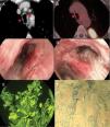
We report the case of a 67-year-old man, former smoker, referred for a flexible bronchoscopy (FB), in view of radiological findings indicative of lung cancer. A detailed study of the imaging tests showed the existence of a lung mass and increased density in both lateral walls of the middle third of the trachea and the main carina with no pathological uptake on PET, visible in the flexible bronchoscopy (Fig. 1). Given these findings and the appearance of the lesions on endoscopy, lung mass and tracheal samples were obtained to confirm the endoscopic suspicion of tracheobronchial amyloidosis vs neoplastic infiltration.

Fig. 1.

(A and B) Computed tomography and PET. (C and D) Raised erythematous hypervascular plaques in the right middle third of the trachea and main carina extending along the medial wall of the right main bronchus. (E and F) Presence of acidophilic deposits in the submucosa corresponding to amyloid substance after specific staining with Congo red and thioflavin T.

(0.16MB).

Histopathological studies established the diagnosis of localized primary tracheobronchial amyloidosis and surgically unresectable pulmonary adenocarcinoma (T2bN3M0). Tracheobronchial amyloidosis is a rare disease that, when detected on endoscopy, requires further studies to rule out secondary involvement.1 In the absence of a standardized treatment2 and the absence of tracheobronchial lumen compromise, chemotherapy was started. Endoscopic treatment was initially ruled out and the patient was followed up with endoscopic and clinical examinations. The presence of raised erythematous plaques in the tracheobronchial tree should prompt the endoscopist to consider this disease in the differential diagnosis.

References
[1]
J. Berraondo, L. Novella, F. Sanz, R. Lluch, E. de Casimiro, T. Lloret.
Manejo de la amiloidosis traqueobronquial mediante técnicas broncoscópicas terapéuticas.
Arch Bronconeumol, 49 (2013), pp. 207-209
[2]
V. Almadana, E. Luque, A.J. Wals, T. Montemayor.
External beam radiation therapy: a treatment modality in diffuse tracheobronchial amyloidosis.
Teodoro Montemayor Rubio Arch Bronconeumol, 51 (2015), pp. 602-610

Please cite this article as: de Vega Sánchez B, Disdier Vicente C, Martínez García G. Amiloidosis traqueobronquial como hallazgo incidental en un paciente con neoplasia pulmonar. Arch Bronconeumol. 2019;55:532.

Copyright © 2018. SEPAR
Archivos de Bronconeumología
Article options
Tools

Are you a health professional able to prescribe or dispense drugs?