Journal Information
Vol. 56. Issue 7.
Pages 455 (July 2020)
Clinical Image
Full text access
Squamous Cell Carcinoma of the Chest Wall in Open Thoracostomy
Carcinoma epidermoide de pared torácica en una toracostomía abierta
Visits
1162
Luis Alfonso Sota Yoldia,
Corresponding author
lsotayol@hotmail.com

Corresponding author.
, Laura Vigil Vigilb, Esther Nogales Nievesc
a Servicio de Neumología, Fundación Hospital de Jove, Gijón, Asturias, Spain
b Servicio de Neumología, Hospital Universitario de Cabueñes, Gijón, Asturias, Spain
c Servicio de Medicina Interna, Fundación Hospital de Jove, Gijón, Asturias, Spain
This item has received
Article information
Full Text
Bibliography
Download PDF
Statistics
Figures (1)
Full Text

We report the case of a 56-year-old man who consulted for the appearance of a mass in the orifice of an open-window thoracostomy performed for tuberculous empyema 15 years previously. The patient had severe constitutional symptoms, along with significant cachexia. A mass was observed in the thoracostomy space on physical examination (Fig. 1). Chest CT revealed a large extrapulmonary heterogeneous tumor measuring 13×7×6cm. Core needle biopsy confirmed well-differentiated infiltrating squamous carcinoma.

Fig. 1.

(A) Tumor protruding through the permanent open thoracostomy space. (B) Extensive growth of the tumor occupying the entire space days later. (C and D) Axial CT slices showing thoracostomy and tumor.

(0.16MB).

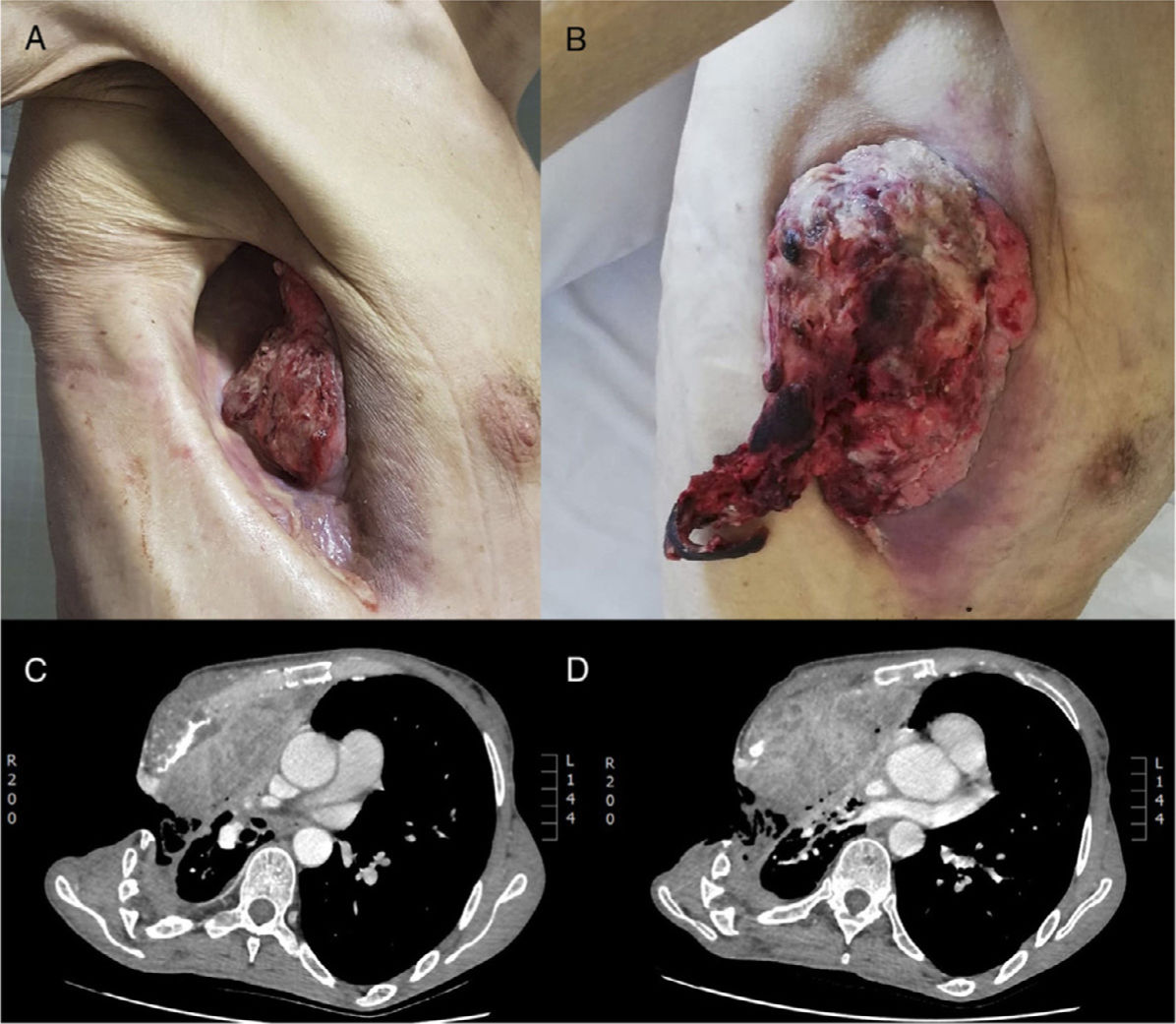
The tumor showed very rapid and aggressive growth occupying the entire thoracostomy space within just a few days. The patient presented serious complications and difficult-to-control pain that led to further clinical deterioration, requiring increasing palliative measures until he finally died.

Chest wall tumors in a thoracostomy are extremely rare, and hardly any cases have been described in the literature.1,2 In all cases, years had passed between surgery and the appearance of the lesion,1,2 hence the importance of monitoring patients with thoracostomies. These tumors are usually squamous,1,2 and difficult to treat because of the unusual scenario in which they develop.

References
[1]
N. Atanaskova Mesinkovska, R.Z. Conic, E.A. Kiracofe, A.T. Vidimos.
Treatment of invasive squamous cell carcinomas in an open clagett window.
Dermatol Surg, 41 (2015), pp. 170-172
[2]
L. Alpay, T. Lacin, A.K. Misirlioglu, I. Yalcinkaya.
Tumor recurrence in an open window thoracostomy space.
Eur J Cardiothorac Surg, 40 (2011), pp. 527

Please cite this article as: Sota Yoldi LA, Vigil Vigil L, Nogales Nieves E. Carcinoma epidermoide de pared torácica en una toracostomía abierta. Arch Bronconeumol. 2020;56:455.

Copyright © 2020. SEPAR
Archivos de Bronconeumología
Article options
Tools

Are you a health professional able to prescribe or dispense drugs?