Insertion of chest tubes into the pleural space is standard therapy for a variety of pleural abnormalities, and is generally considered to be a safe procedure.1 Major thoracic vessel injury is rare, but nevertheless has been previously reported in the literature.2–4
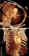
We present the case of a 78-year-old male who was admitted to his local hospital with complaints of thoracic pain and dyspnea after accidentally falling. His past medical history was relevant for a mechanical aortic valve prosthesis implanted 27 years previously, and he was on current treatment with acenocoumarol. At the time of presentation, a chest X-ray revealed a right sided pleural effusion. A 20 F trocar-type chest tube was inserted at the 5th intercostal space in the anterior axillary line. Upon placement of the chest tube more than 1000mL of blood was withdrawn and the patient became severely hypotensive, the chest tube was immediately clamped and a chest X-ray revealed a right massive pleural effusion. The patient was then transferred to our hospital with suspicion of intercostal artery laceration. On arrival, a chest CT scan was performed showing the chest tube inside the main pulmonary artery through the right pulmonary artery (Fig. 1). The patient was immediately transferred to the operating room with the thoracostomy tube clamped. A right antero-lateral thoracotomy was performed through the 4th intercostal space. Pleural adhesions were found and adhesiolysis was performed with cauterization and blunt dissection.
The tube was noted to perforate the right upper lobe, and after following the trajectory the entrance point was noted to be through one of the inferior branches of the right pulmonary artery. The main pulmonary artery was encircled with a vessel loop and the pulmonary circulation was temporally interrupted, the thoracostomy tube was successfully retrieved and the orifice was sutured with monofilament sutures. The vessel loop was released, and after confirmation of bleeding control, sutures were placed at the entrance of the lung parenquima.
New chest tubes were placed and the patient was transferred to the ICU. Following the intervention, chest X-rays showed bilateral infiltrates compatible with ARDS, and over the next few days the patient developed multiorgan failure. Finally, the patient died on the seventh postoperative day.
Several complications have been reported following chest tube insertion, including lung and diaphragm lacerations, intercostals artery bleeding and perforation of intraabdominal organs.1 Damage to cardiac structures has also been reported, such as right atrium perforation described by Meisel, Ram and Priel.5
As previously reported,2 a chest X-ray showing the tip of the catheter passing across the midline and the withdrawal of fresh blood should raise the suspicion of pulmonary artery perforation, as was the case in our patient.
It has been described that pleural adhesions, as also found in this case, could play a role upon misplacing the thoracostomy tube, probably by leading to perforating the lung parenchyma or lacerating vascular structures.3
For the management of pulmonary artery perforation, different approaches have been described2–4 including the progressive withdrawal of the chest tube during several days with no surgical intervention. However, it is accepted that the best option is to keep the tube clamped when there is suspicion of a great vessel rupture until the patient arrives to the operating room. It has been reported that the retrieval of the chest tube prior to arrival to the operating room may lead to a fatal outcome.5
Finally, chest tube insertion is a procedure that saves lives and is commonly performed in everyday clinical practice. However, as in any medical/surgical procedure, complications may occur. In order to achieve the best treatment, it is important to quickly recognize them and to choose the most suitable treatment for each patient. In the case we present we believe that the inmediate surgical approach may be the best choice.
Please cite this article as: Jauregui A, Deu M, Persiva O. Perforación de la arteria pulmonar tras la inserción de un drenaje torácico. Arch Bronconeumol. 2016;52:568–569.


