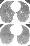
During a follow-up computed tomography (CT) in a 46-year-old patient with Niemann-Pick type B disease (NPBD) diagnosed during childhood, we detected bilateral pulmonary cysts and interstitial lung disease consisting of diffuse thickening of the interlobular septa and areas of ground glass attenuation (Fig. 1A). The pulmonary cysts were small, and located mainly in the middle and lower fields of both lungs (Fig. 1B). From a clinical point of view, the patient presented hepatosplenomegaly and dyspnea on exertion, with no neurological involvement. Lung function tests were normal, except for a slight decrease in CO diffusing capacity.
(A) Axial CT image of the chest (pulmonary parenchymal window) showing thickening of interlobular septa (short arrows) and areas of ground glass attenuation (arrows). (B) Minimum intensity projection (MinIP) axial CT image of the chest (pulmonary parenchymal window) showing small lung cysts in the basal segments of both lower lobes (arrows).
NPBD is a rare hereditary lysosomal storage disease (autosomal recessive) caused by sphingomyelinase deficiency. Clinical features generally include hepatosplenomegaly, pancytopenia, and characteristically, progressive interstitial lung involvement. The most common radiological findings in NPBD patients are thickening of the interlobular septa and ground glass opacities, with an apicobasal gradient (more marked in the lung bases than in the apices).1 We have found only 1 single case of pulmonary cysts in an adult patient with NPBD who, like our patient, presented cysts predominantly in the lung bases.2 We believe that pulmonary cysts are part of the spectrum of pulmonary involvement in NPBD, and that this phenomenon should be included in the differential diagnosis of patients with pulmonary cysts and interstitial lung disease, especially in the case of concomitant hepatosplenic and neurological involvement.
Please cite this article as: Gorospe L, Chinea-Rodríguez A, Villarrubia-Espinosa J, Arrieta P. Enfermedad de Niemann-Pick tipo B: una causa rara de quistes pulmonares. Arch Bronconeumol. 2019;55:100.


