Lipomas are the most common benign soft tissue tumor found in humans, and occur in approximately 1% of the population. They are generally subcutaneous, and appear only rarely in the viscera.1 Pulmonary lipomas are uncommon, most being endobronchial lesions accounting for 0.15%–0.5% of lung tumors. Pulmonary lipomas within the peripheral parenchyma are exceedingly rare.2,3 We report such a case.
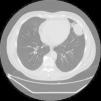
A 58-year-old man presented with pain in the left hemithorax radiating to the left shoulder and arm. Chest X-ray revealed an undefined lesion in the upper left lobe of the lung. Multislice computed tomography showed a rounded peripheral intraparenchymal pulmonary nodule measuring 53×54mm, located in the periphery of the upper left lobe lingula. The lesion was in contact with the diaphragm, the pericardium and the parietal pleura (Fig. 1).
Thoracotomy was performed and intraoperative inspection revealed a tumor in the lingula, adhered to the diaphragm and the pericardium. No hilar or mediastinal lymphadenopathies were found. Unilateral left upper lobectomy was performed and the sample was sent for pathology analysis.
The gross description was a soft, pale brown, round tumor in the lingula, with defined borders, measuring 35×25×25mm. Histological analysis reported a tumor consisting of mature adipose cells with interspersed areas of thin collagen stroma. The tumor nucleus was necrotic. It was separated by a fibrous capsule from the rest of the lung parenchyma and the visceral pleural on one side and the adipose and muscle tissue (considered part of the pericardium and the diaphragm) on the other. The diagnosis, according to appearance on histology, was lipoma. The patient remains well, 4 months after surgery.
Intraparenchymal pulmonary lipomas do not appear to favor any lung lobe or side, and appear in both men and women aged from 26 to 81 years of age. Tumors previously described range in size from 1.3cm to 7cm in diameter.2–4
The clinical course of these tumors is benign and they are generally asymptomatic. In rare cases such as ours, however, they present with paresthesia of the arm, mild dyspnea and lung dysfunction.3,4
Peripheral pulmonary lipomas are indistinguishable from malignant tumors on chest X-ray. Computed tomography is thought to assist diagnosis, although it remains difficult for radiologists to determine the biological nature of the lesion.3
Treatment of solitary pulmonary nodules (including pulmonary lipomas) remains a topic for debate, because in none of the reported cases could malignancy be definitively ruled out. In general, they are surgically resected, the most common procedure being lobectomy.5
In our case, multislice computed tomography and intraoperative inspection of the lung showed an intraparenchymal pulmonary lesion in contact with the diaphragm, the pericardium and the parietal pleura, clinically mimicking a malignant tumor, so lobectomy was selected as treatment of choice. Despite their rarity, intraparenchymal pulmonary lipomas should be considered in the differential diagnosis of pulmonary nodules located in the lung periphery, for careful planning and adaptation of the surgical intervention.
Acknowledgements and FundingThe manuscript was written in the Dubrava University Hospital, Zagreb. No external funding was received. Our thanks to Stela Bulimbašić and Arijana Pačić for their advice during the preparation of this paper.
Please cite this article as: Bacalja J, Nikolić I, Brčić L. Lipoma pulmonar intraparenquimatoso con comportamiento clínico de neoplasia maligna. Arch Bronconeumol. 2015;51:302-303.


