array:24 [
  "pii" => "S157921291400041X"
  "issn" => "15792129"
  "doi" => "10.1016/j.arbr.2014.02.006"
  "estado" => "S300"
  "fechaPublicacion" => "2014-03-01"
  "aid" => "802"
  "copyright" => "SEPAR"
  "copyrightAnyo" => "2013"
  "documento" => "simple-article"
  "crossmark" => 0
  "subdocumento" => "cor"
  "cita" => "Arch Bronconeumol. 2014;50:126-7"
  "abierto" => array:3 [
    "ES" => false
    "ES2" => false
    "LATM" => false
  ]
  "gratuito" => false
  "lecturas" => array:2 [
    "total" => 3299
    "formatos" => array:3 [
      "EPUB" => 103
      "HTML" => 2506
      "PDF" => 690
    ]
  ]
  "Traduccion" => array:1 [
    "es" => array:19 [
      "pii" => "S0300289613002056"
      "issn" => "03002896"
      "doi" => "10.1016/j.arbres.2013.07.003"
      "estado" => "S300"
      "fechaPublicacion" => "2014-03-01"
      "aid" => "802"
      "copyright" => "SEPAR"
      "documento" => "simple-article"
      "crossmark" => 0
      "subdocumento" => "cor"
      "cita" => "Arch Bronconeumol. 2014;50:126-7"
      "abierto" => array:3 [
        "ES" => true
        "ES2" => true
        "LATM" => true
      ]
      "gratuito" => true
      "lecturas" => array:2 [
        "total" => 6084
        "formatos" => array:3 [
          "EPUB" => 112
          "HTML" => 4992
          "PDF" => 980
        ]
      ]
      "es" => array:11 [
        "idiomaDefecto" => true
        "cabecera" => "<span class="elsevierStyleTextfn">Carta al Director</span>"
        "titulo" => "Tuberculosis endobronquial como simulador de c&#225;ncer de pulm&#243;n"
        "tienePdf" => "es"
        "tieneTextoCompleto" => "es"
        "paginas" => array:1 [
          0 => array:2 [
            "paginaInicial" => "126"
            "paginaFinal" => "127"
          ]
        ]
        "titulosAlternativos" => array:1 [
          "en" => array:1 [
            "titulo" => "Endobronchial Tuberculosis Simulating Lung Cancer"
          ]
        ]
        "contieneTextoCompleto" => array:1 [
          "es" => true
        ]
        "contienePdf" => array:1 [
          "es" => true
        ]
        "resumenGrafico" => array:2 [
          "original" => 0
          "multimedia" => array:7 [
            "identificador" => "fig0005"
            "etiqueta" => "Figura 1"
            "tipo" => "MULTIMEDIAFIGURA"
            "mostrarFloat" => true
            "mostrarDisplay" => false
            "figura" => array:1 [
              0 => array:4 [
                "imagen" => "gr1.jpeg"
                "Alto" => 1359
                "Ancho" => 1300
                "Tamanyo" => 222951
              ]
            ]
            "descripcion" => array:1 [
              "es" => "<p id="spar0005" class="elsevierStyleSimplePara elsevierViewall">Im&#225;genes de TAC y broncoscopia antes del tratamiento&#46;</p>"
            ]
          ]
        ]
        "autores" => array:1 [
          0 => array:2 [
            "autoresLista" => "Julen Cadi&#241;anos Loidi, Daniel Abad P&#233;rez, Rosa de Miguel Buckley"
            "autores" => array:3 [
              0 => array:2 [
                "nombre" => "Julen"
                "apellidos" => "Cadi&#241;anos Loidi"
              ]
              1 => array:2 [
                "nombre" => "Daniel"
                "apellidos" => "Abad P&#233;rez"
              ]
              2 => array:2 [
                "nombre" => "Rosa"
                "apellidos" => "de Miguel Buckley"
              ]
            ]
          ]
        ]
      ]
      "idiomaDefecto" => "es"
      "Traduccion" => array:1 [
        "en" => array:9 [
          "pii" => "S157921291400041X"
          "doi" => "10.1016/j.arbr.2014.02.006"
          "estado" => "S300"
          "subdocumento" => ""
          "abierto" => array:3 [
            "ES" => false
            "ES2" => false
            "LATM" => false
          ]
          "gratuito" => false
          "lecturas" => array:1 [
            "total" => 0
          ]
          "idiomaDefecto" => "en"
          "EPUB" => "https://multimedia.elsevier.es/PublicationsMultimediaV1/item/epub/S157921291400041X?idApp=UINPBA00003Z"
        ]
      ]
      "EPUB" => "https://multimedia.elsevier.es/PublicationsMultimediaV1/item/epub/S0300289613002056?idApp=UINPBA00003Z"
      "url" => "/03002896/0000005000000003/v1_201403010227/S0300289613002056/v1_201403010227/es/main.assets"
    ]
  ]
  "itemSiguiente" => array:19 [
    "pii" => "S1579212914000342"
    "issn" => "15792129"
    "doi" => "10.1016/j.arbr.2013.06.009"
    "estado" => "S300"
    "fechaPublicacion" => "2014-03-01"
    "aid" => "803"
    "copyright" => "SEPAR"
    "documento" => "simple-article"
    "crossmark" => 0
    "subdocumento" => "cor"
    "cita" => "Arch Bronconeumol. 2014;50:127"
    "abierto" => array:3 [
      "ES" => false
      "ES2" => false
      "LATM" => false
    ]
    "gratuito" => false
    "lecturas" => array:2 [
      "total" => 2694
      "formatos" => array:3 [
        "EPUB" => 103
        "HTML" => 1988
        "PDF" => 603
      ]
    ]
    "en" => array:10 [
      "idiomaDefecto" => true
      "cabecera" => "<span class="elsevierStyleTextfn">Letter to the Editor</span>"
      "titulo" => "Severe Asthma Exacerbation in an Intermediate Respiratory Care Unit&#58; Fact or Controversy&#63;"
      "tienePdf" => "en"
      "tieneTextoCompleto" => "en"
      "paginas" => array:1 [
        0 => array:1 [
          "paginaInicial" => "127"
        ]
      ]
      "titulosAlternativos" => array:1 [
        "es" => array:1 [
          "titulo" => "Agudizaci&#243;n grave de asma en una unidad de cuidados respiratorios intermedios&#58; &#191;realidad o controversia&#63;"
        ]
      ]
      "contieneTextoCompleto" => array:1 [
        "en" => true
      ]
      "contienePdf" => array:1 [
        "en" => true
      ]
      "autores" => array:1 [
        0 => array:2 [
          "autoresLista" => "Javier Navarro Esteva, Antonio M&#46; Esquinas Rodr&#237;guez"
          "autores" => array:2 [
            0 => array:2 [
              "nombre" => "Javier"
              "apellidos" => "Navarro Esteva"
            ]
            1 => array:2 [
              "nombre" => "Antonio M&#46;"
              "apellidos" => "Esquinas Rodr&#237;guez"
            ]
          ]
        ]
      ]
    ]
    "idiomaDefecto" => "en"
    "Traduccion" => array:1 [
      "es" => array:9 [
        "pii" => "S0300289613002068"
        "doi" => "10.1016/j.arbres.2013.06.005"
        "estado" => "S300"
        "subdocumento" => ""
        "abierto" => array:3 [
          "ES" => true
          "ES2" => true
          "LATM" => true
        ]
        "gratuito" => true
        "lecturas" => array:1 [
          "total" => 0
        ]
        "idiomaDefecto" => "es"
        "EPUB" => "https://multimedia.elsevier.es/PublicationsMultimediaV1/item/epub/S0300289613002068?idApp=UINPBA00003Z"
      ]
    ]
    "EPUB" => "https://multimedia.elsevier.es/PublicationsMultimediaV1/item/epub/S1579212914000342?idApp=UINPBA00003Z"
    "url" => "/15792129/0000005000000003/v1_201403120155/S1579212914000342/v1_201403120155/en/main.assets"
  ]
  "itemAnterior" => array:19 [
    "pii" => "S1579212914000408"
    "issn" => "15792129"
    "doi" => "10.1016/j.arbr.2014.02.005"
    "estado" => "S300"
    "fechaPublicacion" => "2014-03-01"
    "aid" => "797"
    "copyright" => "SEPAR"
    "documento" => "simple-article"
    "crossmark" => 0
    "subdocumento" => "cor"
    "cita" => "Arch Bronconeumol. 2014;50:125"
    "abierto" => array:3 [
      "ES" => false
      "ES2" => false
      "LATM" => false
    ]
    "gratuito" => false
    "lecturas" => array:2 [
      "total" => 2520
      "formatos" => array:3 [
        "EPUB" => 115
        "HTML" => 1921
        "PDF" => 484
      ]
    ]
    "en" => array:11 [
      "idiomaDefecto" => true
      "cabecera" => "<span class="elsevierStyleTextfn">Letter to the Editor</span>"
      "titulo" => "Mucoepidermoid Carcinoma in a Bone Marrow Transplant Patient"
      "tienePdf" => "en"
      "tieneTextoCompleto" => "en"
      "paginas" => array:1 [
        0 => array:1 [
          "paginaInicial" => "125"
        ]
      ]
      "titulosAlternativos" => array:1 [
        "es" => array:1 [
          "titulo" => "Tumor mucoepidermoide en paciente trasplantada de m&#233;dula &#243;sea"
        ]
      ]
      "contieneTextoCompleto" => array:1 [
        "en" => true
      ]
      "contienePdf" => array:1 [
        "en" => true
      ]
      "resumenGrafico" => array:2 [
        "original" => 0
        "multimedia" => array:7 [
          "identificador" => "fig0005"
          "etiqueta" => "Fig&#46; 1"
          "tipo" => "MULTIMEDIAFIGURA"
          "mostrarFloat" => true
          "mostrarDisplay" => false
          "figura" => array:1 [
            0 => array:4 [
              "imagen" => "gr1.jpeg"
              "Alto" => 767
              "Ancho" => 976
              "Tamanyo" => 88982
            ]
          ]
          "descripcion" => array:1 [
            "en" => "<p id="spar0005" class="elsevierStyleSimplePara elsevierViewall">Chest computed axial tomography slice showing an occupation of the segmental and some subsegmental bronchi by soft tissue in the left lung base&#44; probably related to endobronchial tumor disease&#46;</p>"
          ]
        ]
      ]
      "autores" => array:1 [
        0 => array:2 [
          "autoresLista" => "Mar&#237;a del Valle Somiedo Guti&#233;rrez, Rosa Gir&#243;n Moreno, Ram&#243;n Moreno Balsalobre"
          "autores" => array:3 [
            0 => array:2 [
              "nombre" => "Mar&#237;a del Valle"
              "apellidos" => "Somiedo Guti&#233;rrez"
            ]
            1 => array:2 [
              "nombre" => "Rosa"
              "apellidos" => "Gir&#243;n Moreno"
            ]
            2 => array:2 [
              "nombre" => "Ram&#243;n"
              "apellidos" => "Moreno Balsalobre"
            ]
          ]
        ]
      ]
    ]
    "idiomaDefecto" => "en"
    "Traduccion" => array:1 [
      "es" => array:9 [
        "pii" => "S0300289613001877"
        "doi" => "10.1016/j.arbres.2013.06.002"
        "estado" => "S300"
        "subdocumento" => ""
        "abierto" => array:3 [
          "ES" => true
          "ES2" => true
          "LATM" => true
        ]
        "gratuito" => true
        "lecturas" => array:1 [
          "total" => 0
        ]
        "idiomaDefecto" => "es"
        "EPUB" => "https://multimedia.elsevier.es/PublicationsMultimediaV1/item/epub/S0300289613001877?idApp=UINPBA00003Z"
      ]
    ]
    "EPUB" => "https://multimedia.elsevier.es/PublicationsMultimediaV1/item/epub/S1579212914000408?idApp=UINPBA00003Z"
    "url" => "/15792129/0000005000000003/v1_201403120155/S1579212914000408/v1_201403120155/en/main.assets"
  ]
  "en" => array:15 [
    "idiomaDefecto" => true
    "cabecera" => "<span class="elsevierStyleTextfn">Letter to the Editor</span>"
    "titulo" => "Endobronchial Tuberculosis Simulating Lung Cancer"
    "tieneTextoCompleto" => true
    "saludo" => "To the Editor&#58;"
    "paginas" => array:1 [
      0 => array:2 [
        "paginaInicial" => "126"
        "paginaFinal" => "127"
      ]
    ]
    "autores" => array:1 [
      0 => array:4 [
        "autoresLista" => "Julen Cadi&#241;anos Loidi, Daniel Abad P&#233;rez, Rosa de Miguel Buckley"
        "autores" => array:3 [
          0 => array:2 [
            "nombre" => "Julen"
            "apellidos" => "Cadi&#241;anos Loidi"
          ]
          1 => array:4 [
            "nombre" => "Daniel"
            "apellidos" => "Abad P&#233;rez"
            "email" => array:1 [
              0 => "danielabadperez&#64;hotmail&#46;com"
            ]
            "referencia" => array:1 [
              0 => array:2 [
                "etiqueta" => "<span class="elsevierStyleSup">¿</span>"
                "identificador" => "cor0005"
              ]
            ]
          ]
          2 => array:2 [
            "nombre" => "Rosa"
            "apellidos" => "de Miguel Buckley"
          ]
        ]
        "afiliaciones" => array:1 [
          0 => array:2 [
            "entidad" => "Servicio de Medicina Interna&#44; Hospital Universitario de la Princesa&#44; Madrid&#44; Spain"
            "identificador" => "aff0005"
          ]
        ]
        "correspondencia" => array:1 [
          0 => array:3 [
            "identificador" => "cor0005"
            "etiqueta" => "&#8270;"
            "correspondencia" => "Corresponding author&#46;"
          ]
        ]
      ]
    ]
    "titulosAlternativos" => array:1 [
      "es" => array:1 [
        "titulo" => "Tuberculosis endobronquial como simulador de c&#225;ncer de pulm&#243;n"
      ]
    ]
    "resumenGrafico" => array:2 [
      "original" => 0
      "multimedia" => array:7 [
        "identificador" => "fig0005"
        "etiqueta" => "Fig&#46; 1"
        "tipo" => "MULTIMEDIAFIGURA"
        "mostrarFloat" => true
        "mostrarDisplay" => false
        "figura" => array:1 [
          0 => array:4 [
            "imagen" => "gr1.jpeg"
            "Alto" => 1358
            "Ancho" => 1301
            "Tamanyo" => 236735
          ]
        ]
        "descripcion" => array:1 [
          "en" => "<p id="spar0005" class="elsevierStyleSimplePara elsevierViewall">Bronchoscopy and CT images before treatment&#46;</p>"
        ]
      ]
    ]
    "textoCompleto" => "<span class="elsevierStyleSections"><p id="par0005" class="elsevierStylePara elsevierViewall">Tuberculosis &#40;TB&#41; still has an annual incidence in Spain of 18&#46;2&#47;100&#44;000 inhabitants&#46;<a class="elsevierStyleCrossRef" href="#bib0005"><span class="elsevierStyleSup">1</span></a> We report a case of endobronchial tuberculosis with lymph node and skin involvement&#44; an unusual form in our setting&#46;</p><p id="par0010" class="elsevierStylePara elsevierViewall">A 45-year-old woman who had been resident in India until 8 months previously&#44; presented with clinical symptoms for the last 3 months of daily fever &#40;up to 38<span class="elsevierStyleHsp" style=""></span>&#176;C&#41;&#44; anorexia&#44; weight loss&#44; dry cough&#44; and dyspnea that did not improve after 7 days of treatment with amoxicillin&#8211;clavulanic acid&#46; She had good general health upon examination&#44; the only remarkable finding being two mobile&#44; rubbery subcutaneous nodules sized 1<span class="elsevierStyleHsp" style=""></span>cm in the left upper quadrant that were slightly painful but with no signs of inflammation&#44; and a similar one in the left hand&#46; Blood tests revealed anemia&#44; leukocytosis and thrombocytosis&#46; Chest radiography showed a mass in the anterior mediastinum described in the computed tomography &#40;CT&#41; report as &#8220;mass in the left upper lobe and ipsilateral hilar region&#44; with anterior mediastinal infiltration causing stenosis of the bronchial lumen&#59; ipsilateral subcarinal and paratracheal lymphadenopathies&#44; accompanied by multiple independent nodules in the left lung&#44; pleural implants&#44; and subcutaneous abdominal nodules&#8221;&#46; Bronchoscopy revealed inflamed mucosa with implants of tumor-like appearance and left bronchial stenosis&#46; Malignancy was ruled out after biopsy&#44; but both bronchial biopsies and one of the abdominal nodes showed non-necrotizing granulomas&#46; Cultures were initially negative&#44; but then multi-susceptible <span class="elsevierStyleItalic">Mycobacterium tuberculosis</span> grew in sputum&#46; Accordingly&#44; the patient was diagnosed with disseminated TB with tumor-type endobronchial and skin involvement&#46; The patient improved rapidly with TB therapy and corticosteroids&#46; Bronchoscopy was normal at 6 months&#44; while the CT showed only residual thickening of soft tissues &#40;<a class="elsevierStyleCrossRef" href="#fig0005">Fig&#46; 1</a>&#41;&#46;</p><elsevierMultimedia ident="fig0005"></elsevierMultimedia><p id="par0015" class="elsevierStylePara elsevierViewall">Endobronchial TB occurs in 5&#37;&#8211;40&#37; of active pulmonary TB&#44; although it is underdiagnosed&#46;<a class="elsevierStyleCrossRefs" href="#bib0010"><span class="elsevierStyleSup">2&#44;3</span></a> The cause is implantation directly from an adjacent cavity&#44; a focus of tuberculosis or a mediastinal lymph node&#44; or by bloodborne or lymphatic spread&#46;<a class="elsevierStyleCrossRefs" href="#bib0015"><span class="elsevierStyleSup">3&#44;4</span></a> Symptoms include cough &#40;71&#37;&#8211;100&#37;&#41;&#44; fever &#40;50&#37;&#41;&#44; weight loss &#40;30&#37;&#41;&#44; hemoptysis &#40;18&#37;&#8211;25&#37;&#41;&#44; dyspnea&#44; chest pain and anorexia&#46;<a class="elsevierStyleCrossRefs" href="#bib0010"><span class="elsevierStyleSup">2&#44;4&#44;5</span></a> Both the duration of symptoms and the disease progression are variable and can range from complete resolution to the appearance of bronchiectasis&#44; bronchial obstruction or atelectasis&#46; The most useful microbiological test is bronchoalveolar lavage&#44; which outperforms sputum&#46;<a class="elsevierStyleCrossRefs" href="#bib0010"><span class="elsevierStyleSup">2&#8211;5</span></a> X-ray may show alveolar infiltrates &#40;35&#37;&#8211;43&#37;&#41;&#44; nodules &#40;25&#37;&#41;&#44; cavitated lesions &#40;12&#37;&#41;&#44; pleural effusion &#40;9&#37;&#41; or hilar thickening &#40;7&#37;&#41;&#44; but it may be normal in 10&#37;&#8211;20&#37; of cases&#46;<a class="elsevierStyleCrossRefs" href="#bib0010"><span class="elsevierStyleSup">2&#44;3</span></a> CT is useful for assessment of disease extension&#44; degree of bronchial obstruction and progress&#44; and may replace bronchoscopy during follow-up&#44;<a class="elsevierStyleCrossRef" href="#bib0020"><span class="elsevierStyleSup">4</span></a> although it is the imaging study of choice during diagnosis&#46;</p><p id="par0020" class="elsevierStylePara elsevierViewall">Chung and Lee classify TB into seven subtypes<a class="elsevierStyleCrossRef" href="#bib0015"><span class="elsevierStyleSup">3</span></a>&#58; caseating &#40;12&#37;&#8211;43&#37;&#41;&#44; edematous-hyperemic &#40;14&#37;&#8211;44&#37;&#41;&#44; fibrostenotic &#40;6&#37;&#8211;10&#46;5&#37;&#41;&#44; tumoral &#40;10&#46;5&#37;&#8211;30&#37;&#41;&#44;<a class="elsevierStyleCrossRefs" href="#bib0015"><span class="elsevierStyleSup">3&#8211;5</span></a> granular&#44; ulcerative&#44; and nonspecific bronchitis type&#46; The first four have a poorer prognosis&#44; because of associated bronchial stenosis&#46;<a class="elsevierStyleCrossRef" href="#bib0015"><span class="elsevierStyleSup">3</span></a> These types seem to represent different stages of the same disease&#44; starting with granulomas and submucosal inflammatory lesions that progress to masses&#44; fibrosis and airway stenosis&#46;<a class="elsevierStyleCrossRefs" href="#bib0020"><span class="elsevierStyleSup">4&#44;5</span></a> The tumoral subtype has endobronchial masses&#44; with a hemorrhagic surface and a necrotic outer layer&#44; simulating squamous carcinoma&#46; Risk factors for residual bronchial stenosis are age over 45 years&#44; fibrostenotic type&#44; and late diagnosis&#46;<a class="elsevierStyleCrossRefs" href="#bib0010"><span class="elsevierStyleSup">2&#44;5</span></a> Treatment includes endoscopic dilation&#44; mechanical resection&#44; or stenting combined with corticosteroids&#46; The latter seems to be effective in the early stages and the caseating&#47;tumoral forms&#44;<a class="elsevierStyleCrossRef" href="#bib0020"><span class="elsevierStyleSup">4</span></a> so for this reason they were used in our patient&#44; who achieved total recovery&#46;</p><p id="par0025" class="elsevierStylePara elsevierViewall">In the presence of fever and endobronchial lesions or pulmonary mass&#44; consideration of TB at diagnosis is mandatory&#44; especially in immigrants during the early years after arrival&#46;</p></span>"
    "pdfFichero" => "main.pdf"
    "tienePdf" => true
    "NotaPie" => array:1 [
      0 => array:2 [
        "etiqueta" => "&#9734;"
        "nota" => "<p class="elsevierStyleNotepara" id="npar0005">Please cite this article as&#58; Cadi&#241;anos Loidi J&#44; Abad P&#233;rez D&#44; de Miguel Buckley R&#46; Tuberculosis endobronquial como simulador de c&#225;ncer de pulm&#243;n&#46; Arch Bronconeumol&#46; 2014&#59;50&#58;126&#8211;127&#46;</p>"
      ]
    ]
    "multimedia" => array:1 [
      0 => array:7 [
        "identificador" => "fig0005"
        "etiqueta" => "Fig&#46; 1"
        "tipo" => "MULTIMEDIAFIGURA"
        "mostrarFloat" => true
        "mostrarDisplay" => false
        "figura" => array:1 [
          0 => array:4 [
            "imagen" => "gr1.jpeg"
            "Alto" => 1358
            "Ancho" => 1301
            "Tamanyo" => 236735
          ]
        ]
        "descripcion" => array:1 [
          "en" => "<p id="spar0005" class="elsevierStyleSimplePara elsevierViewall">Bronchoscopy and CT images before treatment&#46;</p>"
        ]
      ]
    ]
    "bibliografia" => array:2 [
      "titulo" => "References"
      "seccion" => array:1 [
        0 => array:2 [
          "identificador" => "bibs0005"
          "bibliografiaReferencia" => array:5 [
            0 => array:3 [
              "identificador" => "bib0005"
              "etiqueta" => "1"
              "referencia" => array:1 [
                0 => array:1 [
                  "referenciaCompleta" => "Informe sobre la situaci&#243;n de la tuberculosis&#46; Red Nacional de Vigilancia Epidemiol&#243;gica&#46; Espa&#241;a&#44; 2007&#46; Centro Nacional de Epidemiolog&#237;a&#46; 18 de marzo de 2009&#46; Available from&#58; <a id="intr0010" class="elsevierStyleInterRef" href="http://genmico.unizar.es/PDF/TB_informe%202007_CNE.pdf">http&#58;&#47;&#47;genmico&#46;unizar&#46;es&#47;PDF&#47;TB&#95;informe&#37;202007&#95;CNE&#46;pdf</a> &#91;accessed 18&#46;10&#46;11&#93;&#46;"
                ]
              ]
            ]
            1 => array:3 [
              "identificador" => "bib0010"
              "etiqueta" => "2"
              "referencia" => array:1 [
                0 => array:2 [
                  "contribucion" => array:1 [
                    0 => array:2 [
                      "titulo" => "Endobronchial tuberculosis revisited"
                      "autores" => array:1 [
                        0 => array:2 [
                          "etal" => false
                          "autores" => array:4 [
                            0 => "M&#46;S&#46; Ip"
                            1 => "S&#46;Y&#46; So"
                            2 => "W&#46;K&#46; Lam"
                            3 => "C&#46;K&#46; Mok"
                          ]
                        ]
                      ]
                    ]
                  ]
                  "host" => array:1 [
                    0 => array:1 [
                      "Revista" => array:6 [
                        "tituloSerie" => "Chest"
                        "fecha" => "1986"
                        "volumen" => "89"
                        "paginaInicial" => "727"
                        "paginaFinal" => "730"
                        "link" => array:1 [
                          0 => array:2 [
                            "url" => "https://www.ncbi.nlm.nih.gov/pubmed/3698702"
                            "web" => "Medline"
                          ]
                        ]
                      ]
                    ]
                  ]
                ]
              ]
            ]
            2 => array:3 [
              "identificador" => "bib0015"
              "etiqueta" => "3"
              "referencia" => array:1 [
                0 => array:2 [
                  "contribucion" => array:1 [
                    0 => array:2 [
                      "titulo" => "Bronchoscopic assessment of the evolution of endobronchial tuberculosis"
                      "autores" => array:1 [
                        0 => array:2 [
                          "etal" => false
                          "autores" => array:2 [
                            0 => "H&#46;S&#46; Chung"
                            1 => "J&#46;H&#46; Lee"
                          ]
                        ]
                      ]
                    ]
                  ]
                  "host" => array:1 [
                    0 => array:1 [
                      "Revista" => array:6 [
                        "tituloSerie" => "Chest"
                        "fecha" => "2000"
                        "volumen" => "117"
                        "paginaInicial" => "385"
                        "paginaFinal" => "392"
                        "link" => array:1 [
                          0 => array:2 [
                            "url" => "https://www.ncbi.nlm.nih.gov/pubmed/10669679"
                            "web" => "Medline"
                          ]
                        ]
                      ]
                    ]
                  ]
                ]
              ]
            ]
            3 => array:3 [
              "identificador" => "bib0020"
              "etiqueta" => "4"
              "referencia" => array:1 [
                0 => array:2 [
                  "contribucion" => array:1 [
                    0 => array:2 [
                      "titulo" => "Computed tomography and bronchoscopy in endobronquial tuberculosis"
                      "autores" => array:1 [
                        0 => array:2 [
                          "etal" => false
                          "autores" => array:5 [
                            0 => "H&#46; Yanardag"
                            1 => "C&#46; Tetikkurt"
                            2 => "S&#46; Tetikkurt"
                            3 => "S&#46; Demirci"
                            4 => "T&#46; Karayel"
                          ]
                        ]
                      ]
                    ]
                  ]
                  "host" => array:1 [
                    0 => array:1 [
                      "Revista" => array:7 [
                        "tituloSerie" => "Can Respir J"
                        "fecha" => "2003"
                        "volumen" => "10"
                        "paginaInicial" => "445"
                        "paginaFinal" => "448"
                        "link" => array:1 [
                          0 => array:2 [
                            "url" => "https://www.ncbi.nlm.nih.gov/pubmed/14679409"
                            "web" => "Medline"
                          ]
                        ]
                        "itemHostRev" => array:3 [
                          "pii" => "S147444220570140X"
                          "estado" => "S300"
                          "issn" => "14744422"
                        ]
                      ]
                    ]
                  ]
                ]
              ]
            ]
            4 => array:3 [
              "identificador" => "bib0025"
              "etiqueta" => "5"
              "referencia" => array:1 [
                0 => array:2 [
                  "contribucion" => array:1 [
                    0 => array:2 [
                      "titulo" => "Predictors of persistent airway stenosis in patients with endobronchial tuberculosis"
                      "autores" => array:1 [
                        0 => array:2 [
                          "etal" => true
                          "autores" => array:6 [
                            0 => "S&#46;W&#46; Um"
                            1 => "Y&#46;S&#46; Yoon"
                            2 => "S&#46;M&#46; Lee"
                            3 => "J&#46;J&#46; Yim"
                            4 => "C&#46;G&#46; Yoo"
                            5 => "H&#46;S&#46; Chung"
                          ]
                        ]
                      ]
                    ]
                  ]
                  "host" => array:1 [
                    0 => array:1 [
                      "Revista" => array:5 [
                        "tituloSerie" => "Int J Tuberc Lung Dis"
                        "fecha" => "2007"
                        "volumen" => "11"
                        "paginaInicial" => "57"
                        "paginaFinal" => "62"
                      ]
                    ]
                  ]
                ]
              ]
            ]
          ]
        ]
      ]
    ]
  ]
  "idiomaDefecto" => "en"
  "url" => "/15792129/0000005000000003/v1_201403120155/S157921291400041X/v1_201403120155/en/main.assets"
  "Apartado" => array:4 [
    "identificador" => "14576"
    "tipo" => "SECCION"
    "es" => array:2 [
      "titulo" => "Letters to the editor"
      "idiomaDefecto" => true
    ]
    "idiomaDefecto" => "es"
  ]
  "PDF" => "https://static.elsevier.es/multimedia/15792129/0000005000000003/v1_201403120155/S157921291400041X/v1_201403120155/en/main.pdf?idApp=UINPBA00003Z&text.app=https://archbronconeumol.org/"
  "EPUB" => "https://multimedia.elsevier.es/PublicationsMultimediaV1/item/epub/S157921291400041X?idApp=UINPBA00003Z"
]
Share
Journal Information
Vol. 50. Issue 3.
Pages 126-127 (March 2014)
Vol. 50. Issue 3.
Pages 126-127 (March 2014)
Letter to the Editor
Full text access
Endobronchial Tuberculosis Simulating Lung Cancer
Tuberculosis endobronquial como simulador de cáncer de pulmón
Visits
8324
Julen Cadiñanos Loidi, Daniel Abad Pérez
Corresponding author
danielabadperez@hotmail.com

Corresponding author.
, Rosa de Miguel Buckley
Servicio de Medicina Interna, Hospital Universitario de la Princesa, Madrid, Spain
This item has received
Article information
Full Text
Bibliography
Download PDF
Statistics
Figures (1)
Full Text
To the Editor:

Tuberculosis (TB) still has an annual incidence in Spain of 18.2/100,000 inhabitants.1 We report a case of endobronchial tuberculosis with lymph node and skin involvement, an unusual form in our setting.

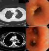
A 45-year-old woman who had been resident in India until 8 months previously, presented with clinical symptoms for the last 3 months of daily fever (up to 38°C), anorexia, weight loss, dry cough, and dyspnea that did not improve after 7 days of treatment with amoxicillin–clavulanic acid. She had good general health upon examination, the only remarkable finding being two mobile, rubbery subcutaneous nodules sized 1cm in the left upper quadrant that were slightly painful but with no signs of inflammation, and a similar one in the left hand. Blood tests revealed anemia, leukocytosis and thrombocytosis. Chest radiography showed a mass in the anterior mediastinum described in the computed tomography (CT) report as “mass in the left upper lobe and ipsilateral hilar region, with anterior mediastinal infiltration causing stenosis of the bronchial lumen; ipsilateral subcarinal and paratracheal lymphadenopathies, accompanied by multiple independent nodules in the left lung, pleural implants, and subcutaneous abdominal nodules”. Bronchoscopy revealed inflamed mucosa with implants of tumor-like appearance and left bronchial stenosis. Malignancy was ruled out after biopsy, but both bronchial biopsies and one of the abdominal nodes showed non-necrotizing granulomas. Cultures were initially negative, but then multi-susceptible Mycobacterium tuberculosis grew in sputum. Accordingly, the patient was diagnosed with disseminated TB with tumor-type endobronchial and skin involvement. The patient improved rapidly with TB therapy and corticosteroids. Bronchoscopy was normal at 6 months, while the CT showed only residual thickening of soft tissues (Fig. 1).

Fig. 1.

Bronchoscopy and CT images before treatment.

(0.23MB).

Endobronchial TB occurs in 5%–40% of active pulmonary TB, although it is underdiagnosed.2,3 The cause is implantation directly from an adjacent cavity, a focus of tuberculosis or a mediastinal lymph node, or by bloodborne or lymphatic spread.3,4 Symptoms include cough (71%–100%), fever (50%), weight loss (30%), hemoptysis (18%–25%), dyspnea, chest pain and anorexia.2,4,5 Both the duration of symptoms and the disease progression are variable and can range from complete resolution to the appearance of bronchiectasis, bronchial obstruction or atelectasis. The most useful microbiological test is bronchoalveolar lavage, which outperforms sputum.2–5 X-ray may show alveolar infiltrates (35%–43%), nodules (25%), cavitated lesions (12%), pleural effusion (9%) or hilar thickening (7%), but it may be normal in 10%–20% of cases.2,3 CT is useful for assessment of disease extension, degree of bronchial obstruction and progress, and may replace bronchoscopy during follow-up,4 although it is the imaging study of choice during diagnosis.

Chung and Lee classify TB into seven subtypes3: caseating (12%–43%), edematous-hyperemic (14%–44%), fibrostenotic (6%–10.5%), tumoral (10.5%–30%),3–5 granular, ulcerative, and nonspecific bronchitis type. The first four have a poorer prognosis, because of associated bronchial stenosis.3 These types seem to represent different stages of the same disease, starting with granulomas and submucosal inflammatory lesions that progress to masses, fibrosis and airway stenosis.4,5 The tumoral subtype has endobronchial masses, with a hemorrhagic surface and a necrotic outer layer, simulating squamous carcinoma. Risk factors for residual bronchial stenosis are age over 45 years, fibrostenotic type, and late diagnosis.2,5 Treatment includes endoscopic dilation, mechanical resection, or stenting combined with corticosteroids. The latter seems to be effective in the early stages and the caseating/tumoral forms,4 so for this reason they were used in our patient, who achieved total recovery.

In the presence of fever and endobronchial lesions or pulmonary mass, consideration of TB at diagnosis is mandatory, especially in immigrants during the early years after arrival.

References
[1]
Informe sobre la situación de la tuberculosis. Red Nacional de Vigilancia Epidemiológica. España, 2007. Centro Nacional de Epidemiología. 18 de marzo de 2009. Available from: http://genmico.unizar.es/PDF/TB_informe%202007_CNE.pdf [accessed 18.10.11].
[2]
M.S. Ip, S.Y. So, W.K. Lam, C.K. Mok.
Endobronchial tuberculosis revisited.
Chest, 89 (1986), pp. 727-730
[3]
H.S. Chung, J.H. Lee.
Bronchoscopic assessment of the evolution of endobronchial tuberculosis.
Chest, 117 (2000), pp. 385-392
[4]
H. Yanardag, C. Tetikkurt, S. Tetikkurt, S. Demirci, T. Karayel.
Computed tomography and bronchoscopy in endobronquial tuberculosis.
Can Respir J, 10 (2003), pp. 445-448
[5]
S.W. Um, Y.S. Yoon, S.M. Lee, J.J. Yim, C.G. Yoo, H.S. Chung, et al.
Predictors of persistent airway stenosis in patients with endobronchial tuberculosis.
Int J Tuberc Lung Dis, 11 (2007), pp. 57-62

Please cite this article as: Cadiñanos Loidi J, Abad Pérez D, de Miguel Buckley R. Tuberculosis endobronquial como simulador de cáncer de pulmón. Arch Bronconeumol. 2014;50:126–127.

Copyright © 2013. SEPAR
Archivos de Bronconeumología
Article options
Tools

Are you a health professional able to prescribe or dispense drugs?