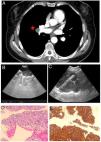
We report the case of a 59-year-old woman with a diagnosis of uterine leiomyosarcoma (T1bN0, stage IB) undergoing hysterectomy and double adnexectomy, referred for profiling of right hilar adenopathy (corresponding to lymph node station 11R) of pathological appearance in a computed axial tomography performed during oncological follow-up (Fig. 1a).
A) Computed tomography image with adenopathy in right hilar lymph node station. B) Image of lymph node station 11R from endobronchial ultrasound. C) Transvascular puncture by endobronchial ultrasound. D) Histological image at amplification ×20 of hematoxylin-eosin staining showing spindle cell proliferation with atypical forms. E) Histological image at amplification ×20 showing positivity for specific muscle actin.
Once the patient had been fully informed, endobronchial ultrasound was performed, and a single lymphadenopathy measuring 12.2 × 14 mm was found in lymph node station 11R. Given the presence of ultrasound characteristics predictive of malignancy1 (hypoechoic appearance, homogeneous, rounded shape, presence of easily distinguishable margins, and absence of hilar center [Fig. 1B]), we performed a 2-pass transvascular puncture (EBUS-TVNA) using a Cook medical needle ECHO-HD-22-EBUS-P®, with a 5 cm aspiration, with no immediate complications (Fig. 1C). Cytohistological diagnosis of the lesion demonstrated the presence of fusiform cells with hyperchromatic elongated nuclei, marked atypia and cell expression of specific muscle actin, histologically consistent with leiomyosarcoma metastases (Figs. 1D and E).
Although the use of this approach is rare, EBUS-TVNA has demonstrated a 71% yield in units with extensive experience in endoscopic lymphadenopathy management and the absence of major complications requiring subsequent therapeutic interventions2. In spite of the limited information available in the literature, this technique should be considered as a diagnostic method provided that the appropriate physical infrastructure and personnel are on hand to mitigate possible complications.
Please cite this article as: de Vega Sánchez B, Disdier Vicente C, Borrego Pintado H. Diagnóstico de metástasis intratorácica de leiomiosarcoma uterino mediante punción transvascular guiada por ecobroncoscopia. Arch Bronconeumol. 2021;57:765.


