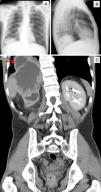
A 45-year-old man was admitted to the pulmonology department with a 2-week history of cough, sputum, hemoptysis and pleuritic chest pain on the right side. He reported anorexia, weight loss of about 6kg and excessive night sweating for the previous 2 months. For several months he had had recurrent episodes of dysuria, pollakiuria, nocturia and bilateral lumbar pain radiating to the groin. On physical examination, there were slightly decreased breath sounds in the right lung base and slight right flank pain on deep palpation. Chest X-ray (Fig. 1) revealed a rounded opacity in the right lung base, and thoracoabdominal computed tomography (CT) (Fig. 1) confirmed the presence of a large abdominal mass invading the thorax (see figure legend). Pathological examination of the lesion revealed poorly differentiated carcinoma of apparently urothelial origin. The patient started chemotherapy, but died about 8 months later due to disease progression.
(A and B) Chest X-ray (posteroanterior and lateral aspects, respectively) revealing a rounded opacity in the right lung base. (C) Thoracoabdominal CT (coronal plane) showing a large abdominal mass of about 13cm×11cm×8.3cm, apparently originating in the right kidney, invading the right lobe of the liver, the diaphragm and the lower lobe of the right lung (red arrow).
The authors declare that no funding was received for this paper.
Conflict of InterestsThe authors declare that there is no conflict of interests.
Please cite this article as: Dabó H, Damas C. Una causa inusual de hemoptisis. Arch Bronconeumol. 2016;52:275.


