We report the case of a 63-year-old woman with diabetes,1 who presented in the emergency department with painful swallowing and inability to swallow solids that was diagnosed as acute tonsillitis. She received a 10-day course of azithromycin due to a history of penicillin allergy. In view of her severe clinical picture (deterioration of her general status associated with mucocutaneous pallor) and laboratory results (leukocytosis and increased C-reactive protein), we decided to perform a cervical and thoracic CT with intravenous contrast to rule out complications associated with tonsillitis.
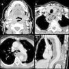
The study revealed a small abscess in the sinus of the right palatine tonsil (Fig. 1a) communicating with the ipsilateral visceral infrahyoid space through a descending fistula crossing the parapharyngeal space. A collection then formed, following a pretracheal route, crossing the midline in the fat plane underlying the superficial layer of the deep cervical fascia, then taking a perithyroidal route, and returning to enter the chest through the upper left thoracic inlet (Fig. 1b). It reached its maximum dimension at the entry point, presenting gas bubbles, and took on a tubular form in the middle and posterior mediastinum, almost completely surrounding the descending thoracic aorta along its entire length until the thoracoabdominal transition (Fig. 1c and d).
(a) Cervical CT in axial plane at the level of the epiglottis, showing an abscess measuring millimeters in the lower part of the right palatine tonsil (arrow); (b) cervical CT in axial plane at the level of the thyroid, revealing a collection with gas that surrounds the left thyroid lobe in an anterior direction, and then subsequently progresses in an posterior direction, passing into the chest through the upper left thoracic inlet (arrow); (c) chest CT in axial plane at the level of the carina: the collection is displayed in the middle and posterior mediastinum surrounding the thoracic aorta by more than 180°. This acts as a mass on the middle esophagus (asterisk); and (d) chest CT oblique sagittal plane, showing the collection extending along the entire length of the descending thoracic aorta. A small amount of pleural effusion can be observed.
This case is unique in 2 ways: the unusual path followed by the collection while making its way toward the chest through the anterior cervical planes, when the deep planes are the more common route2 (retropharyngeal route); and access of the collection to the middle and posterior mediastinum, when the anterior mediastinum would be more expected.
We thank Luis Gorospe Sarasua and Agustina Vicente Bártulos of the Radiodiagnostic Department, Hospital Universitario Ramón y Cajal, Madrid, Spain.
Please cite this article as: López-Frías López-Jurado A, Pecharromán de las Heras I, Pérez Templado Ladrón de Guevara J. Un caso raro de mediastinitis necrosante descendente. Arch Bronconeumol. 2019;55:435.


