
We present an 85-year-old patient with a history of 40 pack-years of smoking, hypertension, dyslipidemia, and ischaemic heart disease.
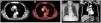
The patient's admission was prompted by symptoms including respiratory failure, asthenia, and atypical chest pain persisting for a week. A chest X-ray revealed mediastinal widening. In response to these findings, a computed tomography (CT) scan was performed, unveiling the presence of a solid mass in the mediastinal region and the right superior hilar area. This mass exhibited close proximity to the right tracheal margin, the right brachiocephalic trunk, the right main pulmonary artery, as well as the ipsilateral superior lobar pulmonary arteries and veins, suggesting possible infiltration (Fig. 1). Consequently, an endobronchial ultrasound-guided transbronchial needle aspiration (EBUS-TBNA) was conducted, enabling the diagnosis of the mass as lung adenocarcinoma and the identification of partial thrombosis within the superior vena cava, as illustrated in Video A.

It is noteworthy that venous thromboembolic disease affects approximately 20% of cancer patients.1 Although the utilization of EBUS for diagnosing central pulmonary embolism in patients with contrast allergies has been documented, there are no reported instances of superior vena cava thrombosis.2 This underscores the importance of bronchoscopists acquiring proficiency in recognizing such findings.
Conflict of InterestsThe authors state that they have no conflict of interests.


