


Congenital lung malformations (CLMs) encompass a range of developmental anomalies, including congenital pulmonary airway malformation (CPAM), bronchopulmonary sequestration (BPS), congenital lobar overinflation (CLO), bronchogenic cysts (BC), and bronchial atresia (BA). These lesions present with varying severity, from incidental findings to severe neonatal respiratory distress. Advances in prenatal imaging, such as ultrasound and fetal magnetic resonance image (MRI), have significantly improved early detection, aiding in better management planning.
CLMs arise from abnormalities during specific stages of lung development. CPAMs are categorized by cyst size and histological type, while BPS is characterized by systemic arterial supply. CLO and BA are associated with air trapping and hyperinflation, and BCs are typically fluid-filled, well defined, and compressive. Postnatal diagnosis is based on high-resolution computed tomography (CT) and occasionally MRI for detailed evaluation.
Surgical resection is recommended for symptomatic lesions, while management of asymptomatic cases remains debated. Elective surgery may help prevent complications like infection or rare malignancy, particularly in CPAM Types 1 and 4 and BCs. However, some lesions remain stable or regress, supporting conservative management in selected cases.
Long-term outcomes are generally positive, with children undergoing early resection often maintaining good lung function due to compensatory lung growth, though subtle functional deficits may persist. Risk stratification using imaging and genetic markers, such as DICER1 mutations, is gaining importance for guiding treatment decisions.
Management should be individualized, involving a multidisciplinary approach and shared decision-making with families. Further research is needed to clarify the natural history of CLMs, optimize the timing of interventions, and standardize long-term surveillance strategies.

Congenital anomalies of the lung are a heterogeneous group of developmental anomalies that originate during fetal lung development [1]. They are typically classified into three main categories: pulmonary growth anomalies, which include pulmonary agenesis, pulmonary hypoplasia, and pulmonary hyperplasia; diffuse congenital malformations of genetic origin, such as congenital surfactant deficiency, alveolar capillary dysplasia, and acinar dysplasia; and localized congenital lung malformations (CLMs), which encompass congenital pulmonary airway malformations (CPAMs), bronchopulmonary sequestration (BPS), either intralobar (ILS) or extralobar (ELS), congenital hybrid pulmonary lesions (CHPL), congenital lobar overinflation (CLO), bronchogenic cyst (BC), and congenital bronchial atresia (CBA) [1,2].
Among these anomalies, CLMs are the most commonly diagnosed in medical and surgical practice, largely owing to significant advances in both prenatal and postnatal imaging techniques [1]. CLMs are increasingly detected during routine prenatal imaging, with an estimated incidence ranging from 1 in 2500 to 1 in 8000 live births [1]. The pathogenesis of CLMs involves disruptions in key developmental signaling pathways during critical windows of lung and airway morphogenesis, particularly those governing epithelial–mesenchymal interactions [3]. Disrupted airway branching morphogenesis, impaired bronchial canalization, and anomalous pulmonary vascular development have been proposed as key mechanisms underlying the formation of these congenital lesions [3,4].
Clinically, the spectrum of presentations is heterogeneous [3]. While a substantial proportion of infants remains asymptomatic, others may manifest with respiratory distress, recurrent pulmonary infections, or, in rare instances, malignant transformation later in life [2,4,5]. Ongoing advancements in fetal imaging and perinatal care have significantly enhanced early detection and improved survival outcomes; nevertheless, the optimal management approach, particularly in asymptomatic cases, continues to be a matter of clinical uncertainty and active investigation [1–9].
This narrative review explores the current knowledge and clinical management of the most common CLMs. It covers pathophysiology, clinical presentation, diagnosis, and treatment, integrating recent advances with established practices to offer a clear and practical guide for clinicians. A comprehensive literature search was conducted in PubMed in May 2025, using the terms “congenital pulmonary malformations”, “congenital lung malformations”, “congenital lung abnormalities”, “congenital pulmonary airway malformations”, “congenital lung masses”, “congenital lung lesions”, “congenital bronchopulmonary malformations”, “bronchopulmonary sequestration”, “pulmonary sequestration”, “congenital lobar emphysema”, “congenital lobar overinflation”, “bronchogenic cyst”, “congenital lung cysts”, and “congenital bronchial atresia”, without restrictions on date or language. Relevant studies were critically selected for inclusion in this review.
PathophysiologyCLMs result from defects in the complex and tightly regulated process of fetal lung development. This development occurs in five overlapping stages: embryonic, pseudoglandular, canalicular, saccular, and alveolar, starting around the 4th week of gestation and continuing into early childhood [1,3,10]. Each malformation type is thought to result from an insult occurring at a specific stage, leading to unique anatomic and functional abnormalities [10].
CPAMs, the most common type of CLMs (1 per 8300–35,000 live births), are characterized by abnormal proliferation of terminal bronchioles and impaired alveolar differentiation [3]. The lesions are mainly unilateral and confined to a single lobe, maintaining communication with the tracheobronchial tree and receiving blood supply from the pulmonary artery [1–3,5]. According to the updated Stocker JT classification, CPAMs are divided into five types (0–4) based on their histological and anatomical characteristics, each corresponding to a specific airway level and stage of lung development, Table 1[11–13]. Type 0 is the rarest form of CPAM, originating from tracheal or bronchial tissue [5]. It is a diffuse malformation affecting the entire lung, is now recognized as a variant of acinar dysplasia, and is incompatible with life [5]. Types 1 and 2 are the most commonly observed [5]. Type 1 CPAM originates from distal bronchi or proximal bronchioles and is characterized by large cysts, whereas type 2 arises from bronchiolar structures and consists of multiple smaller cysts [13]. Type 2 CPAM can occur associated with other congenital anomalies in 60% of cases [13]. Type 3 typically presents as a large mass composed of numerous small cysts, and may involve an entire lobe or multiple lobes [5]. Type 4, also known as pleuropulmonary blastoma Type 1 or Type 1r (“regressed Type 1”), has acinar origin and has a high malignant potential [5,11–13]. This type is strongly associated with DICER1 related disorders [14]. DICER1 Familial Tumour Predisposition Syndrome (OMIM 606241, 601200) is a rare hereditary condition that significantly increases the risk of developing various types of tumors, particularly during childhood and adolescence [15]. It is caused by pathogenic variants in the DICER1 gene, located on chromosome 14q32.13, which plays a crucial role in regulating gene expression and controlling essential cellular processes such as growth and differentiation [15].
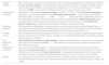
Classification and characteristics of congenital pulmonary airway malformations (CPAMs) [13].
| Stocker classification | Proportion | Origin | Embriologic origin | Cysts size | Genetic associations | Characteristics |
|---|---|---|---|---|---|---|
| Type 0(acinar dysplasia) | 1–3% | Tracheal or bronchial | Pseudoglandular period or earlier | <0.5cm | TBX4-FGF10Pathway (germline) | Diffuse malformation affecting entire both lungs; lungs do not aerate after birth; ciliated pseudostratified epithelium; lethal |
| Type 1 | 60–70% | Distal bronchial or proximal bronchioles | Pseudoglandular period | 2–10cm | KRAS(somatic) | Single or multilocular thin-walled cyst; ciliated pseudostratified columnar epithelium; one-third contains mucus-producing cells; in 95% of the cases a single lobe is involved; rarely bilateral; course depends on cyst size; mediastinal shift and respiratory distress at birth can occur; modest risk of malignancy – bronchioloalveolar carcinoma; can be difficult to distinguish from type 4; good prognosis overall |
| Type 2(bronchial atresia related cystic lung disease) | 10–15% | Terminal bronchioles | Pseudoglandular period | 0.5–2cm small mixed solid-cystic lesions | – | 80–90% have associated bronchial atresia; ciliated columnar or cuboidal epithelium; may have associated intra and extralobar sequestration; 60% associated with other congenital anomaliesa; no risk of malignancy; variable prognosis |
| Type 3 | 5–10% | Alveolar duct | Canalicular period | <0.5cm and solid appearing areas or completely solid appearance | KRAS or BRAF(somatic) | Most severe form; usually big masses; entire lobe or several lobes affected; non-ciliated cuboidal epithelium; fetal hydrops with lung hypoplasia may develop; severe respiratory distress at birth; no risk of malignancy; male predominance; poor prognosis |
| Type 4(pleuropulmonary blastoma tipe I/Ir) | 10–15% | Distal acinar | Saccular period | <7cm | DICER1 (germline) | Large thin-walled cysts; flattened alveolar epithelium overlying loose mesenchymal tissue; may be bilateral and multifocal or single large air-filled cyst; can present as spontaneous pneumothorax; can be radiographically indistinguishable from type 1; strong malignant potential, considered a form of pleuropulmonary blastoma; may have family history of DICER1 predisposition syndrome; good prognosis with timely surgery |
BPS, is a non-functioning lung tissue that lacks communication with the normal tracheobronchial tree and receives its blood supply from one or more aberrant systemic arteries, typically arising from the descending aorta, although other systemic arteries may also be involved [1]. BPS are the second commonest CLM [16]. It is classified into ILS and ELS forms, distinguished by their pleural coverage and the timing of their embryological separation from the developing lung bud. ILS shares the visceral pleura with the surrounding lung, whereas ELS is enclosed within its own separate pleural covering. A key difference is their venous drainage: ILS drains into the pulmonary veins, whereas ILS typically drains into systemic veins such as the azygos, hemiazygos, or portal veins [1,17]. There is broad consensus that ILS can be either congenital or acquired, is always located within the lung parenchyma, and typically presents as an isolated malformation. In contrast, ELS is usually congenital, may have ectopic extrapulmonary locations, such as the neck, mediastinum, retroperitoneal, intra-abdominal cavity, intra-diaphragmatic or pericardium, and is associated with other congenital anomalies in 50% of the cases, including diaphragmatic hernia, pulmonary hypoplasia, scimitar syndrome, CPAM (particularly Type 2), pulmonary artery sling, and renal abnormalities [2,4,18,19].
CHPL refer to malformations that combine morphological and/or vascular features of different entities, most notably BPS and CPAM [20]. These lesions often exhibit systemic arterial supply typical of sequestration, alongside cystic areas characteristic of CPAM, suggesting a possible overlap within the spectrum of pulmonary developmental anomalies [20]. Accurate identification of these lesions is essential for therapeutic planning, which is generally surgical [20,21].
CLO primarily results from focal airway obstruction due to intrinsic factors, such as bronchial cartilage deficiency, but can also be caused by extrinsic factors like vascular compression [1]. This obstruction leads to air trapping and overinflation of a pulmonary lobe. The condition most commonly affects the left upper lobe, followed by the right upper lobe, lower lobes, and less frequently the middle lobe. Although typically involving a single lobe, bilobar or even multilobar cases have been reported and are associated with a poorer prognosis [22–24]. It may causes compression of adjacent lung parenchyma and mediastinal shift [1,22–24].
BC results from abnormal budding of the ventral foregut during the embryonic stage [1]. These lesions are typically lined with ciliated epithelium and may contain cartilage and smooth muscle [1]. Bronchogenic cysts are most commonly located in the mediastinum or within the lung parenchyma and can cause symptoms either from compression of adjacent structures or secondary infection, which occurs in 25–35% of cases; malignant transformation is rare, occurring in less than 1% [1,2,25].
BA is a rare anomaly characterized by interruption of a segmental or subsegmental bronchus, typically occurring during the pseudoglandular stage of lung development [2]. This interruption leads to mucus accumulation (mucocele) distal to the atretic segment and hyperinflation of the affected lung segment due to collateral ventilation. BA is often asymptomatic and discovered incidentally, although it may be associated with other CLMs [2,26,27].
A thorough understanding of the CLMs is essential for accurate diagnosis, informed clinical decision-making, and the development of tailored management strategies throughout the perinatal period and beyond.
Prenatal diagnosis and managementThe widespread use of prenatal ultrasound has significantly improved the detection rate of CLMs [28]. Most CLMs are identified as space-occupying echogenic lesions within the fetal thorax, typically detected between 18 and 24 weeks of gestation [1,2]. Although ultrasound remains the primary imaging modality for prenatal screening, fetal magnetic resonance imaging (MRI) is increasingly employed as a complementary tool, particularly in complex or equivocal cases. MRI offers high sensitivity and specificity, reaching at least 95% for pulmonary lesions, and provides detailed anatomical information without exposing the fetus to ionizing radiation, Table 2[29]. However, its diagnostic and prognostic advantages over ultrasound remain limited, as both modalities demonstrate comparable accuracy in the identification of isolated malformations [30].
Prenatal imaging differential diagnosis of congenital pulmonary malformations.
| Malformation | Fetal ultrasound findings | Fetal MRI findings | Comments |
|---|---|---|---|
| Congenital pulmonary airway malformations (CPAM) | Cystic or mixed mass | Lesion with liquid or solid content | May regress spontaneously; it can be mistaken for congenital diaphragmatic hernia |
| Bronchopulmonary sequestration | Homogeneous mass with systemic arterial flow | Extrapulmonary vascular enhancement | Confirmed with Doppler; infradiaphragmatic masses, which may be confused with neuroblastomas of the suprarenal region, can be diagnosed as sequestration if a systemic vessel characteristic and pathognomonic is identified |
| Congenital lobar overinflation | Lobar overinflation | Hyperlucent lobe, mediastinal shift | Difficult to differentiate without postnatal CT |
| Bronchogenic cyst | Single cystic mass | Well-defined lesion | Can be confused with CPAM type 1, as well as with foregut duplications |
| Bronchial atresia | Rare prenatal diagnosis | Segmental hyperinflation | Usually diagnosed postnatally |
CT, computed tomography; MRI, magnetic resonance image.
CPAM lesions are typically unilateral and appear more echogenic than the adjacent normal lung parenchyma on prenatal ultrasound imaging [1]. They are classified into macrocystic (cysts>5mm) and microcystic (solid-appearing or containing small cysts) subtypes [31]. A critical prognostic indicator is the CPAM volume ratio (CVR), calculated by dividing the lesion volume (length×width×height×0.52) by the head circumference. A CVR greater than 1.6 is strongly associated with the development of fetal hydrops and poor clinical outcomes [1,32]. In high-risk cases, prenatal interventions such as maternal corticosteroid therapy or thoracoamniotic shunting may be considered [33]. Corticosteroids are particularly effective in reducing the size of microcystic lesions, whereas macrocystic CPAMs may benefit from cyst decompression procedures, Table 3[33].
Congenital pulmonary airway malformations (CPAMs): Prenatal diagnosis and management.
| Imaging modality | US – is the primary modality; can be limited due to maternal obesity, oligohydramnios, overlying ribs, and fetal lieColor and power Doppler – to assess arterial supply and venous drainage; if a systemic feeding vessel is note, the lesion is considered a bronchopulmonary sequestration if it is solid, or an hybrid lesion if it is both cystic and solidMRI – used to better characterize the mass and to assess residual lung volume |
| Postdiagnostic evaluation | Assessment for associated congenital anomalies – present in 10–20% of CPAMsFetal echocardiography – evaluation of cardiac anomalies and functionQuantitative measurements – CPAM volume ratio (CVR) by dividing the lesion volume (length×width×height×0.52) by the head circumference; CVR ≥1.6 before 26 weeks of gestation is associated with a high risk of developing hydropsMicroarray genetic test – should be offered in cases of associated congenital anomalies and/or nonimmune hydropsExome and genome sequencing – second-tier tests for patients with other congenital anomalies |
| Diferential diagnosis | Bronchopulmonary sequestration, congenital lobar overinflation, bronchogenic cyst, diaphragmatic hernia, congenital high airway obstruction, neurenteric/enteric cyst, mediastinal cystic teratoma, rare fetal pulmonary neoplasms (congenital peribronchial myofibroblastic tumor, fetal lung interstitial tumor-FLIT) |
| Prenatal course | Most CPAMs demonstrate rapid progressive growth from 20 to 26 weeks, peaking at approximately 25 weeks and then growth plateaus and often regress. If hydrops has not developed by 28 weeks, it is unlikely to develop and the prognosis is excellent |
| Poor prognostic factors | Hydrops with abnormal fetal echocardiography – hydrops associated with poor heart function is associated with a high risk for perinatal deathCVR>1.6 is predictive of risk for hydrops, respiratory distress at birth and need for early surgery |
| Management of hydrops | Fetus over 32–34 weeks of gestation – delivery with immediate postnatal resectionFetus 20–32 weeks of gestation with microcystic lesions – maternal betamethasone (2–3 courses may be needed, with one week interval)Fetus 20–32 weeks with macrocystic lesions – cyst drainage and placement of a thoracoamniotic shuntOpen resection in selected cases is the most invasive option, with risks for both, mother and fetus |
| Delivery | Small masses are not an indication for early delivery or C-sectionFor fetuses with large masses that cause mediastinal shift and/or hydrops, delivery should be planned for a tertiary care center with pediatric surgery and ECMO |
MRI, magnetic resonance image; US, ultrasound.
BPS typically appears as a homogeneous, echogenic mass, most often located in the lower lobes, especially the left lower lobe, and is best identified by the presence of a systemic feeding artery on color Doppler ultrasound [34,35]. Although many BPS lesions regress spontaneously, larger lesions associated with hydrops may require intervention [34,35]. Thoracoamniotic shunting, or in selected cases, laser ablation of the feeding vessel, has been used to decompress the thoracic cavity and prevent fetal compromise [35,36].
CLO is a rare diagnosis that is often made postnatally due to the absence of distinct ultrasound features during gestation [37]. When identified prenatally, it typically appears as a homogeneously echogenic and enlarged lobe, lacking systemic blood supply or obvious cystic changes [37,38]. In this context, it is not uncommon for CLO to be misdiagnosed as BA or CPAM on prenatal ultrasound. MRI can aid in differentiating CLH from CPAM or BPS when ultrasound findings are inconclusive [37].
BC is typically identified prenatally as a well-circumscribed, unilocular, fluid-filled mass. BC is commonly located in the mediastinum and can exert a mass effect, potentially leading to esophageal or tracheobronchial compression, polyhydramnios, or mediastinal shift [39]. In rare cases, fetal aspiration or thoracoamniotic shunting has been employed to relieve compression and prevent the development of hydrops [40].
BA is infrequently diagnosed in utero [41]. When visualized, it typically appears as a hyperinflated lung segment associated with an adjacent fluid-filled bronchocele [42]. Prenatally, BA is often misdiagnosed as CPAM, and only an elective postnatal CT scan allows for a definitive differentiation between the two entities. Although often asymptomatic, larger lesions causing significant mass effect may occasionally require fetal intervention [41,42].
A multidisciplinary evaluation at specialized fetal centers is recommended for fetuses presenting with large or symptomatic lung lesions. Management strategies should be individualized based on lesion size, growth dynamics, the presence of hydrops, and gestational age, with the goal of optimizing perinatal outcomes while minimizing intervention-related risks [1].
Postnatal presentation and diagnosisThe postnatal presentation of CLMs is highly variable, ranging from incidental findings in asymptomatic infants to severe respiratory distress at birth, Table 4[1]. The clinical course is influenced by multiple factors, including lesion size, location, and the extent of mass effect on adjacent structures [1,2,40]. While many lesions identified prenatally remain asymptomatic after birth, approximately one-third of patients may develop symptoms within the first months or years of life [3].
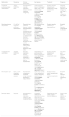
Congenital pulmonary malformations – comparative table.
| Malformation | Prevalence | Clinical presentation | Key features | Treatment | Prognosis |
|---|---|---|---|---|---|
| Congenital pulmonary airway malformations | 1:8300–35,000 live births | Asymptomatic or neonatal respiratory distress; recurrent infections; malignancy | Cystic/multicystic lesion; 5 histological types (Type 1 most common, best prognosis)Airway connection-yesBlood supply-pulmonary arteryTypical location-any lobeMalignancy risk-yesCystic appearance-yes | Surgical resection (lobectomy) for symptomatic or large asymptomatic lesions | Generally good post-resection; low malignancy risk (Type 1), high malignancy risk (Type 4) |
| Bronchopulmonary sequestration | 0.1–6.4% of congenital pulmonary malformations | Recurrent lung infections; dyspnea; cardiac decompensation; can be asymptomatic.ILS typically present later in childhood or adulthood with recurrent pulmonary infections, whereas ELS are usually diagnosed in infancy due to respiratory distress or mass effect | Non-functional lung tissueAirway connection-noBlood supply-systemic artery (Aorta)Typical location-posterior/inferior lobesMalignancy risk-very lowCystic appearance-may be present (hybrid lesions) | Surgical resection (lobectomy or segmentectomy) | Excellent post-resection; complications if untreated |
| Congenital lobar overinflation | 1:20,000–30,000 live births | Neonatal respiratory distress; respiratory symptoms in early infancy. May be asymptomatic | Hyperinflation of a pulmonary lobe; partial unidirectional valve effectAirway connection-yesBlood supply-pulmonary arteryTypical location-upper lobes (L>R; commonly left upper)Malignancy risk-noCystic appearance-absent | Surgical resection (lobectomy) in significant symptomatic cases | Very good post-treatment; mild to moderate symptomatic cases may be observed |
| Bronchogenic cyst | Rare (∼1–2% of pediatric mediastinal masses) | Asymptomatic; compressive symptoms (cough, dyspnea, dysphagia) if large; infection | Cystic lesion usually in the mediastinum; lined by respiratory epithelium with cartilageAirway connection-noBlood supply-pulmonary arteryTypical location-mediastinal or centralMalignancy risk-yesCystic appearance-unilocular cyst | Surgical excision (even if asymptomatic due to infection/complication risk) | Excellent post-resection; very low malignancy risk |
| Bronchial atresia | Very rare | Asymptomatic or recurrent respiratory infections; often incidental finding | Congenital obstruction of segmental bronchus; distal lobe may show mucocele and hyperinflationAirway connection-noBlood supply-pulmonary arteryTypical location-segmental bronchiMalignancy risk-noneCystic appearance-mucocele with hyperinflation | Observation for mild cases; surgery if recurrent symptoms | Good in most cases; depends on extent of obstruction |
CPAMs can present with respiratory distress or pneumothorax in the neonatal period, particularly when the lesion causes significant mass effect or air trapping [1,2,8]. However, many cases remain asymptomatic and are discovered incidentally on chest imaging [1]. CPAMs can lead to recurrent localized pulmonary infections, and certain types carry a risk of malignant transformation [1,2,8]. Postnatal radiographs may appear normal or may reveal findings such as hyperlucency, mediastinal shift, or air-filled cysts. Definitive diagnosis relies on chest computed tomography (CT), which provides detailed characterization of the cystic architecture and enables differentiation from other thoracic anomalies. In some cases, monitoring by ultrasound is possible [1,2,8].
CHPL share the clinical features of CPAMs but are characterized by the presence of a systemic arterial supply [1]. This entity underlines the importance of performing contrast-enhanced CT angiography in all suspected cases of CPAM and BPS, and more broadly in the evaluation of congenital pulmonary malformations, in order to accurately delineate the vascular supply.
BPS often presents similarly to CPAM but is distinguished by its systemic arterial blood supply [1]. The clinical presentation is highly variable and often asymptomatic or nonspecific, ranging from recurrent pneumonia, chronic cough, fever, sputum production, and hemoptysis, to incidental findings on imaging or prenatal diagnosis in cases of ELS [1–4]. Contrast-enhanced CT angiography is essential to identify the aberrant feeding arteries, which typically originates from the thoracic or abdominal aorta. Additionally, Doppler ultrasonography may aid in vascular identification when CT is not immediately available. BPS can grow rapidly due to a shunt effect, causing a symptomatic mass [1,4].
CLO is a condition that typically presents in the postnatal period, with symptoms of progressive respiratory distress caused by air trapping and lobar overinflation [43]. Approximately 50–75% of CLO cases become symptomatic by the end of the first month of life, and up to 85–90% show symptoms by six months of age [43]. Physical examination may reveal diminished breath sounds and overexpansion of the affected hemithorax. A chest X-ray typically shows hyperinflation of a single lobe with compression of adjacent lung tissue. High-resolution CT imaging is useful for confirming the diagnosis and excluding extrinsic causes of bronchial obstruction, while bronchoscopy plays a key role in evaluating bronchial anatomy and providing diagnostic confirmation [43].
BC may remain asymptomatic or present later in life with symptoms related to mass effect, such as wheezing, dysphagia, or recurrent respiratory infections [2]. On imaging, these lesions typically appear as well-defined, fluid-filled cystic structures, most commonly located in the mediastinum [44]. Both CT and MRI are valuable imaging modalities for evaluating lesion size, internal composition, and anatomical relationships with adjacent structures [45]. A BC primarily contains mucoid or serous fluid, giving it a smooth appearance. However, the cyst's contents can vary; in some cases, they may contain air or an air-fluid level, especially if a connection forms with the bronchial tree due to infection or rupture. Some cysts also contain proteinaceous material or blood, which can appear as solid material on imaging.
CBA is often an incidental finding; however, in some cases, it may present with localized wheezing or segmental hyperinflation [46]. Radiographic features typically include a mucocele (bronchocele) at the site of the atretic bronchus and hyperlucency of the corresponding lung segment due to collateral ventilation [46]. CT imaging plays a crucial role in confirming the diagnosis and differentiating bronchial atresia from other cystic lung lesions [2,47]. Bronchoscopy and bronchography can also aid in confirming the diagnosis by demonstrating agenesis or atresia of a lobar, segmental, or subsegmental bronchus [48].
Postnatal imaging plays a central role in characterizing CLMs [49]. While chest radiographs are useful for initial evaluation, high-resolution CT, preferably with angiography, is considered the gold standard for accurate delineation of lesion morphology and vascular supply. In asymptomatic neonates with a prenatal diagnosis, the need for an immediate chest X-ray after delivery remains debated, with some centers deferring imaging until further follow-up to avoid unnecessary radiation exposure.
In certain cases, MRI may be considered a radiation-free alternative; however, its availability and use in neonates remain limited [49].
Postnatal managementThe management of CLMs after birth depends on clinical presentation, lesion type and size, risk of complications, and institutional experience [1]. Symptomatic patients, particularly those presenting with respiratory distress, recurrent infections, or complications such as pneumothorax, are typically offered surgical resection, which is generally curative [50].
CPAMs are among the most controversial and debated conditions regarding management strategies [3]. While surgical resection is clearly indicated in symptomatic cases, the optimal approach for asymptomatic patients remains controversial. Arguments in favor of early elective resection include prevention of infection, reduction of long-term risk of malignant transformation, improved compensatory lung growth when surgery is performed during infancy, and absence of radiation accumulation over the years [33,50–52]. Arguments in favor of conservative management with surveillance include the fact that early surgery for asymptomatic CPAM may not be necessary, as many lesions decrease in size spontaneously, the risk of malignancy is low, and careful monitoring enables timely intervention if complications develop [33,50–52]. Although lobectomy remains the preferred surgical approach for CPAMs, segmentectomies and other lung-preserving procedures have been performed in some cases; however, no evidence has demonstrated their superiority over lobectomy [33,53]. For asymptomatic infants, lobectomy is generally recommended in high-risk cases (e.g., large lesions, bilateral cysts, or DICER1 mutations), as it may support lung development, although supporting evidence remains limited [26,32]. For low-risk patients, both surgical resection (typically performed between 6 and 12 months of age) and observation are considered reasonable options, depending on family preferences. Observation requires close clinical and radiologic follow-up, although spontaneous regression is rare and not well documented. There is currently no consensus regarding the optimal timing of intervention or standardized imaging protocols [26,32,35,54].
BPS is typically managed through surgical intervention, especially in symptomatic cases or when large systemic feeding arteries are present, as these may lead to cardiac overload [1,55]. Even in asymptomatic cases, surgical resection is often recommended to prevent infections and hemorrhagic complications [1,55,56]. Thoracoscopic resection represents a viable and increasingly favored approach, particularly in high-volume or experienced centers [57]. In selected cases, embolization of the feeding vessel may be considered as an alternative or adjunctive therapy, particularly in the setting of significant left-to-left shunting or cardiac failure [58]. Conservative observation may be appropriate for asymptomatic extralobar sequestrations without significant shunting, as these lesions have the potential to regress or even resolve spontaneously over time [59].
CLO often necessitates surgical intervention in neonates presenting with significant respiratory compromise due to air trapping [1]. Lobectomy remains the standard treatment approach, although conservative management may be appropriate in asymptomatic patients or those with only mild symptoms [1,60,61]. Some authors recommend a preoperative echocardiogram due to the known association with cardiac anomalies [1]. Serial imaging with x-ray and/or CT is essential for monitoring lesion behavior over time [1].
BC, although often asymptomatic, carry a significant risk of infection, compression of adjacent structures, and, in rare cases, malignant transformation [39,53]. Surgical excision, generally via a thoracoscopic approach, is recommended even in asymptomatic patients, particularly when the cyst is located in the mediastinum or parahilar region [39].
CBA is typically diagnosed incidentally and is usually managed conservatively unless recurrent infections or other complications arise [62]. Surgical resection may be considered in cases of persistent mucocele or documented infection within the affected segment [63].
While surgical resection remains the prevailing standard of care for symptomatic CLMs, there is ongoing debate regarding the optimal management of asymptomatic patients [3]. The decision must carefully weigh the potential benefits of early resection, such as avoiding future complications and limiting radiation exposure from serial imaging, against the inherent risks of surgery. A multidisciplinary approach involving pediatric surgeons, neonatologists, pulmonologists, radiologists, and the patient's family is essential to ensure individualized and balanced care [3].
Long-term outcomesThe long-term outcomes of CLMs are influenced by several factors, including the lesion type, the timing and extent of surgical intervention, and the presence or absence of symptoms. While short-term surgical outcomes are well documented, data regarding long-term respiratory function, growth, and malignancy risks remain limited and are occasionally controversial [1,50,64,65].
Pulmonary function and compensatory growthFollowing surgical resection, particularly lobectomy, total lung capacity (TLC) is often preserved, indicating the potential for compensatory lung growth [50]. Studies have demonstrated that alveolar multiplication continues into early childhood, supporting the theory that early resection, particularly within the first two years of life, optimizes compensatory mechanisms [50,66]. However, several studies also report that, although TLC is maintained, subtle impairments in forced expiratory volume in one second (FEV1) and ventilation distribution may persist [50]. Residual lung hyperinflation may obscure underlying ventilation inefficiency, suggesting that anatomical growth does not necessarily result in fully functional tissue [50].
In children who undergo early resections, the maintenance of adequate exercise tolerance and a satisfactory quality of life is often observed, despite minor declines in pulmonary function, as indicated by formal testing [50].
Age at surgery and extent of resectionThe question of whether earlier surgery leads to superior lung function remains a subject of debate [67]. Early elective resection of CLM prior to four months of age is feasible and not associated with increased operative risk [67]. Some authors advocate for resection before the age of one year in order to maximize compensatory growth [50,51]. However, other studies suggest that lung function outcomes are similar, regardless of whether surgery is performed before or after the age of two [50]. Furthermore, parenchyma-sparing techniques, such as segmentectomy or wedge resection, are increasingly being considered to preserve lung tissue, although concerns about incomplete resection and recurrence continue to persist [53].
In rare cases where pneumonectomy is required, patients face an increased risk of musculoskeletal complications, such as scoliosis and post-pneumonectomy syndrome [50].
Conservative management and spontaneous resolutionA subset of CLMs, particularly small, asymptomatic lesions, may regress spontaneously. Serial imaging studies have documented cases of partial or complete resolution [1,68]. However, truly complete regression, without any residual abnormal lung tissue, is rare. This raises concerns about the long-term safety of observation, particularly with respect to the potential risks of infection, malignancy, and exposure to ionizing radiation from repeated imaging procedures [68].
Risk of developing symptoms in asymptomatic lesionsAmong asymptomatic prenatally diagnosed CLMs, studies have reported that 3–13% of patients eventually develop symptoms, such as infection, respiratory distress, or pneumothorax, typically within the first two years of life [50]. Later symptoms development, although less common, has also been reported in adolescents and adults [50].
Patients who initially undergo conservative management but later develop symptoms have a higher rate of postoperative complications compared to those who undergo elective early surgery [69].
Many authors recommend elective resection of all CPAMs, BCs, and BPS due to the potential risk of complications, including infection, hemorrhage, pneumothorax, sudden respiratory compromise, and malignant transformation [69]. Elective lobectomy is generally well tolerated and remains the most commonly performed surgical procedure. Thoracoscopic surgery has been shown to be both safe and effective. However, the optimal timing for surgery in asymptomatic patients remains unclear, with recommendations varying from one month to two years of age. Some authors suggest surgery between six and 12 months, as the risks associated with anesthesia and surgery are lower in the first few months of life [69].
Malignancy riskConcerns regarding the potential for malignancy remain a central argument in favor of elective resection for CLMs, even in asymptomatic patients [1–3,70]. CPAM Type 1 lesions have been linked to bronchioalveolar carcinoma, while Type 4 lesions raise concerns about pleuropulmonary blastoma (PPB), a rare but aggressive pediatric neoplasm [70]. Notably, a study by Feinberg et al. proposed a clinical algorithm for surgical decision-making based on features that predict malignancy [71]. The study identified specific characteristics linked to PPB, such as pneumothorax at presentation, bilateral or multifocal cysts, and the presence of DICER1 gene mutations. These factors were used to identify high-risk patients who may benefit from early resection [70]. Conversely, for infants with asymptomatic CPAM who lack high-risk features, a conservative approach with close clinical and radiological follow-up is considered feasible, with surgery reserved for cases of lesion growth or symptom development [1].
Although rare, malignant transformation has been documented in BCs, including cases of rhabdomyosarcoma, pulmonary blastoma, and other malignant mesenchymal tumors, as identified in histopathological analysis of resected specimens. This further supports the recommendation for surgical excision of asymptomatic BCs, due to diagnostic uncertainty and the potential for late complications [1].
Quality of lifeIn general, children who undergo surgical resection, whether for symptomatic or asymptomatic lesions, experience a good quality of life and are able to fully participate in physical activities [50]. Even among those with mild pulmonary function deficits, clinical repercussions are uncommon [1,50].
Nevertheless, some studies suggest that these patients may present a higher risk of developing certain respiratory complications, such as recurrent wheezing or reduced pulmonary reserve, particularly in long-term follow-up. These findings underline the importance of ongoing surveillance and counseling for families, even in cases with initially favorable postoperative outcomes [64].
Conclusions and future directionsCLMs encompass a range of developmental anomalies. Advances in prenatal imaging and perinatal care have significantly improved early detection and outcomes, transforming conditions once frequently fatal into manageable clinical entities. Surgical resection remains the cornerstone of management for symptomatic patients, supported by strong evidence of excellent postoperative recovery and preserved long-term lung function. However, the optimal approach for asymptomatic patients remains a subject of debate.
Although early elective surgery is justified by the risks of infection, pneumothorax, and malignant transformation, particularly in CPAMs and BCs, a conservative approach may be reasonable in selected cases, provided there is rigorous monitoring and well-defined criteria for intervention.
Long-term follow-up studies suggest that most children, whether treated surgically or conservatively, exhibit normal respiratory function and quality of life. However, subtle impairments in pulmonary mechanics and rare instances of malignancy require careful, individualized decision-making. Future research should focus on standardizing risk stratification tools, such as integrating clinical, radiological, and genetic markers (e.g., DICER1 mutations), to more accurately identify patients who would benefit most from early resection versus observation.
There is an urgent need for large prospective cohort studies with long-term follow-up into adulthood. Such studies are essential for clarifying the natural history of unresected lesions and validating management algorithms. Emerging techniques, including minimally invasive and lung-sparing surgeries, continue to improve the balance between therapeutic efficacy and preservation of pulmonary function.
Ultimately, a multidisciplinary, individualized approach remains paramount, ensuring that each patient's management plan is tailored not only to the anatomical characteristics of the malformation but also to their clinical course, parental preferences, and emerging evidence.
Contribution of each authorJoão Faria Dias [1] conducted the literature review, selected the most relevant scientific articles cited in the bibliography, and drafted the manuscript. Gustavo Rocha [1], a neonatologist with expertise in pulmonology, and Mariana Borges Dias [3], a pediatric surgeon, performed a comprehensive revision of the manuscript. All authors reviewed and approved the final version.
Artificial intelligence involvementNo part of the manuscript was produced with the help of artificial intelligence software or tools.
Funding of the researchThis research did not receive any specific grant from funding agencies in the public, commercial, or not-for-profit sectors.
Conflicts of interestThe authors declare not to have any conflicts of interest that may be considered to influence directly or indirectly the content of the manuscript.


