


Pleomorphic carcinoma of the lung is a subtype of sarcomatoid carcinoma of the lung. These are rare neoplasms, accounting for less than 1% of lung tumors. Pleomorphic carcinomas have no characteristic radiological features, although they present more frequently as solid peripheral masses predominantly in the upper lobes with invasion of the chest wall that may include a necrotic component.1
We report 2 cases of male patients aged 64 and 57 years, who consulted due to hemoptysis. Computed tomography showed pulmonary masses with similar radiological characteristics in the form of subsolid nodules with a central solid cavitary component and an extensive area of surrounding ground glass opacity (Fig. 1A and B). In the PET study, the solid component had more intense uptake (A: 8 SUV, B: 6.2 SUV) and the ground glass component had lower uptake (A: 2 SUV).
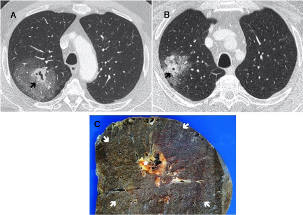
(A and B) Chest CT with IV contrast: lung window. Mass with central solid cavitary component (arrow) and extensive surrounding ground glass opacities. (C) Macroscopic specimen of lobectomy from case A, showing the central cavitary nodular lesion (star) surrounded by extensive perilesional hemorrhage (arrows) corresponding to the ground glass area on CT.
Transthoracic fine needle aspiration and biopsy (FNAB) was performed, accessing the peripheral part. The result showed hemorrhage and the pathology diagnosis of the surgical specimen was pleomorphic carcinoma of the lung with extensive peritumoral hemorrhage (Fig. 1C).
Cases of pleomorphic carcinoma with surrounding ground glass opacities have been reported in the literature, but these were related to the component associated with lung adenocarcinomas or infiltration of inflammatory cells.2
In the cases described, material obtained in the transthoracic FNAB was from the ground glass region, which determined the false negative result on cytology, as it corresponded to bleeding. For this reason, in a patient with hemoptysis, bleeding should always be considered as a cause of the ground glass component of a subsolid nodule.
Please cite this article as: Gómez Herrero H, Amat Villegas I. Carcinoma pleomorfo pulmonar con presentación como masa subsólida por extensa hemorragia peritumoral: a propósito de 2 casos. Arch Bronconeumol. 2020;56:458.


