
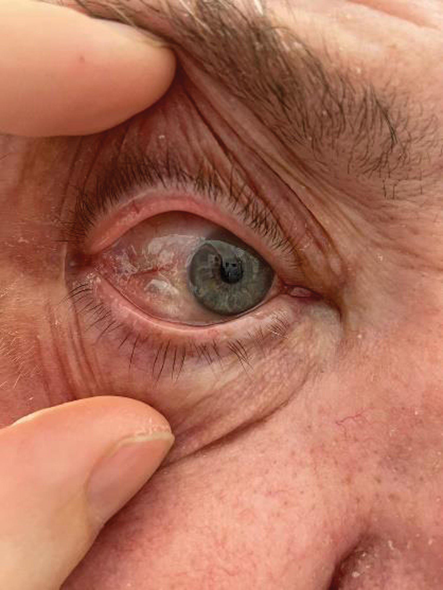
We report the case of a 70 year-old male patient, former smoker with a history of hypertension, chronic obstructive pulmonary disease and remarkable stenosis of the right carotid artery. Throughout a syncope study, a 2.9cm pulmonary nodule on the left upper lobe was spotted, suggesting malignancy. Pulmonary function test revealed a forced expiratory volume in the first second of 54.2% and diffusing capacity for carbon monoxide of 63.2%. Given the comorbidity and the impaired pulmonary function, a video-thoracoscopic lingular segmentectomy was performed. On the second postoperative day, the patient developed subcutaneous emphysema on his face, neck and thorax secondary to air leak. Bilateral subconjunctival emphysema was also noted (Fig. 1). No vision complications were accounted for, and eye motility remained unaffected. Aspiration was applied to the chest drain and skin incisions were performed to decompress the air trapped in subcutaneous space and improve patient comfort. Air leak was solved on tenth postoperative day after autologous blood patch pleurodesis while subconjunctival emphysema resolved with no additional intervention.

Subconjunctival emphysema is a rare condition commonly related to ophthalmological surgery and blunt traumas.1 Nonetheless, few cases following thoracic surgery have been documented.2 Notably, all reported cases were successfully managed through conservative measures.
FundingNone.
Conflict of interestsThe authors declare that they have no conflict of interest related directly or indirectly to the contents of the manuscript.


