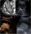
We report the case of a 61-year-old man diagnosed with right basal pneumonia and parapneumonic effusion requiring pleural drainage. Due to his poor clinical course and the low drainage yield, a chest CT was performed, which showed significant pleural effusion with regular parietal pleural enhancement suggesting exudate (Fig. 1A). A chest ultrasound with 2.4 ml contrast agent was performed to complete the study (Sonovue, Rovi, Spain). Ultrasound showed a pleural effusion with countless echoes and internal septa, but no separate loculation (Fig. 1B and C). Following administration of the ultrasound contrast agent, a pattern of enhancement consistent with the known pneumonia and hyperenhancing thickening of the pleural layers were observed, which, together with the ultrasound appearance of the fluid, suggested empyema.
(A–C) Bacterial pleural empyema. (A) CT axial image showing pleural effusion with thickening and hyperenhancement of pleural layers (white arrows) consistent with the split pleura sign. (B) Chest ultrasound showing multiple echoes and septa in pleural fluid, with no obvious loculations (asterisk), findings not visible on previous CT. (C) Chest contrast-enhanced ultrasound. The image on the left shows the contrast-enhanced ultrasound scan, 70 s after administration, and the image on the right shows the standard procedure. The contrast-enhanced image shows thickening and hyperenhancement of the parietal and visceral pleural layers (white arrows), that can be superimposed on those seen in Figure A.
Contrast-enhanced ultrasound has proven to be useful in multiple diseases.1 The split pleura sign consists of hyperenhancing thickening of the visceral and parietal layers of the pleura, separated by a collection of fluid, visualized on intravenous contrast-enhanced CT.2 In our case, contrast-enhanced ultrasound was able to detect this typical CT sign and, together with other findings, led to the diagnosis of suspected empyema.
Please cite this article as: Jiménez Serrano S, Radalov I, Vollmer I. Diagnóstico del signo de la pleura hendida mediante ecografía con contraste. Arch Bronconeumol. 2021;57:431.


