

A young woman with no history of interest attended the emergency department complaining of a 5-day history of dyspnea, dry cough, and pleuritic pain in the left costal region. She also reported a 1-month history of right shoulder pain and slight weight loss.
Physical examination revealed tachypnea, chest indrawing, and absent breath sounds in the left hemitorax. Lactate dehydrogenase (LDH) and alkaline phosphatase (AP) were elevated.
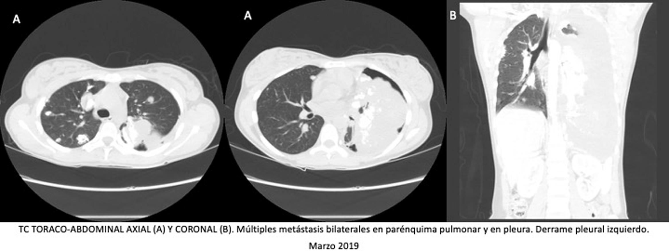
Chest X-ray and computed tomography showed multiple bilateral lung and pleural masses containing calcium, left pleural effusion, and a blastic lesion on the right humeral head (Fig. 1). Pathology study of a lung mass biopsy confirmed the diagnosis of osteosarcoma metastases.
 and chest-abdominal CT coronal slice (B) at diagnosis, showing massive left pleural effusion, right humeral head blastic lesion, and multiple calcium density images in bilateral pulmonary parenchyma and pleura. Chest CT axial slice (C1 and C2) after drainage of pleural effusion, significant for multiple bilateral metastases in lung parenchyma and pleura, and left pneumothorax. Plain PA and lateral chest X-rays (D.1 and D.2) at 10 months after diagnosis, showing an increase in the number and size of bilateral pulmonary and pleural nodules and masses, left pleural effusion, and an increase in the size of the primary lesion in the right humeral head.")
Plain PA chest X-ray (A) and chest-abdominal CT coronal slice (B) at diagnosis, showing massive left pleural effusion, right humeral head blastic lesion, and multiple calcium density images in bilateral pulmonary parenchyma and pleura. Chest CT axial slice (C1 and C2) after drainage of pleural effusion, significant for multiple bilateral metastases in lung parenchyma and pleura, and left pneumothorax. Plain PA and lateral chest X-rays (D.1 and D.2) at 10 months after diagnosis, showing an increase in the number and size of bilateral pulmonary and pleural nodules and masses, left pleural effusion, and an increase in the size of the primary lesion in the right humeral head.
She received doxorubicin and cisplatin and achieved a partial response. Thirteen months later, progression was observed and the patient presented with lower respiratory tract infection and partial respiratory failure, causing her death.
Primary bone tumors account for less than 0.2% of malignant neoplasms, most frequently osteosarcoma1. Lung metastases are the most common and, along with elevated LDH and AP, are indicators of poor prognosis. The aggressiveness of the lung involvement of this tumor is illustrated in this clinical image2.
The following is Supplementary data to this article:

Please cite this article as: Martínez Barroso K, Borregón Rivilla M, Mazariegos Rubí M, Medina Martínez J. Afectación pulmonar y pleural severa por osteosarcoma en mujer de 24 años. La manifestación visceral más agresiva de un tumor óseo. Arch Bronconeumol. 2021;57:771.


