array:22 [
  "pii" => "S0300289624003788"
  "issn" => "03002896"
  "doi" => "10.1016/j.arbres.2024.09.018"
  "estado" => "S200"
  "fechaPublicacion" => "2024-10-21"
  "aid" => "3667"
  "copyright" => "SEPAR"
  "copyrightAnyo" => "2024"
  "documento" => "article"
  "crossmark" => 0
  "subdocumento" => "sco"
  "abierto" => array:3 [
    "ES" => false
    "ES2" => false
    "LATM" => false
  ]
  "gratuito" => false
  "lecturas" => array:1 [
    "total" => 0
  ]
  "itemSiguiente" => array:17 [
    "pii" => "S0300289624003703"
    "issn" => "03002896"
    "doi" => "10.1016/j.arbres.2024.09.009"
    "estado" => "S200"
    "fechaPublicacion" => "2024-10-21"
    "aid" => "3658"
    "copyright" => "SEPAR"
    "documento" => "simple-article"
    "crossmark" => 0
    "subdocumento" => "crp"
    "abierto" => array:3 [
      "ES" => false
      "ES2" => false
      "LATM" => false
    ]
    "gratuito" => false
    "lecturas" => array:1 [
      "total" => 0
    ]
    "en" => array:8 [
      "idiomaDefecto" => true
      "cabecera" => "<span class="elsevierStyleTextfn">Scientific Letter</span>"
      "titulo" => "Individual and Structural Factors Influencing Participation to Low-Dose Computed Tomography Screening in a Chinese Centralized Lung Cancer Screening Cohort"
      "tienePdf" => "en"
      "tieneTextoCompleto" => "en"
      "contieneTextoCompleto" => array:1 [
        "en" => true
      ]
      "contienePdf" => array:1 [
        "en" => true
      ]
      "autores" => array:1 [
        0 => array:2 [
          "autoresLista" => "Shenglin Zhao, Bibo Li, Zhikai Yu, Jia Du, Hong Zhou, Mei He"
          "autores" => array:6 [
            0 => array:2 [
              "nombre" => "Shenglin"
              "apellidos" => "Zhao"
            ]
            1 => array:2 [
              "nombre" => "Bibo"
              "apellidos" => "Li"
            ]
            2 => array:2 [
              "nombre" => "Zhikai"
              "apellidos" => "Yu"
            ]
            3 => array:2 [
              "nombre" => "Jia"
              "apellidos" => "Du"
            ]
            4 => array:2 [
              "nombre" => "Hong"
              "apellidos" => "Zhou"
            ]
            5 => array:2 [
              "nombre" => "Mei"
              "apellidos" => "He"
            ]
          ]
        ]
      ]
    ]
    "idiomaDefecto" => "en"
    "EPUB" => "https://multimedia.elsevier.es/PublicationsMultimediaV1/item/epub/S0300289624003703?idApp=UINPBA00003Z"
    "url" => "/03002896/unassign/S0300289624003703/v1_202410210415/en/main.assets"
  ]
  "itemAnterior" => array:16 [
    "pii" => "S0300289612000968"
    "issn" => "03002896"
    "doi" => "10.1016/j.arbres.2012.02.017"
    "estado" => "S200"
    "fechaPublicacion" => "2013-05-08"
    "aid" => "621"
    "documento" => "simple-article"
    "crossmark" => 0
    "subdocumento" => "ret"
    "abierto" => array:3 [
      "ES" => false
      "ES2" => false
      "LATM" => false
    ]
    "gratuito" => false
    "lecturas" => array:2 [
      "total" => 348
      "formatos" => array:2 [
        "HTML" => 82
        "PDF" => 266
      ]
    ]
    "en" => array:8 [
      "idiomaDefecto" => true
      "titulo" => "WITHDRAWN&#58; Respiratory Muscle Assessment in Predicting Extubation Outcome in Patients With Stroke"
      "tienePdf" => "en"
      "tieneTextoCompleto" => 0
      "tieneResumen" => "en"
      "contieneResumen" => array:1 [
        "en" => true
      ]
      "contienePdf" => array:1 [
        "en" => true
      ]
      "autores" => array:1 [
        0 => array:2 [
          "autoresLista" => "Antonio A&#46;M&#46; Castro, Felipe Cortopassi, Russell Sabbag, Luis Torre-Bouscoulet, Claudia K&#252;mpel, Elias Ferreira Porto"
          "autores" => array:6 [
            0 => array:2 [
              "nombre" => "Antonio A&#46;M&#46;"
              "apellidos" => "Castro"
            ]
            1 => array:2 [
              "nombre" => "Felipe"
              "apellidos" => "Cortopassi"
            ]
            2 => array:2 [
              "nombre" => "Russell"
              "apellidos" => "Sabbag"
            ]
            3 => array:2 [
              "nombre" => "Luis"
              "apellidos" => "Torre-Bouscoulet"
            ]
            4 => array:2 [
              "nombre" => "Claudia"
              "apellidos" => "K&#252;mpel"
            ]
            5 => array:2 [
              "nombre" => "Elias Ferreira"
              "apellidos" => "Porto"
            ]
          ]
        ]
      ]
    ]
    "idiomaDefecto" => "en"
    "EPUB" => "https://multimedia.elsevier.es/PublicationsMultimediaV1/item/epub/S0300289612000968?idApp=UINPBA00003Z"
    "url" => "/03002896/unassign/S0300289612000968/v2_201305131254/en/main.assets"
  ]
  "en" => array:12 [
    "idiomaDefecto" => true
    "cabecera" => "<span class="elsevierStyleTextfn">Clinical Image</span>"
    "titulo" => "Pulmonary Chondroscarcoma"
    "tieneTextoCompleto" => true
    "autores" => array:1 [
      0 => array:4 [
        "autoresLista" => "Shashank Kumar, Manoj Kumar Panigrahi, Pritinanda Mishra"
        "autores" => array:3 [
          0 => array:3 [
            "nombre" => "Shashank"
            "apellidos" => "Kumar"
            "referencia" => array:1 [
              0 => array:2 [
                "etiqueta" => "<span class="elsevierStyleSup">a</span>"
                "identificador" => "aff0005"
              ]
            ]
          ]
          1 => array:4 [
            "nombre" => "Manoj Kumar"
            "apellidos" => "Panigrahi"
            "email" => array:1 [
              0 => "panigrahimanoj75@gmail.com"
            ]
            "referencia" => array:2 [
              0 => array:2 [
                "etiqueta" => "<span class="elsevierStyleSup">a</span>"
                "identificador" => "aff0005"
              ]
              1 => array:2 [
                "etiqueta" => "<span class="elsevierStyleSup">&#42;</span>"
                "identificador" => "cor0005"
              ]
            ]
          ]
          2 => array:3 [
            "nombre" => "Pritinanda"
            "apellidos" => "Mishra"
            "referencia" => array:1 [
              0 => array:2 [
                "etiqueta" => "<span class="elsevierStyleSup">b</span>"
                "identificador" => "aff0010"
              ]
            ]
          ]
        ]
        "afiliaciones" => array:2 [
          0 => array:3 [
            "entidad" => "Department of Pulmonary and Critical Care Medicine&#44; AIIMS Bhubaneswar&#44; Odisha&#44; India"
            "etiqueta" => "a"
            "identificador" => "aff0005"
          ]
          1 => array:3 [
            "entidad" => "Department of Pathology &#38; Lab Medicine&#44; AIIMS Bhubaneswar&#44; Odisha&#44; India"
            "etiqueta" => "b"
            "identificador" => "aff0010"
          ]
        ]
        "correspondencia" => array:1 [
          0 => array:3 [
            "identificador" => "cor0005"
            "etiqueta" => "&#8270;"
            "correspondencia" => "Corresponding author&#46;"
          ]
        ]
      ]
    ]
    "resumenGrafico" => array:2 [
      "original" => 0
      "multimedia" => array:7 [
        "identificador" => "fig0005"
        "etiqueta" => "Fig&#46; 1"
        "tipo" => "MULTIMEDIAFIGURA"
        "mostrarFloat" => true
        "mostrarDisplay" => false
        "figura" => array:1 [
          0 => array:4 [
            "imagen" => "gr1.jpeg"
            "Alto" => 630
            "Ancho" => 1250
            "Tamanyo" => 134094
          ]
        ]
        "descripcion" => array:1 [
          "en" => "<p id="spar0005" class="elsevierStyleSimplePara elsevierViewall">CT thorax &#40;A and B &#8211; coronal section&#41; showing a large heterogenous lung mass in the right hemithorax with focal areas of calcification &#40;red arrow&#41; and necrosis &#40;green arrow&#41;&#44; shifting mediastinum to the left&#46; USG guided biopsy of the lung mass was done and the HE-stained images showed the chondrocytes arranged in small clusters&#44; and singly in a lacuna &#40;yellow arrows&#41;&#46; There is moderate increase in cellularity with multinucleation&#44; and mild atypia &#40;C &#8211; 20&#215;&#59; D &#8211; 40&#215;&#41;&#46; Image E &#40;40&#215;&#41; shows areas of calcification within the tumour &#40;white arrow&#41;&#46;</p>"
        ]
      ]
    ]
    "textoCompleto" => "<span class="elsevierStyleSections"><p id="par0005" class="elsevierStylePara elsevierViewall">A 37-year-old man presented with dyspnea&#44; dry cough and right-sided chest pain for 3 months&#46; Physical examination revealed diminished breath sounds on the right hemithorax with mediastinal shift to left&#46; Chest radiograph showed a right middle and lower zone opacity&#44; and contrast-enhanced computed tomography showed a large heterogenous mass with multiple foci of calcification and necrosis &#40;<a class="elsevierStyleCrossRef" href="#fig0005">Fig&#46; 1A and B</a>&#41;&#46; An ultrasound-guided biopsy showed features of a cartilage-forming tumor with chondrocytes arranged in lacunae with atypia&#44; features consistent with pulmonary chondrosarcoma &#40;<a class="elsevierStyleCrossRef" href="#fig0005">Fig&#46; 1C and D</a>&#41;&#46; He progressively worsened and passed away during the hospital stay before any surgery or palliative treatment could be initiated&#46; Pulmonary chondrosarcomas are rare lung tumors that represent less than 1&#37; of primary lung tumors&#46; They present as enlarged&#44; lobulated&#44; or cystic calcified masses&#46; The average age is between 50 and 70 years&#46; It is of two types &#8211; tracheobronchial and lung&#46; Immunohistochemistry is positive for S-100&#44; particularly in the chondromatous component&#46;<a class="elsevierStyleCrossRef" href="#bib0015"><span class="elsevierStyleSup">1</span></a> Symptoms depend on the size and location of the lesion&#46; Treatment is primarily surgical&#44; with complete resection rates of over 80&#37;&#46; Adjuvant chemotherapy and radiotherapy are recommended for high-grade lesions or incomplete resection&#46; In cases of unresectable tumours&#44; surgery is the primary treatment&#46;<a class="elsevierStyleCrossRef" href="#bib0020"><span class="elsevierStyleSup">2</span></a></p><elsevierMultimedia ident="fig0005"></elsevierMultimedia><span id="sec0005" class="elsevierStyleSection elsevierViewall"><span class="elsevierStyleSectionTitle" id="sect0005">Declaration of Generative AI and AI-Assisted Technologies in the Writing Process</span><p id="par0010" class="elsevierStylePara elsevierViewall">There was no involvement of AI in preparation of this manuscript&#46;</p></span><span id="sec0010" class="elsevierStyleSection elsevierViewall"><span class="elsevierStyleSectionTitle" id="sect0010">Funding</span><p id="par0015" class="elsevierStylePara elsevierViewall">This research did not receive any specific grant from funding agencies in the public&#44; commercial&#44; or not-for-profit sectors&#46;</p></span><span id="sec0015" class="elsevierStyleSection elsevierViewall"><span class="elsevierStyleSectionTitle" id="sect0015">Conflicts of Interest</span><p id="par0020" class="elsevierStylePara elsevierViewall">The authors declare not to have any conflicts of interest that may be considered to influence directly or indirectly the content of the manuscript&#46;</p></span></span>"
    "textoCompletoSecciones" => array:1 [
      "secciones" => array:4 [
        0 => array:2 [
          "identificador" => "sec0005"
          "titulo" => "Declaration of Generative AI and AI-Assisted Technologies in the Writing Process"
        ]
        1 => array:2 [
          "identificador" => "sec0010"
          "titulo" => "Funding"
        ]
        2 => array:2 [
          "identificador" => "sec0015"
          "titulo" => "Conflicts of Interest"
        ]
        3 => array:1 [
          "titulo" => "References"
        ]
      ]
    ]
    "pdfFichero" => "main.pdf"
    "tienePdf" => true
    "multimedia" => array:1 [
      0 => array:7 [
        "identificador" => "fig0005"
        "etiqueta" => "Fig&#46; 1"
        "tipo" => "MULTIMEDIAFIGURA"
        "mostrarFloat" => true
        "mostrarDisplay" => false
        "figura" => array:1 [
          0 => array:4 [
            "imagen" => "gr1.jpeg"
            "Alto" => 630
            "Ancho" => 1250
            "Tamanyo" => 134094
          ]
        ]
        "descripcion" => array:1 [
          "en" => "<p id="spar0005" class="elsevierStyleSimplePara elsevierViewall">CT thorax &#40;A and B &#8211; coronal section&#41; showing a large heterogenous lung mass in the right hemithorax with focal areas of calcification &#40;red arrow&#41; and necrosis &#40;green arrow&#41;&#44; shifting mediastinum to the left&#46; USG guided biopsy of the lung mass was done and the HE-stained images showed the chondrocytes arranged in small clusters&#44; and singly in a lacuna &#40;yellow arrows&#41;&#46; There is moderate increase in cellularity with multinucleation&#44; and mild atypia &#40;C &#8211; 20&#215;&#59; D &#8211; 40&#215;&#41;&#46; Image E &#40;40&#215;&#41; shows areas of calcification within the tumour &#40;white arrow&#41;&#46;</p>"
        ]
      ]
    ]
    "bibliografia" => array:2 [
      "titulo" => "References"
      "seccion" => array:1 [
        0 => array:2 [
          "identificador" => "bibs0015"
          "bibliografiaReferencia" => array:2 [
            0 => array:3 [
              "identificador" => "bib0015"
              "etiqueta" => "1"
              "referencia" => array:1 [
                0 => array:2 [
                  "contribucion" => array:1 [
                    0 => array:2 [
                      "titulo" => "Primary pulmonary chondrosarcoma and a fast-growing mass that accidentally mimicked teratoma"
                      "autores" => array:1 [
                        0 => array:2 [
                          "etal" => false
                          "autores" => array:4 [
                            0 => "J&#46; Jiang"
                            1 => "Q&#46; Shen"
                            2 => "W&#46; Ding"
                            3 => "J&#46; Zhou"
                          ]
                        ]
                      ]
                    ]
                  ]
                  "host" => array:1 [
                    0 => array:2 [
                      "doi" => "10.21037/jtd.2016.09.18"
                      "Revista" => array:7 [
                        "tituloSerie" => "J Thorac Dis"
                        "fecha" => "2016"
                        "volumen" => "8"
                        "numero" => "9"
                        "paginaInicial" => "E947"
                        "paginaFinal" => "E951"
                        "link" => array:1 [
                          0 => array:2 [
                            "url" => "https://www.ncbi.nlm.nih.gov/pubmed/27747033"
                            "web" => "Medline"
                          ]
                        ]
                      ]
                    ]
                  ]
                ]
              ]
            ]
            1 => array:3 [
              "identificador" => "bib0020"
              "etiqueta" => "2"
              "referencia" => array:1 [
                0 => array:2 [
                  "contribucion" => array:1 [
                    0 => array:2 [
                      "titulo" => "A rare primary pulmonary chondrosarcoma"
                      "autores" => array:1 [
                        0 => array:2 [
                          "etal" => false
                          "autores" => array:5 [
                            0 => "S&#46; Nasri"
                            1 => "Y&#46; Amane"
                            2 => "I&#46; Alloubi"
                            3 => "N&#46; Aichouni"
                            4 => "I&#46; Skiker"
                          ]
                        ]
                      ]
                    ]
                  ]
                  "host" => array:1 [
                    0 => array:1 [
                      "Revista" => array:4 [
                        "tituloSerie" => "Cureus"
                        "fecha" => "2021"
                        "volumen" => "13"
                        "numero" => "7"
                      ]
                    ]
                  ]
                ]
              ]
            ]
          ]
        ]
      ]
    ]
  ]
  "idiomaDefecto" => "en"
  "url" => "/03002896/unassign/S0300289624003788/v1_202410210415/en/main.assets"
  "Apartado" => null
  "PDF" => "https://static.elsevier.es/multimedia/03002896/unassign/S0300289624003788/v1_202410210415/en/main.pdf?idApp=UINPBA00003Z&text.app=https://archbronconeumol.org/"
  "EPUB" => "https://multimedia.elsevier.es/PublicationsMultimediaV1/item/epub/S0300289624003788?idApp=UINPBA00003Z"
]
Share
Journal Information
Share
Share
Download PDF
More article options
Visits
60
Clinical Image
Full text access
Available online 21 October 2024
Pulmonary Chondroscarcoma
Visits
60
Shashank Kumara, Manoj Kumar Panigrahia,
Corresponding author
panigrahimanoj75@gmail.com

Corresponding author.
, Pritinanda Mishrab
a Department of Pulmonary and Critical Care Medicine, AIIMS Bhubaneswar, Odisha, India
b Department of Pathology & Lab Medicine, AIIMS Bhubaneswar, Odisha, India
This item has received
Article information
Full Text
Bibliography
Download PDF
Statistics
Figures (1)
Full Text

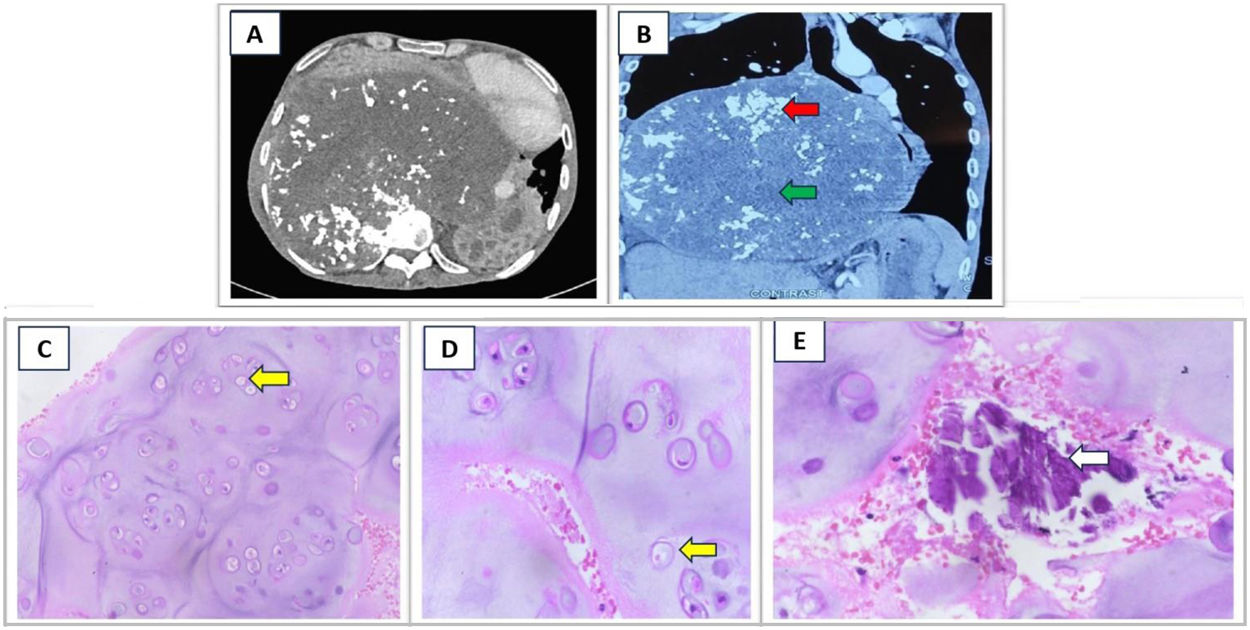
A 37-year-old man presented with dyspnea, dry cough and right-sided chest pain for 3 months. Physical examination revealed diminished breath sounds on the right hemithorax with mediastinal shift to left. Chest radiograph showed a right middle and lower zone opacity, and contrast-enhanced computed tomography showed a large heterogenous mass with multiple foci of calcification and necrosis (Fig. 1A and B). An ultrasound-guided biopsy showed features of a cartilage-forming tumor with chondrocytes arranged in lacunae with atypia, features consistent with pulmonary chondrosarcoma (Fig. 1C and D). He progressively worsened and passed away during the hospital stay before any surgery or palliative treatment could be initiated. Pulmonary chondrosarcomas are rare lung tumors that represent less than 1% of primary lung tumors. They present as enlarged, lobulated, or cystic calcified masses. The average age is between 50 and 70 years. It is of two types – tracheobronchial and lung. Immunohistochemistry is positive for S-100, particularly in the chondromatous component.1 Symptoms depend on the size and location of the lesion. Treatment is primarily surgical, with complete resection rates of over 80%. Adjuvant chemotherapy and radiotherapy are recommended for high-grade lesions or incomplete resection. In cases of unresectable tumours, surgery is the primary treatment.2

Fig. 1.

CT thorax (A and B – coronal section) showing a large heterogenous lung mass in the right hemithorax with focal areas of calcification (red arrow) and necrosis (green arrow), shifting mediastinum to the left. USG guided biopsy of the lung mass was done and the HE-stained images showed the chondrocytes arranged in small clusters, and singly in a lacuna (yellow arrows). There is moderate increase in cellularity with multinucleation, and mild atypia (C – 20×; D – 40×). Image E (40×) shows areas of calcification within the tumour (white arrow).

(0.13MB).
Declaration of Generative AI and AI-Assisted Technologies in the Writing Process

There was no involvement of AI in preparation of this manuscript.

Funding

This research did not receive any specific grant from funding agencies in the public, commercial, or not-for-profit sectors.

Conflicts of Interest

The authors declare not to have any conflicts of interest that may be considered to influence directly or indirectly the content of the manuscript.

References
[1]
J. Jiang, Q. Shen, W. Ding, J. Zhou.
Primary pulmonary chondrosarcoma and a fast-growing mass that accidentally mimicked teratoma.
J Thorac Dis, 8 (2016), pp. E947-E951
[2]
S. Nasri, Y. Amane, I. Alloubi, N. Aichouni, I. Skiker.
A rare primary pulmonary chondrosarcoma.
Cureus, 13 (2021),
Copyright © 2024. SEPAR
Archivos de Bronconeumología
Article options
Tools

Are you a health professional able to prescribe or dispense drugs?