


Patients with hereditary hemorrhagic telangiectasia (HHT) and pulmonary arteriovenous malformation (PAVM) face higher risk of embolic complications. It is not clear whether poor outcomes are related to PAVM severity or pulmonary symptoms. Furthermore, there are currently no available data on HHT patients in Argentina. We conducted a cross sectional study in a teaching hospital in Buenos Aires, Argentina. We describe baseline characteristics of HHT and compare the prevalence of embolic complications in patients with significant PAVM compared to patients without significant PAVM. One hundred and eight consecutive patients were included. Significant PAVM was defined as: contrast echocardiography grade 2 or greater; bilateral PAVM or feeding artery bigger than 3mm; or previous PAVM treatment. Primary composite outcome was defined as: cerebrovascular accident, cerebral abscess or peripheral embolism. 20% of the participants had embolic complications, and the most frequent one was stroke. Embolic complications were associated with significant PAVM and respiratory symptoms.
Los pacientes con telangiectasia hemorrágica hereditaria (THH) y malformación arteriovenosa pulmonar (MAVP) afrontan un riesgo más elevado de complicaciones embólicas. No está claro si la mala evolución clínica está relacionada con la gravedad de la MAVP o con los síntomas pulmonares. Además, en la actualidad no disponemos de datos de pacientes con THH en Argentina. Llevamos a cabo un estudio transversal en un hospital universitario de Buenos Aires, Argentina. Describimos aquí las características basales de la THH y comparamos la prevalencia de complicaciones embólicas en pacientes con una MAVP significativa frente a la de los pacientes sin una MAVP significativa. Un total de 108 pacientes consecutivos fueron incluidos en el estudio. La MAVP significativa se definió de la siguiente forma: ecocardiografía con contraste de grado 2 o superior; MAVP bilateral o aferencia de más de 3mm, o tratamiento previo de la MAVP. La variable de valoración combinada primaria se definió como: accidente cerebrovascular, absceso cerebral o embolia periférica. Un 20% de los participantes presentó complicaciones embólicas, la más frecuente de las cuales fue el ictus. Las complicaciones embólicas se asociaron a una MAVP significativa y síntomas respiratorios.
Hereditary hemorrhagic telangiectasia (HHT) is a dominant autosomic vascular dysplasia with an estimated prevalence of 1 in 5000.1 Almost 50% of the HHT patients have pulmonary arteriovenous malformations (PAVM) and the development of embolic complications has been widely described.1,2
No data have been published on HHT patients in Argentina. Moreover, it is not clear if poor outcomes with embolic complications are related to PAVM severity or pulmonary symptoms.
This cross-sectional study describes the clinical characteristics of patients with HHT referred to our teaching hospital in Buenos Aires, Argentina. We also evaluated the association between significant PAVM, pulmonary symptoms and embolic complications.
MethodsA cross-sectional study was performed on data from the institutional records of HHT in the Hospital Italiano, a tertiary teaching hospital in Buenos Aires, Argentina.3 A total of 108 consecutive patients were evaluated in the HHT unit between 2010 and 2012 and included in the study after informed consent was obtained. Participants had a definitive clinical diagnosis of HHT (defined as three or more Curacao criteria: epistaxis, telangiectasia, visceral arteriovenous malformation [AVM] or family history). The study was approved by the internal review committee of the Hospital Italiano.
Significant PAVM (exposure) was defined as the presence of at least one of the following factors: contrast echocardiography grade 2 or greater,4 bilateral PAVM or feeding artery >3mm, or previous PAVM treatment. The primary composite outcome was defined as: cerebrovascular accident, transient ischemic accident, brain abscess or peripheral embolism.
The baseline variables were compared, depending on the presence or absence of significant PAVM. A t-test was used for unpaired data, a Wilcoxon rank test for continuous variables and the Pearson χ2 for discrete variables. The association between significant PAVM (or pulmonary symptoms) and embolic complications was evaluated using Fisher's exact test. A logistic regression was used to adjust for the effects of potential confounding factors.
Finally, to evaluate the possible bias derived from missing PAVM data, a conservative sensitivity analysis was performed assuming that all patients from the missing data group had significant PAVM, and the change in prevalence of the primary outcome variable in this hypothetical situation was evaluated.
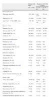
ResultsThe patients’ baseline characteristics are summarized in Table 1. A total of 35 patients (39.8%) had a significant PAVM. Embolic complications occurred in 17 participants (19.3%); the most common complication was stroke.
Baseline Characteristics of Patients With Hereditary Hemorrhagic Telangiectasia.
| Patients with HHT | Diagnosis of PAVM available | ||
| (No.=108) | Yes (No.=88) | No (No.=20) | |
| Demographic factor | |||
| Mean age, years (SD) | 45.5 (16.8) | 46.4 (15.8) | 42 (4.6) |
| Male sex, No. (%) | 33 (30.6) | 25 (28.4) | 8 (40) |
| Age at onset, median (IQR), years | 12 (6–25) | 12 (6.75–25) | 10 (1–19) |
| Curacao criteria | |||
| Epistaxis, No. (%) | 103 (95.4) | 83 (94.3) | 20 (100) |
| Telangiectasia, No. (%) | 107 (99.1) | 88 (100) | 19 (95) |
| Family history, No. (%) | 96 (88.9) | 77 (87.5) | 19 (95) |
| AVM, No. (%) | 104 (96.2) | 84 (95.5) | 11 (55) |
| Arteriovenous malformation | |||
| PAVM, No. (%)a | 67 (62) | 67 (76.1) | ND |
| Hepatic AVM, No. (%) | 54 (50) | 53 (60.2) | ND |
| Gastrointestinal AVM, No. (%) | 41 (38) | 36 (40.9) | 5 (25) |
| CNS AVM, No. (%) | 22 (20.4) | 19 (21.6) | 3 (15) |
| Pulmonary symptoms | |||
| Any symptom, No. (%) | 28 (25.9) | 25 (28.4) | 3 (15) |
| Dyspnea, No. (%) | 14 (13) | 12 (13.6) | 2 (10) |
| Hemoptysis, No. (%) | 8 (7.4) | 8 (9.1) | 0 (0) |
| Orthodeoxia, No. (%) | 8 (7.4) | 7 (8) | 1 (5) |
| Pulmonary hypertension, No. (%)b | 4 (3.7) | 4 (4.5) | ND |
| Blood disorders | |||
| Anemia, No. (%) | 66 (61) | 55 (62.5) | 11 (55) |
| Venous thromboembolism, No. (%) | 8 (7.4) | 7 (8%) | 1 (5) |
| Quality of life | |||
| VAS, median (IQR) | 70 (60–80) | 70 (60–80) | 69.1 (6.7) |
| Embolic complications, No. (%) | 18 (16.7) | 17 (19.3) | 1 (5) |
| Stroke/TIA, No. (%) | 14 (13) | 13 (14.8) | 1 (5) |
| Brain abscess, No. (%) | 3 (2.8) | 3 (3.4) | 0 (0) |
| Focal abscess, distal infectious embolism or bacteremia, No. (%) | 3 (2.8) | 3 (3.4) | 0 (0) |
| Peripheral thrombotic embolism, No. (%) | 1 (0.9) | 1 (1.1) | 0 (0) |
TIA: transient ischemic accident; SD: standard deviation; VAS: visual analog scale; AVM: arteriovenous malformation; PAVM: pulmonary AVM; No.: absolute number; IQR: interquartile range; CNS: central nervous system; HHT: hereditary hemorrhagic telangiectasia.
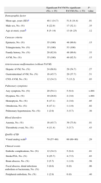
The characteristics of patients with or without significant PAVM are shown in Table 2. Patients with PAVM were significantly younger (P=.01), had more pulmonary symptoms (60% versus 11%, P<.001) and more embolic complications (34.3% versus 9.4%, P=.006). This difference remained significant in the sensitivity analysis. Furthermore, patients with pulmonary symptoms had a significantly higher rate of embolic events compared to patients without pulmonary symptoms (91.7% versus 8.3%, P=.003).
Baseline and Clinical Characteristics of Patients With and Without Significant Pulmonary Arteriovenous Malformation.
| Significant PAVM (No.=35) | No significant PAVM (No.=53) | P-value | |
| Demographic factor | |||
| Mean age, years (SD)a | 40.1 (14.7) | 51.8 (14.4) | .01 |
| Male sex, No. (%) | 8 (22.9) | 17 (32.1) | .35 |
| Age at onset, yearsb | 8 (5–14) | 13 (8–25) | .12 |
| Curacao criteria | |||
| Epistaxis, No. (%) | 35 (100) | 48 (90.6) | .15 |
| Telangiectasia, No. (%) | 35 (100) | 53 (100) | |
| Family history, No. (%) | 29 (82.9) | 48 (90.6) | .33 |
| AVM, No. (%) | 35 (100) | 49 (92.5) | .15 |
| Arteriovenous malformation (without PAVM) | |||
| Hepatic AVM, No. (%) | 24 (68.6) | 29 (54.7) | .27 |
| Gastrointestinal AVM, No. (%) | 16 (45.7) | 20 (37.7) | .51 |
| CNS AVM, No. (%) | 12 (34.3) | 7 (13.2) | .03 |
| Pulmonary symptoms | |||
| Any symptom, No. (%) | 20 (54.1) | 5 (9.4) | <.001 |
| Dyspnea, No. (%) | 10 (28.6) | 2 (3.8) | <.001 |
| Hemoptysis, No. (%) | 6 (17.1) | 2 (3.8) | .05 |
| Ortodeoxia, No. (%) | 6 (17.1) | 1 (1.9) | .02 |
| Pulmonary hypertension, No. (%) | 1 (2.9) | 3 (5.7) | .30 |
| Blood disorders | |||
| Anemia, No. (%) | 16 (45.7) | 39 (73.6) | .01 |
| Thrombotic event, No. (%) | 4 (11.4) | 3 (5.7) | .43 |
| Quality of life | |||
| Visual analog scaleb | 70 (57–80) | 60 (60–80) | .29 |
| Clinical events | |||
| Embolic complications, No. (%) | 12 (34.3) | 5 (9.4) | .01 |
| Stroke/TIA, No. (%) | 9 (25.7) | 4 (7.5) | .03 |
| Brain abscess, No. (%) | 2 (5.7) | 1 (1.9) | .56 |
| Focal abscess, distal infectious embolism or bacteremia, No. (%) | 3 (8.6) | 0 (0) | .06 |
| Peripheral embolism, No. (%) | 1 (2.9) | 0 (0) | .40 |
TIA: transient ischemic accident; SD: standard deviation; AVM: arteriovenous malformation; PAVM: pulmonary AVM; No.: absolute number; IQR: interquartile range; CNS: central nervous system; HHT: hereditary hemorrhagic telangiectasia. Hypothesis test (two-tailed alpha of 0.05): Fisher exact test.
In the unadjusted analysis, the odds ratio (OR) for embolic complications in patients with significant PAVM was 6.3 (95% CI 1.8–21.8). In the final model, that included age, sex, age at onset and anemia, the OR rose to 7 (95% CI 1.8–27.3).
DiscussionPatients with HHT seen in our hospital are relatively young. Onset occurs at an early age and their quality of life is moderately affected. Almost 25% of patients with PAVM have pulmonary symptoms, and up to 20% suffer embolic complications. Compared to previous studies in other geographical areas, the age at onset of patients seen in our hospital is younger and there is a greater prevalence of AVM, but they also have several characteristics in common.
Our study shows that patients with significant PAVM have a higher risk of developing embolic complications than those without significant PAVM. This association is marginally influenced by the covariables of age, sex, anemia and age at onset. While Shovlin et al. suggested that pulmonary symptoms were not related with embolic complications,5 in our study these symptoms were closely associated with such complications. However, the data may be biased, since patients with PAVM and embolic complications may be more likely to describe pulmonary symptoms.
The strengths of the study lie in the fact that it is the first of its kind to address the issue of HHT patients in Argentina, and a close association was detected between significant PAVM, pulmonary symptoms and embolic complications. This can be added to the body of evidence underlining the importance of PAVM in poor clinical outcome. On the other hand, the study also has some limitations. Firstly, the use of electronic clinical records means that variable may be measured and handled differently between the two groups, making them incomparable. Moreover, missing data on exposure may lead to the exclusion of patients that differ in some way from those finally included. Another possible limitation is the fact that patients with grade 2 echocardiography were classified as cases of significant PAVM even though they appear to constitute a heterogeneous group that could include patients with or without significant PAVM. Finally the initial selection of patients may undermine the external validity of our study, since patients referred to our tertiary hospital may be more likely to have a serious disease.
To conclude, significant PAVM and pulmonary symptoms may be associated with poor outcome in terms of embolic complications, a finding that would justify further prospective studies.
AuthorshipFA: Study design, data collection and analysis. Preparation of preliminary manuscript.
BLF: Study design, data collection and analysis. Preparation of preliminary manuscript.
EJW: Study design, data interpretation. Manuscript review.
MMS: Study design, baseline patient evaluation and data interpretation. Manuscript review.
All authors read and approved the final manuscript.
Conflict of InterestsThe authors declare no conflict of interests.
The authors would like to thank the Methods in Epidemiological, Clinical and Operations Research (MECOR) course of the American Thoracic Society.
Please cite this article as: Angriman F, Ferreyro BL, Wainstein EJ, Serra MM. Malformaciones arteriovenosas pulmonares y complicaciones embólicas en pacientes con telangiectasia hemorrágica hereditaria. Arch Bronconeumol. 2014;50:301–304.


