array:19 [
  "pii" => "13053341"
  "issn" => "15792129"
  "estado" => "S300"
  "fechaPublicacion" => "2003-11-01"
  "documento" => "article"
  "crossmark" => 0
  "subdocumento" => "fla"
  "cita" => "Arch Bronconeumol. 2003;39:507-13"
  "abierto" => array:3 [
    "ES" => false
    "ES2" => false
    "LATM" => false
  ]
  "gratuito" => false
  "lecturas" => array:2 [
    "total" => 8907
    "formatos" => array:3 [
      "EPUB" => 159
      "HTML" => 7777
      "PDF" => 971
    ]
  ]
  "itemSiguiente" => array:15 [
    "pii" => "13053342"
    "issn" => "15792129"
    "estado" => "S300"
    "fechaPublicacion" => "2003-11-01"
    "documento" => "article"
    "crossmark" => 0
    "subdocumento" => "fla"
    "cita" => "Arch Bronconeumol. 2003;39:514-23"
    "abierto" => array:3 [
      "ES" => false
      "ES2" => false
      "LATM" => false
    ]
    "gratuito" => false
    "lecturas" => array:2 [
      "total" => 3988
      "formatos" => array:3 [
        "EPUB" => 139
        "HTML" => 2985
        "PDF" => 864
      ]
    ]
    "en" => array:9 [
      "idiomaDefecto" => true
      "titulo" => "Recommendations on the Treatment of Tobacco Dependence"
      "tienePdf" => "en"
      "tieneTextoCompleto" => "en"
      "paginas" => array:1 [
        0 => array:2 [
          "paginaInicial" => "514"
          "paginaFinal" => "523"
        ]
      ]
      "titulosAlternativos" => array:1 [
        "es" => array:1 [
          "titulo" => "Recomendaciones para el tratamiento del tabaquismo"
        ]
      ]
      "contieneTextoCompleto" => array:1 [
        "en" => true
      ]
      "contienePdf" => array:1 [
        "en" => true
      ]
      "autores" => array:1 [
        0 => array:2 [
          "autoresLista" => "CA Jiménez-Ruiz, JI de Granda Orive, S Solano Reina, F Carrión Valero, P Romero Palacios, M Barrueco Ferrero"
          "autores" => array:6 [
            0 => array:2 [
              "Iniciales" => "CA"
              "apellidos" => "Jiménez-Ruiz"
            ]
            1 => array:2 [
              "Iniciales" => "JI"
              "apellidos" => "de Granda Orive"
            ]
            2 => array:2 [
              "Iniciales" => "S"
              "apellidos" => "Solano Reina"
            ]
            3 => array:2 [
              "Iniciales" => "F"
              "apellidos" => "Carrión Valero"
            ]
            4 => array:2 [
              "Iniciales" => "P"
              "apellidos" => "Romero Palacios"
            ]
            5 => array:2 [
              "Iniciales" => "M"
              "apellidos" => "Barrueco Ferrero"
            ]
          ]
        ]
      ]
    ]
    "idiomaDefecto" => "en"
    "EPUB" => "https://multimedia.elsevier.es/PublicationsMultimediaV1/item/epub/13053342?idApp=UINPBA00003Z"
    "url" => "/15792129/0000003900000011/v0_201307090858/13053342/v0_201307090859/en/main.assets"
  ]
  "itemAnterior" => array:15 [
    "pii" => "13053340"
    "issn" => "15792129"
    "estado" => "S300"
    "fechaPublicacion" => "2003-11-01"
    "documento" => "article"
    "crossmark" => 0
    "subdocumento" => "fla"
    "cita" => "Arch Bronconeumol. 2003;39:501-6"
    "abierto" => array:3 [
      "ES" => false
      "ES2" => false
      "LATM" => false
    ]
    "gratuito" => false
    "lecturas" => array:2 [
      "total" => 5752
      "formatos" => array:3 [
        "EPUB" => 130
        "HTML" => 4781
        "PDF" => 841
      ]
    ]
    "en" => array:11 [
      "idiomaDefecto" => true
      "titulo" => "Factors Associated With Complications Caused by Bronchoscopy in Pediatric Patients"
      "tienePdf" => "en"
      "tieneTextoCompleto" => "en"
      "tieneResumen" => array:2 [
        0 => "en"
        1 => "es"
      ]
      "paginas" => array:1 [
        0 => array:2 [
          "paginaInicial" => "501"
          "paginaFinal" => "506"
        ]
      ]
      "titulosAlternativos" => array:1 [
        "es" => array:1 [
          "titulo" => "Factores asociados a complicaciones debidas a la realización de broncoscopia en pacientes pediátricos"
        ]
      ]
      "contieneResumen" => array:2 [
        "en" => true
        "es" => true
      ]
      "contieneTextoCompleto" => array:1 [
        "en" => true
      ]
      "contienePdf" => array:1 [
        "en" => true
      ]
      "autores" => array:1 [
        0 => array:2 [
          "autoresLista" => "C Rodríguez Martínez, MP Sossa"
          "autores" => array:2 [
            0 => array:2 [
              "Iniciales" => "C"
              "apellidos" => "Rodríguez Martínez"
            ]
            1 => array:2 [
              "Iniciales" => "MP"
              "apellidos" => "Sossa"
            ]
          ]
        ]
      ]
    ]
    "idiomaDefecto" => "en"
    "EPUB" => "https://multimedia.elsevier.es/PublicationsMultimediaV1/item/epub/13053340?idApp=UINPBA00003Z"
    "url" => "/15792129/0000003900000011/v0_201307090858/13053340/v0_201307090858/en/main.assets"
  ]
  "en" => array:11 [
    "idiomaDefecto" => true
    "titulo" => "New Approaches to the Surgical Treatment of Severe Thoracic Cage Diseases With Respiratory Repercussions"
    "tieneTextoCompleto" => true
    "paginas" => array:1 [
      0 => array:2 [
        "paginaInicial" => "507"
        "paginaFinal" => "513"
      ]
    ]
    "autores" => array:1 [
      0 => array:3 [
        "autoresLista" => "A Molina, M Ramírez, O García-Casas, Ll Puig, E Cáceres-Palou, J Gea, J Bago"
        "autores" => array:7 [
          0 => array:3 [
            "Iniciales" => "A"
            "apellidos" => "Molina"
            "referencia" => array:1 [
              0 => array:2 [
                "etiqueta" => "<span class="elsevierStyleSup">a</span>"
                "identificador" => "affa"
              ]
            ]
          ]
          1 => array:3 [
            "Iniciales" => "M"
            "apellidos" => "Ram&#237;rez"
            "referencia" => array:1 [
              0 => array:2 [
                "etiqueta" => "<span class="elsevierStyleSup">a</span>"
                "identificador" => "affa"
              ]
            ]
          ]
          2 => array:3 [
            "Iniciales" => "O"
            "apellidos" => "Garc&#237;a-Casas"
            "referencia" => array:1 [
              0 => array:2 [
                "etiqueta" => "<span class="elsevierStyleSup">a</span>"
                "identificador" => "affa"
              ]
            ]
          ]
          3 => array:3 [
            "nombre" => "Ll"
            "apellidos" => "Puig"
            "referencia" => array:1 [
              0 => array:2 [
                "etiqueta" => "<span class="elsevierStyleSup">a</span>"
                "identificador" => "affa"
              ]
            ]
          ]
          4 => array:3 [
            "Iniciales" => "E"
            "apellidos" => "C&#225;ceres-Palou"
            "referencia" => array:1 [
              0 => array:2 [
                "etiqueta" => "<span class="elsevierStyleSup">a</span>"
                "identificador" => "affa"
              ]
            ]
          ]
          5 => array:3 [
            "Iniciales" => "J"
            "apellidos" => "Gea"
            "referencia" => array:1 [
              0 => array:2 [
                "etiqueta" => "<span class="elsevierStyleSup">b</span>"
                "identificador" => "affb"
              ]
            ]
          ]
          6 => array:3 [
            "Iniciales" => "J"
            "apellidos" => "Bago"
            "referencia" => array:1 [
              0 => array:2 [
                "etiqueta" => "<span class="elsevierStyleSup">c</span>"
                "identificador" => "affc"
              ]
            ]
          ]
        ]
        "afiliaciones" => array:3 [
          0 => array:3 [
            "entidad" => "Servicio de Cirugía Ortopédica y Traumatología (COT), Hospital del Mar, IMAS, Barcelona, Spain."
            "etiqueta" => "<span class="elsevierStyleSup">a</span>"
            "identificador" => "affa"
          ]
          1 => array:3 [
            "entidad" => "Servicio de Neumología, Hospital del Mar, IMAS, Barcelona, Spain."
            "etiqueta" => "<span class="elsevierStyleSup">b</span>"
            "identificador" => "affb"
          ]
          2 => array:3 [
            "entidad" => "Servicio de Cirugía Ortopédica y Traumatología (COT), Hospital Vall d'Hebron, Universidad Autónoma de Barcelona, Barcelona, Spain."
            "etiqueta" => "<span class="elsevierStyleSup">c</span>"
            "identificador" => "affc"
          ]
        ]
      ]
    ]
    "titulosAlternativos" => array:1 [
      "es" => array:1 [
        "titulo" => "Nuevas perspectivas en el tratamiento quir&#250;rgico de las enfermedades graves de la caja tor&#225;cica que cursan con afectaci&#243;n respiratoria"
      ]
    ]
    "textoCompleto" => "<p class="elsevierStylePara"><span class="elsevierStyleBold">Introduction</span></p><p class="elsevierStylePara"> Scoliosis is defined as lateral curvature of the spine with vertebral rotation&#44; meaning that it is a 3-dimensional deformity&#46; Scoliosis leads to thoracic deformity if the spinal alterations occur in that zone &#40;Figure 1&#41;&#46;</p><p class="elsevierStylePara"><img src="260v39n11-13053341tab01.gif"></img></p><p class="elsevierStylePara">Figure&#46; 1&#46; Thoracoabdominal x-ray showing posteroanterior &#40;a&#41; and lateral &#40;b&#41; views of a spine with scoliosis&#46;</p><p class="elsevierStylePara"> Scoliosis is classified as either idiopathic or secondary&#44; regardless of what part of the spine is affected&#46; Idiopathic scoliosis is currently thought almost certainly to arise from multiple factors&#46; The most common causes are disordered development of the central nervous system or genetically determined familial disorders&#46;<span class="elsevierStyleSup">1&#44;2</span></p><p class="elsevierStylePara">The prevalence of idiopathic scoliosis in the population at risk &#40;individuals between 10 and 16 years of age&#41; is approximately 2&#37; to 3&#37;&#46; As the magnitude of the curve increases&#44; the incidence decreases&#44; and it is estimated that the percentage of patients who require surgery does not exceed 0&#46;1&#37;&#46;<span class="elsevierStyleSup">3</span></p><p class="elsevierStylePara">The decision of how to treat patients with spinal deformity should be based on an understanding of the natural history of the process&#46; Any decisions taken should have as their objective the modification of that natural history&#46; The main factors influencing therapeutic decisions are the type and magnitude of the curve&#44; age&#44; skeletal maturity&#44; and sex&#46; The most important factors at the time of diagnosis are age and curve magnitude&#46;<span class="elsevierStyleSup">4&#44;5</span> Age is especially important because it is related to elasticity<span class="elsevierStyleSup">3</span> and on elasticity will depend the possibility of reducing the angle of curvature and the patient&#39;s muscular response to it&#46;</p><p class="elsevierStylePara"><span class="elsevierStyleBold">Respiratory Function and Deformity</span></p><p class="elsevierStylePara"> Respiratory function is mainly affected in cases of thoracic scoliosis&#46; The development of the pulmonary parenchyma ends when a child is 8 years old&#44; and if the onset of deformity occurs at that time or earlier&#44; the process may be accompanied by decreased vital capacity &#40;VC&#41; and total lung capacity &#40;TLC&#41;&#46;<span class="elsevierStyleSup">6</span></p><p class="elsevierStylePara">A direct relation has been found between lung function changes and magnitude of the thoracic curve&#46; The type of alteration seen most often is restriction&#44; which means less lung volume&#44; greater work of breathing&#44; higher energy expenditure&#44; hypoxemia&#44; respiratory acidosis due to hypoventilation&#44; inadequate ventilatory response to hypoxic stimulus&#44; pulmonary artery hypertension&#44; underdevelopment of the pulmonary parenchyma&#44; and an altered relationship between lung function and respiratory muscle mechanics&#46;</p><p class="elsevierStylePara"> Individuals with deformity of this type have reduced VC and&#44; to a lesser degree&#44; reduced forced expiratory volume in 1 second &#40;FEV<span class="elsevierStyleInf">1</span>&#41; even if they are asymptomatic&#46; A higher prevalence of preoperative and postoperative pulmonary problems in these patients has been mentioned in numerous articles&#46;</p><p class="elsevierStylePara">The most marked alterations in lung function have been reported to occur when the Cobb angle of the curve exceeds 70&#186;-80&#186;&#46; In such cases&#44; diaphragm function changes and respiratory function comes to depend more on accessory muscles&#46; Given that accessory muscle activity decreases at night&#44; hypoxemia and&#44; as a consequence&#44; higher red cell counts may develop&#46; Other factors to remember are the location and number of thoracic vertebrae involved&#46;<span class="elsevierStyleSup">7</span> In the long run&#44; only patients with curves exceeding 100&#186; concomitant with other factors develop chronic respiratory insufficiency and cor pulmonale&#46; According to some authors&#44; when VC is less than 40&#37; of the reference value&#44; the risk of pulmonary function impairment is great&#46;<span class="elsevierStyleSup">8</span> When the deformity is no greater than 50&#186; but is associated with hypokyphosis &#40;loss of the natural thoracic kyphosis&#41;&#44; the prognosis worsens&#46; Smoking also worsens the prognosis&#46; Scoliosis in the middle range &#40;40&#186;-60&#186;&#41; in the absence of changes in the normal kyphosis is unlikely to cause severe respiratory problems&#46; In cases of mild thoracic scoliosis &#40;&#60;35&#186;&#41;&#44; lung function can be improved by exercise&#46; Nevertheless&#44; the overall mortality of individuals with idiopathic thoracic scoliosis is comparable to that of the general population&#46;</p><p class="elsevierStylePara"> Little is known of the situation in adulthood of patients who had juvenile scoliosis&#44; whether treated surgically or not&#46;<span class="elsevierStyleSup">6</span> Such individuals usually experience certain limitations when engaging in intense physical activity&#44; such as lifting or carrying heavy objects&#46;</p><p class="elsevierStylePara"><span class="elsevierStyleBold">Treatment</span></p><p class="elsevierStylePara"> Orthopedic approaches &#40;braces&#41; and surgery seek to reduce spinal curvature in order to improve appearance and prevent the likely deterioration of respiratory function&#46; The outcome of treatment depends on the spine&#39;s elasticity&#46; Therefore&#44; the timing of treatment is essential&#46; As will be discussed later&#44; the course of respiratory function is directly related to patient age--whether the patient is an adolescent&#44; considered &#34;more elastic&#44;&#34; or an adult&#44; considered more &#34;rigid&#46;&#34;<span class="elsevierStyleSup">3</span></p><p class="elsevierStylePara"> Orthopedic treatment with rigid braces has been shown to be effective in curvatures of up to 40&#186; and in individuals with elastic deformities&#46; However&#44; recurrence has been observed in some patients&#46; When this treatment is considered&#44; 3 basic questions must be answered&#46; What deformity is going to be treated&#63; Why must it be treated&#63; Can it be successfully corrected by conservative treatment&#63;</p><p class="elsevierStylePara"> Orthopedic treatment is used in adolescents&#44; who usually have a remarkable ability to adapt&#46; Although continuous application of such braces causes problems for thoracic expansion&#44; long-term studies seem to indicate that respiratory function deterioration does not persist&#46;<span class="elsevierStyleSup">9</span></p><p class="elsevierStylePara"> Surgery is used to reduce 3-dimensional vertebral deformity by manipulating vertebrae to achieve a stable arthrodesis that prevents progression&#46;<span class="elsevierStyleSup">4</span> Surgery is indicated when deformity is greater than 60&#186; and symptomatic &#40;pain in the adult&#44; esthetic problems in the child or adolescent&#41;&#44; or greater than 40&#186; during periods of growth&#44; given that curvature has been shown to progress in such cases in spite of orthopedic treatment&#46; Finally&#44; surgery is appropriate if there is a deformity that is unacceptable for the patient&#46;<span class="elsevierStyleSup">4&#44;5</span> No evidence supports surgical intervention solely to treat deterioration in lung function&#46;</p><p class="elsevierStylePara"> Thoracic deformities can be corrected using an anterior approach &#40;by thoracotomy or thoracoscopy&#41;&#44; a posterior approach&#44; or a combination&#46; All approaches enlist the spine&#39;s own elastic properties to reduce curvature&#46; Thus&#44; the approach to the spine will depend not only on technical considerations&#44; but also on the type of curve&#44; elasticity&#44; and number of vertebrae that must be fused&#46; When the effect on appearance is very evident owing to the presence of a gibbus &#40;hump&#41;&#44; thoracoplasty might also be performed for esthetic reasons&#44; with partial resection of the ribs in the most prominent zone&#46; This technique is usually seen in posterior orthopedic interventions&#46; The aims of surgical alteration of the thoracic curve are to correct the defect &#40;preserving the sagittal balance of the spine&#41;&#44; to preserve or improve lung function&#44; to exercise a positive influence on the lumbar spine&#44; to achieve minimum morbidity and pain for the patient&#44; and to maximize return to full function&#46;</p><p class="elsevierStylePara"> Nevertheless&#44; the effect of surgical correction on the lung function of patients with idiopathic scoliosis is not entirely clear&#46; Although several studies have demonstrated that such surgical correction improves some lung function variables&#44;<span class="elsevierStyleSup">10</span> others have observed no such improvement<span class="elsevierStyleSup">11</span> or have even seen functional decline&#46;<span class="elsevierStyleSup">12</span> Such discrepancies may arise from the fact that many studies have enrolled populations that are heterogenous as to age&#44; sex&#44; type and intensity of curvature&#44; and etiology of the scoliosis&#46; Moreover&#44; the techniques used for surgical correction have differed&#44; as have the criteria used to evaluate respiratory function&#46;</p><p class="elsevierStylePara">The study performed by Rizzi et al&#44;<span class="elsevierStyleSup">7</span> in 35 adult patients undergoing spinal surgery between 1978 and 1994 gave mixed outcomes&#46; Seven patients died during the first year&#44; while the rest experienced considerably improved respiratory function and quality of life &#40;72-month follow-up&#41;&#46; The authors concluded that the outcome of spinal surgery may range from &#34;extremely good&#34; &#40;a high percentage of improvement&#41; to &#34;extremely unsatisfactory&#34; &#40;high mortality&#41;&#46;</p><p class="elsevierStylePara"><span class="elsevierStyleBold">Surgical Technique</span></p><p class="elsevierStylePara"> During the last decade&#44; surgical approaches to treating thoracic deformities have changed markedly in terms of indications and pre-&#44; intra-&#44; and postoperative management of the patient&#46;<span class="elsevierStyleSup">4</span> Currently&#44; the objective is to halt the progression of deformity and maintain or even increase respiratory capacity in these patients&#46;</p><p class="elsevierStylePara">As has been mentioned&#44; surgery may involve either posterior or anterior approaches&#44; the latter involving thoracotomy &#40;usually with section of the diaphragm&#41; or thoracoplasty&#46; The two techniques may also be combined&#44; in what is called a &#34;double approach&#34; which includes&#58; <span class="elsevierStyleItalic">a&#41;</span> the anterior thoracic approach to perform diskectomy and insert an implant in the space vacated by the intervertebral disk&#44; and <span class="elsevierStyleItalic">b&#41;</span> the posterior route&#44; with spinal fixation and placement of an implant if necessary&#46; Some specialists refer to this technique as &#34;circumferential arthrodesis&#46;&#34;</p><p class="elsevierStylePara"><span class="elsevierStyleItalic">Thoracoplasty</span></p><p class="elsevierStylePara"> Thoracoplasty consists in a partial resection of the ribs of the convexity of the curve &#40;Figures 2 and 3&#41;&#46; The main aim is to improve appearance&#46; Thoracoplasty has also been described in the context of instrumentation performed through a posterior route&#46; When this technique is used as a complement to vertebral fixation&#44; a high number of peri- and postoperative complications &#40;pneumothorax&#41; occur&#44; with altered respiratory mechanics during the first 2 months &#40;decline in lung volume of 12&#37; to 16&#37;&#41;&#46; Some authors attribute part of this decline to postoperative pain&#46;<span class="elsevierStyleSup">13</span> However&#44; 2 years after surgery&#44; respiratory function has returned to its preoperative status&#46;</p><p class="elsevierStylePara"><img src="260v39n11-13053341tab02.gif"></img></p><p class="elsevierStylePara">Figure 2&#46; Schematic depiction of thoracoplasty&#46;</p><p class="elsevierStylePara"><img src="260v39n11-13053341tab03.gif"></img></p><p class="elsevierStylePara"><span class="elsevierStyleItalic">Posterior Approach</span></p><p class="elsevierStylePara">A posterior approach is chosen if the elasticity of the spine allows the deformity to be modified in this way&#46; The main positive feature of this approach is that it does not alter the structure of muscles that act directly on ventilatory mechanics&#46; The first instruments used were Harrington rods&#44;<span class="elsevierStyleSup">6&#44;8&#44;9&#44;14</span> which did not allow for a natural thoracic kyphosis&#46; As a result&#44; esthetic defects &#40;a flattened spine&#41; and&#44; probably&#44; functional impairment developed&#46; Some authors report that this technique produces a slight gain in VC and static lung volumes&#44; although they do not usually become normal&#46; However&#44; other authors have observed a slight decrease in VC&#44; which they have attributed to the loss of kyphosis&#46; Nevertheless&#44; a very high percentage of such patients had also undergone prolonged immobilization with braces&#44; and that treatment may also have influenced the outcome&#46;</p><p class="elsevierStylePara">The implants used currently with the same approach have undergone marked changes &#40;third generation implants&#41;&#46; The changes introduced have had the aim of producing a repositioning of the thoracic cage that is as anatomically normal as possible by way of a distraction-rotation maneuver&#46; With this technique&#44; applied to elastic thoracic deformities&#44; correction has taken place in the coronal and frontal planes&#44; in the deformity of the rib cage&#44; and in the asymmetry of the thoracic cage &#40;normal kyphosis in the lateral plane&#44; the normal axis in the frontal plane and derotations in the coronal plane&#41; &#40;Figure 4&#41;&#46;</p><p class="elsevierStylePara"><img src="260v39n11-13053341tab04.gif"></img></p><p class="elsevierStylePara">Figure 4&#46; X-ray of a scoliotic thoracic spine after surgical treatment&#44; in anteroposterior &#40;a&#41; and lateral &#40;b&#41; views&#46;</p><p class="elsevierStylePara"> Adolescent patients treated by this approach and with third generation implants have experienced improved VC and forced expiratory volume in 1 second&#46; Some authors report that this improvement endures&#44; whereas others have seen it disappear after 1 year&#46;<span class="elsevierStyleSup">12</span></p><p class="elsevierStylePara"><span class="elsevierStyleItalic">Anterior Approach&#58; Thoracotomy</span></p><p class="elsevierStylePara"> Thoracotomy is considered an alternative to the posterior approach&#46;<span class="elsevierStyleSup">15</span> The objective is to obtain greater reduction of spinal curvature&#44; to involve the least number of vertebrae in the fixation procedure and prevent pseudoarthrosis&#46; Nevertheless&#44; this approach leads to marked changes in elements directly involved in ventilatory mechanics&#44; such as the large dorsal muscles&#44; the diaphragm&#44; and the intercostal muscles&#46; Moreover&#44; it is difficult to correct the loss of thoracic kyphosis&#46; The main drawback to the anterior approach is that the lung on the side through which the spine is approached may collapse&#44; probably leading to posterior expansion&#46; The most frequent complications of this type of intervention are atelectasis and decreased lung volume&#46; In general the patients experience impaired lung function for 3 postoperative months&#44; although function has recovered by 2 years later&#46;</p><p class="elsevierStylePara">A retrospective study by Grossfeld et al<span class="elsevierStyleSup">16</span> in 599 children operated on between 1967 and 1991 identified a set of risk factors in spinal interventions&#58; use of thoracotomy&#44; age over 14 years&#44; male sex&#44; presence of a kyphotic curve&#44; a Cobb angle greater than 100&#186; and a VC less than 40&#37; of the reference value&#46; The most important risk factors for &#34;serious&#34; postoperative complications were age and marked angulation&#46; However&#44; a study by Rawlins et al<span class="elsevierStyleSup">17</span> in 32 patients aged between 7 and 17 years found that the results of using thoracotomy to access the spine can be good&#44; even when the patient&#39;s VC is less than 40&#37; of the predicted value&#44; provided interdisciplinary medical support is effective &#40;supervision by pneumologists and internists&#41;&#46;</p><p class="elsevierStylePara"><span class="elsevierStyleItalic">Anterior Approach&#58; Thoracoscopy</span></p><p class="elsevierStylePara"> Minimal access spinal surgery has been moving forward in recent years and will therefore be covered in more detail in this review&#46; Thoracoscopy &#40;Figure 5&#41; has been introduced as a new therapeutic approach for treating spinal pathology&#46; The endoscope was first used in surgery on the spine in 1993&#44; when it was considered an alternative to &#34;open surgery&#46;&#34; At first it was used mainly in patients considered to be at high risk from thoracotomy&#44; given that the minimal procedure is less traumatic for the chest wall&#46;<span class="elsevierStyleSup">18</span> Time has shown&#44; however&#44; that the deformity must be elastic and the angle greater than 75&#186; if maximum benefit is to be derived from thoracoscopy&#46; The technique should not be used in patients in whom a lung must be collapsed&#44; who have severe respiratory insufficiency&#44; who require high positive pressures to maintain adequate ventilation&#44; or who have pleural adhesions&#46; For the past 7 years&#44; an anterior endoscopic approach has been used for spinal release surgery &#40;with the purpose of increasing flexibility&#41;&#44; for anterior arthrodesis&#44; for epiphysiodesis &#40;to prevent a flat back or the crankshaft problem&#41;&#44; and for disk resections&#46; It has also been used to treat certain primary tumors&#44; spinal metastases&#44; and fractured or herniated disks&#46;</p><p class="elsevierStylePara"><img src="260v39n11-13053341tab05.gif"></img></p><p class="elsevierStylePara">Figure 5&#46; An artist&#39;s depiction &#40;a&#41; and a surgical image &#40;b&#41; of thoracoscopy&#46;</p><p class="elsevierStylePara"> Thoracoscopy is preferred from the patient&#39;s point of view because it is less invasive and causes less pain and morbidity&#46; However&#44; these advantages are less evident to the spinal surgeon&#46; The minimal access approach requires a longer training period&#44; more time in the operating room and often the help of a thoracic or abdominal surgeon&#46; An argument in its favor&#44; however&#44; is the growing interest in shortening hospital stays and decreasing costs&#46;</p><p class="elsevierStylePara"> Arlet<span class="elsevierStyleSup">19</span> published a meta-analysis of 151 studies that attempted to answer prevailing questions about the indications for thoracoscopy in spinal deformities&#46; Only 57 of the articles found&#44; however&#44; were relatively acceptable&#46; Twenty-one were about spinal fusion&#44; 11 on spinal release&#44; and the rest on complications of thoracoscopy&#46; Of the 57&#44; only 9 were considered optimal sources of evidence &#40;Table&#41;&#46;<span class="elsevierStyleSup">20-28</span> Most cases involved children&#44; in whom the spine is much more flexible than it is in the adult&#46; It is interesting to note&#44; moreover&#44; that the mean angle of curvature was 65&#186; for the cases published&#44; although the anterior approach is widely held to be indicated only for patients with curvatures greater than 75&#186; and with radiologically evident lateral bending of less than 50&#186;&#46; However&#44; thoracoscopy might be justified for preventing the development of the flat-back syndrome&#44; performing epiphysiodesis with the aim of eliminating growth and&#47;or performing disk removal to afford flexibility to the spine&#46; From a technical standpoint&#44; all the reports reviewed approached the spine from the convex side of the curvature&#44; with the patient in lateral decubitus position&#44; with selective collapse of the ipsilateral lung &#40;as is the case in open thoracotomy&#41;&#46; The learning curve rose very gradually for this technique&#44; given that training is needed for the approach phase and for the performance of disk removal itself&#46; An additional problem is the difficulty of evaluating the amount of intervertebral disk removed&#46; The duration of interventions &#40;mean 2&#46;5 hours&#41; was unrelated to type of deformity&#46; When an operation included a period in which a posterior approach was taken&#44; the duration of surgery was much longer &#40;10 hours&#41;&#46; Successful repair of the deformity was achieved in 56&#37; to 63&#37; of cases&#46; The cost of thoracoscopy was 30&#37; higher than that of open thoracotomy&#44;<span class="elsevierStyleSup">14</span> when the learning curve&#44; material&#44; equipment depreciation&#44; hours in the operating room&#44; and the need for immediate availability for conversion to open thoracotomy were all taken into account&#46; The mean duration of hospitalization was 9 days&#46; Complications developed in 20&#37; of the patients &#40;6&#37; being serious&#41;&#44; although they did not affect the final outcomes&#46; The duration of assisted ventilation was seen to be high&#44; more so in patients with neuromuscular problems&#46;</p><p class="elsevierStylePara"><img src="260v39n11-13053341tab06.gif"></img></p><p class="elsevierStylePara"> Newton and Wenger&#44;<span class="elsevierStyleSup">29</span> following a study comparing thoracotomy and thoracoscopy in children and adolescents with spinal deformity&#44; reported that the benefits of one procedure over the other were not significant&#44; and they even found that patients who underwent thoracoscopy needed longer pleural drainage&#46;</p><p class="elsevierStylePara"> From these reports we can conclude that endoscopic surgery on the thoracic spine has important limitations related to the infrastructure required and the ratio between training and yield&#46; For that reason&#44; the indications for thoracoscopy and the hospitals where it is performed should be strictly rationalized&#46;</p><p class="elsevierStylePara"><span class="elsevierStyleItalic"> Indications for Thoracoscopy in Scoliotic Deformity&#46;</span> Endoscopy can be effective for pediatric patients&#44; but the results of repair are similar to those achieved by open procedures&#46; When the deformity is severe or flexibility of the spine is reduced &#40;as in adults&#41;&#44; outcome is less satisfactory&#46;</p><p class="elsevierStylePara"><span class="elsevierStyleItalic"> Indications for Thoracoscopy in Kyphotic Deformity&#46;</span> The consensus is that only kyphoses exceeding 75&#186; should be reduced&#46; The fixation systems currently available allow us to reduce the deformity by facetectomy &#40;posterior resection of articulations&#41;&#44; making an anterior approach unnecessary&#46; This surgical procedure is easier in kyphotic patients because they have a &#34;larger&#34; thoracic cavity&#46;</p><p class="elsevierStylePara"><span class="elsevierStyleBold">Conclusion</span></p><p class="elsevierStylePara">The magnitude of the thoracic curve determines how respiratory function is affected&#44; creating the possibility that a restrictive ventilatory pattern might develop&#46; The presence of a loss of normal kyphosis also influences lung function negatively&#46; Thus&#44; in scoliotic deformities of equal magnitude&#44; those in whom kyphosis is reduced are more likely to develop altered respiratory function&#46; Smoking also aggravates functional impairment &#40;a nonsmoker only develops functional alterations when deformity is between 100&#186; and 120&#186;&#41;&#46;</p><p class="elsevierStylePara"> Scoliotic deformity can be resolved using an anterior &#40;endoscopy or thoracotomy&#41;&#44; posterior&#44; or combined approach&#46; Moreover&#44; thoracoplasty can be considered as a complementary procedure for improving the appearance of the patient&#46; The application of each technique will depend on the indications explained in this review&#46;</p><p class="elsevierStylePara"> Although thoracoplasty initially raised hopes in surgery to repair thoracic spinal deformity&#44; important limitations on its application exist &#40;specific indications&#44; a prolonged learning curve&#44; and moderately positive outcomes&#41;&#46;</p><hr></hr><p class="elsevierStylePara">Correspondence&#58; Dr&#46; A&#46; Molina Ros&#46;<br></br> Servicio de COT&#46; Hospital del Mar&#46;<br></br> Pg&#46; Mar&#237;tim&#44; 27&#46; 08003 Barcelona&#46; Espa&#241;a&#46;<br></br> E-mail&#58; <a href="mailto&#58;20017&#64;imas&#46;imim&#46;es" class="elsevierStyleCrossRefs"> 20017&#64;imas&#46;imim&#46;es</a></p><p class="elsevierStylePara">Manuscript received April 30&#44; 2003&#46;<br></br> Accepted for publication May 13&#44; 2003&#46;</p>"
    "pdfFichero" => "260v39n11a13053341pdf001.pdf"
    "tienePdf" => true
    "multimedia" => array:12 [
      0 => array:8 [
        "identificador" => "tbl1"
        "etiqueta" => "Figure. 1"
        "tipo" => "MULTIMEDIATABLA"
        "mostrarFloat" => true
        "mostrarDisplay" => false
        "copyright" => "Elsevier Espa&#241;a"
        "tabla" => array:1 [
          "tablatextoimagen" => array:1 [
            0 => array:1 [
              "tablaImagen" => array:1 [
                0 => array:4 [
                  "imagenFichero" => "260v39n11-13053341tab01.gif"
                  "imagenAlto" => 359
                  "imagenAncho" => 389
                  "imagenTamanyo" => 39174
                ]
              ]
            ]
          ]
        ]
        "descripcion" => array:1 [
          "en" => "Thoracoabdominal x-ray showing posteroanterior &#40;a&#41; and lateral &#40;b&#41; views of a spine with scoliosis&#46;"
        ]
      ]
      1 => array:8 [
        "identificador" => "tbl2"
        "etiqueta" => "Figure 2"
        "tipo" => "MULTIMEDIATABLA"
        "mostrarFloat" => true
        "mostrarDisplay" => false
        "copyright" => "Elsevier Espa&#241;a"
        "tabla" => array:1 [
          "tablatextoimagen" => array:1 [
            0 => array:1 [
              "tablaImagen" => array:1 [
                0 => array:4 [
                  "imagenFichero" => "260v39n11-13053341tab02.gif"
                  "imagenAlto" => 240
                  "imagenAncho" => 389
                  "imagenTamanyo" => 14854
                ]
              ]
            ]
          ]
        ]
        "descripcion" => array:1 [
          "en" => "Schematic depiction of thoracoplasty&#46;"
        ]
      ]
      2 => array:8 [
        "identificador" => "tbl3"
        "etiqueta" => "Figure 3"
        "tipo" => "MULTIMEDIATABLA"
        "mostrarFloat" => true
        "mostrarDisplay" => false
        "copyright" => "Elsevier Espa&#241;a"
        "tabla" => array:1 [
          "tablatextoimagen" => array:1 [
            0 => array:1 [
              "tablaImagen" => array:1 [
                0 => array:4 [
                  "imagenFichero" => "260v39n11-13053341tab03.gif"
                  "imagenAlto" => 217
                  "imagenAncho" => 615
                  "imagenTamanyo" => 109865
                ]
              ]
            ]
          ]
        ]
        "descripcion" => array:1 [
          "en" => "Thoracoplasty&#44; surgical image&#46;"
        ]
      ]
      3 => array:8 [
        "identificador" => "tbl4"
        "etiqueta" => "Figure 4"
        "tipo" => "MULTIMEDIATABLA"
        "mostrarFloat" => true
        "mostrarDisplay" => false
        "copyright" => "Elsevier Espa&#241;a"
        "tabla" => array:1 [
          "tablatextoimagen" => array:1 [
            0 => array:1 [
              "tablaImagen" => array:1 [
                0 => array:4 [
                  "imagenFichero" => "260v39n11-13053341tab04.gif"
                  "imagenAlto" => 470
                  "imagenAncho" => 585
                  "imagenTamanyo" => 70172
                ]
              ]
            ]
          ]
        ]
        "descripcion" => array:1 [
          "en" => "X-ray of a scoliotic thoracic spine after surgical treatment&#44; in anteroposterior &#40;a&#41; and lateral &#40;b&#41; views&#46;"
        ]
      ]
      4 => array:8 [
        "identificador" => "tbl5"
        "etiqueta" => "Figure 5"
        "tipo" => "MULTIMEDIATABLA"
        "mostrarFloat" => true
        "mostrarDisplay" => false
        "copyright" => "Elsevier Espa&#241;a"
        "tabla" => array:1 [
          "tablatextoimagen" => array:1 [
            0 => array:1 [
              "tablaImagen" => array:1 [
                0 => array:4 [
                  "imagenFichero" => "260v39n11-13053341tab05.gif"
                  "imagenAlto" => 768
                  "imagenAncho" => 388
                  "imagenTamanyo" => 117911
                ]
              ]
            ]
          ]
        ]
        "descripcion" => array:1 [
          "en" => "An artist&#39;s depiction &#40;a&#41; and a surgical image &#40;b&#41; of thoracoscopy&#46;"
        ]
      ]
      5 => array:8 [
        "identificador" => "tbl6"
        "etiqueta" => "TABLE"
        "tipo" => "MULTIMEDIATABLA"
        "mostrarFloat" => true
        "mostrarDisplay" => false
        "copyright" => "Elsevier Espa&#241;a"
        "tabla" => array:1 [
          "tablatextoimagen" => array:1 [
            0 => array:1 [
              "tablaImagen" => array:1 [
                0 => array:4 [
                  "imagenFichero" => "260v39n11-13053341tab06.gif"
                  "imagenAlto" => 248
                  "imagenAncho" => 727
                  "imagenTamanyo" => 18276
                ]
              ]
            ]
          ]
        ]
        "descripcion" => array:1 [
          "en" => "Meta-analysis by Arlet19 on the Use of Thoracoscopy for Surgical Repair of the Spine&#42;"
        ]
      ]
      6 => array:5 [
        "identificador" => "tbl7"
        "tipo" => "MULTIMEDIATABLA"
        "mostrarFloat" => true
        "mostrarDisplay" => false
        "copyright" => "Elsevier Espa&#241;a"
      ]
      7 => array:5 [
        "identificador" => "tbl8"
        "tipo" => "MULTIMEDIATABLA"
        "mostrarFloat" => true
        "mostrarDisplay" => false
        "copyright" => "Elsevier Espa&#241;a"
      ]
      8 => array:5 [
        "identificador" => "tbl9"
        "tipo" => "MULTIMEDIATABLA"
        "mostrarFloat" => true
        "mostrarDisplay" => false
        "copyright" => "Elsevier Espa&#241;a"
      ]
      9 => array:5 [
        "identificador" => "tbl10"
        "tipo" => "MULTIMEDIATABLA"
        "mostrarFloat" => true
        "mostrarDisplay" => false
        "copyright" => "Elsevier Espa&#241;a"
      ]
      10 => array:5 [
        "identificador" => "tbl11"
        "tipo" => "MULTIMEDIATABLA"
        "mostrarFloat" => true
        "mostrarDisplay" => false
        "copyright" => "Elsevier Espa&#241;a"
      ]
      11 => array:5 [
        "identificador" => "tbl12"
        "tipo" => "MULTIMEDIATABLA"
        "mostrarFloat" => true
        "mostrarDisplay" => false
        "copyright" => "Elsevier Espa&#241;a"
      ]
    ]
    "bibliografia" => array:2 [
      "titulo" => "Bibliography"
      "seccion" => array:1 [
        0 => array:1 [
          "bibliografiaReferencia" => array:29 [
            0 => array:3 [
              "identificador" => "bib1"
              "etiqueta" => "1"
              "referencia" => array:1 [
                0 => array:2 [
                  "contribucion" => array:1 [
                    0 => array:3 [
                      "titulo" => "Cause of idiopathic scoliosis&#46;"
                      "idioma" => "en"
                      "autores" => array:1 [
                        0 => array:2 [
                          "etal" => false
                          "autores" => array:1 [
                            0 => "Machida M&#46;"
                          ]
                        ]
                      ]
                    ]
                  ]
                  "host" => array:1 [
                    0 => array:1 [
                      "Revista" => array:6 [
                        "tituloSerie" => "Spine"
                        "fecha" => "1999"
                        "volumen" => "24"
                        "paginaInicial" => "2576"
                        "paginaFinal" => "83"
                        "link" => array:1 [
                          0 => array:2 [
                            "url" => "https://www.ncbi.nlm.nih.gov/pubmed/10635520"
                            "web" => "Medline"
                          ]
                        ]
                      ]
                    ]
                  ]
                ]
              ]
            ]
            1 => array:3 [
              "identificador" => "bib2"
              "etiqueta" => "2"
              "referencia" => array:1 [
                0 => array:2 [
                  "contribucion" => array:1 [
                    0 => array:3 [
                      "titulo" => "Classification and terminology&#46; In&#58; Lippincot-Ravent&#44; editor&#46; Textbook of scoliosis and other spinal deformities&#46; Philadelphia&#58; W&#46;B&#46; Saunders Co&#46;"
                      "idioma" => "en"
                      "autores" => array:1 [
                        0 => array:2 [
                          "etal" => false
                          "autores" => array:1 [
                            0 => "Winter R&#46;"
                          ]
                        ]
                      ]
                    ]
                  ]
                  "host" => array:1 [
                    0 => array:1 [
                      "Libro" => array:4 [
                        "titulo" => "Classification and terminology&#46; In&#58; Lippincot-Ravent&#44; editor&#46; Textbook of scoliosis and other spinal deformities&#46; Philadelphia&#58; W&#46;B&#46; Saunders Co&#46;"
                        "fecha" => "1995"
                        "paginaInicial" => "39"
                        "paginaFinal" => "43"
                      ]
                    ]
                  ]
                ]
              ]
            ]
            2 => array:3 [
              "identificador" => "bib3"
              "etiqueta" => "3"
              "referencia" => array:1 [
                0 => array:2 [
                  "contribucion" => array:1 [
                    0 => array:3 [
                      "titulo" => "Relation of spinal and thoracic cage deformities and their flexibilities with altered pulmonary functions in adolescent idiopathic scoliosis&#46;"
                      "idioma" => "en"
                      "autores" => array:1 [
                        0 => array:2 [
                          "etal" => false
                          "autores" => array:4 [
                            0 => "Upadhyay SS"
                            1 => "Mullaji AB"
                            2 => "Luk KD"
                            3 => "Leong JC&#46;"
                          ]
                        ]
                      ]
                    ]
                  ]
                  "host" => array:1 [
                    0 => array:1 [
                      "Revista" => array:6 [
                        "tituloSerie" => "Spine"
                        "fecha" => "1995"
                        "volumen" => "20"
                        "paginaInicial" => "2415"
                        "paginaFinal" => "20"
                        "link" => array:1 [
                          0 => array:2 [
                            "url" => "https://www.ncbi.nlm.nih.gov/pubmed/8578392"
                            "web" => "Medline"
                          ]
                        ]
                      ]
                    ]
                  ]
                ]
              ]
            ]
            3 => array:3 [
              "identificador" => "bib4"
              "etiqueta" => "4"
              "referencia" => array:1 [
                0 => array:2 [
                  "contribucion" => array:1 [
                    0 => array:3 [
                      "titulo" => "The natural history of idiopathic scoliosis&#46;"
                      "idioma" => "en"
                      "autores" => array:1 [
                        0 => array:2 [
                          "etal" => false
                          "autores" => array:1 [
                            0 => "Bunell W&#46;"
                          ]
                        ]
                      ]
                    ]
                  ]
                  "host" => array:1 [
                    0 => array:1 [
                      "Revista" => array:6 [
                        "tituloSerie" => "Clin Orthop"
                        "fecha" => "1988"
                        "volumen" => "229"
                        "paginaInicial" => "20"
                        "paginaFinal" => "5"
                        "link" => array:1 [
                          0 => array:2 [
                            "url" => "https://www.ncbi.nlm.nih.gov/pubmed/3280198"
                            "web" => "Medline"
                          ]
                        ]
                      ]
                    ]
                  ]
                ]
              ]
            ]
            4 => array:3 [
              "identificador" => "bib5"
              "etiqueta" => "5"
              "referencia" => array:1 [
                0 => array:2 [
                  "contribucion" => array:1 [
                    0 => array:3 [
                      "titulo" => "Scoliosis at less than 30 degrees&#46; Properties of the evolutivity &#40;risk of progression&#41;&#46;"
                      "idioma" => "en"
                      "autores" => array:1 [
                        0 => array:2 [
                          "etal" => false
                          "autores" => array:2 [
                            0 => "Duval-Beaupere G"
                            1 => "Lamireau T&#46;"
                          ]
                        ]
                      ]
                    ]
                  ]
                  "host" => array:1 [
                    0 => array:1 [
                      "Revista" => array:6 [
                        "tituloSerie" => "Spine"
                        "fecha" => "1985"
                        "volumen" => "10"
                        "paginaInicial" => "421"
                        "paginaFinal" => "4"
                        "link" => array:1 [
                          0 => array:2 [
                            "url" => "https://www.ncbi.nlm.nih.gov/pubmed/4049108"
                            "web" => "Medline"
                          ]
                        ]
                      ]
                    ]
                  ]
                ]
              ]
            ]
            5 => array:3 [
              "identificador" => "bib6"
              "etiqueta" => "6"
              "referencia" => array:1 [
                0 => array:2 [
                  "contribucion" => array:1 [
                    0 => array:3 [
                      "titulo" => "Pulmonary function in adolescent idiopathic scoliosis&#58; a 25 year follow up after surgery or start of brace treatment&#46;"
                      "idioma" => "en"
                      "autores" => array:1 [
                        0 => array:2 [
                          "etal" => false
                          "autores" => array:3 [
                            0 => "Pehrsson K"
                            1 => "Danielsson A"
                            2 => "Nachemson A&#46;"
                          ]
                        ]
                      ]
                    ]
                  ]
                  "host" => array:1 [
                    0 => array:1 [
                      "Revista" => array:6 [
                        "tituloSerie" => "Thorax"
                        "fecha" => "2001"
                        "volumen" => "56"
                        "paginaInicial" => "388"
                        "paginaFinal" => "93"
                        "link" => array:1 [
                          0 => array:2 [
                            "url" => "https://www.ncbi.nlm.nih.gov/pubmed/11312408"
                            "web" => "Medline"
                          ]
                        ]
                      ]
                    ]
                  ]
                ]
              ]
            ]
            6 => array:3 [
              "identificador" => "bib7"
              "etiqueta" => "7"
              "referencia" => array:1 [
                0 => array:2 [
                  "contribucion" => array:1 [
                    0 => array:3 [
                      "titulo" => "Adult spinal deformity and respiratory failure&#46; Surgical results in 35 patients&#46;"
                      "idioma" => "en"
                      "autores" => array:1 [
                        0 => array:2 [
                          "etal" => false
                          "autores" => array:5 [
                            0 => "Rizzi PE"
                            1 => "Winter RB"
                            2 => "Lonstein JE"
                            3 => "Denis F"
                            4 => "Perra JH&#46;"
                          ]
                        ]
                      ]
                    ]
                  ]
                  "host" => array:1 [
                    0 => array:1 [
                      "Revista" => array:6 [
                        "tituloSerie" => "Spine"
                        "fecha" => "1997"
                        "volumen" => "22"
                        "paginaInicial" => "2517"
                        "paginaFinal" => "30"
                        "link" => array:1 [
                          0 => array:2 [
                            "url" => "https://www.ncbi.nlm.nih.gov/pubmed/9383859"
                            "web" => "Medline"
                          ]
                        ]
                      ]
                    ]
                  ]
                ]
              ]
            ]
            7 => array:3 [
              "identificador" => "bib8"
              "etiqueta" => "8"
              "referencia" => array:1 [
                0 => array:2 [
                  "contribucion" => array:1 [
                    0 => array:3 [
                      "titulo" => "A prospective evaluation of pulmonary function in patients with adolescent idiopathic scoliosis relative to the surgical approach for spinal arthrodesis&#46;"
                      "idioma" => "en"
                      "autores" => array:1 [
                        0 => array:2 [
                          "etal" => false
                          "autores" => array:5 [
                            0 => "Vedantam R"
                            1 => "Lenke LG"
                            2 => "Bridwell KH"
                            3 => "Linville DL"
                            4 => "Blanke K&#46;"
                          ]
                        ]
                      ]
                    ]
                  ]
                  "host" => array:1 [
                    0 => array:1 [
                      "Revista" => array:6 [
                        "tituloSerie" => "Spine"
                        "fecha" => "2000"
                        "volumen" => "25"
                        "paginaInicial" => "82"
                        "paginaFinal" => "90"
                        "link" => array:1 [
                          0 => array:2 [
                            "url" => "https://www.ncbi.nlm.nih.gov/pubmed/10647165"
                            "web" => "Medline"
                          ]
                        ]
                      ]
                    ]
                  ]
                ]
              ]
            ]
            8 => array:3 [
              "identificador" => "bib9"
              "etiqueta" => "9"
              "referencia" => array:1 [
                0 => array:2 [
                  "contribucion" => array:1 [
                    0 => array:3 [
                      "titulo" => "Lung function in adults with idiopathic scoliosis&#58; a 20 year follow-up&#46;"
                      "idioma" => "en"
                      "autores" => array:1 [
                        0 => array:2 [
                          "etal" => false
                          "autores" => array:4 [
                            0 => "Pehrsson K"
                            1 => "Bake B"
                            2 => "Larssson S"
                            3 => "Nachmenson A&#46;"
                          ]
                        ]
                      ]
                    ]
                  ]
                  "host" => array:1 [
                    0 => array:1 [
                      "Revista" => array:6 [
                        "tituloSerie" => "Thorax"
                        "fecha" => "1991"
                        "volumen" => "46"
                        "paginaInicial" => "474"
                        "paginaFinal" => "8"
                        "link" => array:1 [
                          0 => array:2 [
                            "url" => "https://www.ncbi.nlm.nih.gov/pubmed/1877034"
                            "web" => "Medline"
                          ]
                        ]
                      ]
                    ]
                  ]
                ]
              ]
            ]
            9 => array:3 [
              "identificador" => "bib10"
              "etiqueta" => "10"
              "referencia" => array:1 [
                0 => array:2 [
                  "contribucion" => array:1 [
                    0 => array:3 [
                      "titulo" => "Relief of central airways obstruction following spinal release in a patient with idiopathic scoliosis&#46;"
                      "idioma" => "en"
                      "autores" => array:1 [
                        0 => array:2 [
                          "etal" => false
                          "autores" => array:3 [
                            0 => "Borowitz D"
                            1 => "Armstrong D"
                            2 => "Cerny F&#46;"
                          ]
                        ]
                      ]
                    ]
                  ]
                  "host" => array:1 [
                    0 => array:1 [
                      "Revista" => array:6 [
                        "tituloSerie" => "Pediatric Pulmonology"
                        "fecha" => "2001"
                        "volumen" => "31"
                        "paginaInicial" => "86"
                        "paginaFinal" => "8"
                        "link" => array:1 [
                          0 => array:2 [
                            "url" => "https://www.ncbi.nlm.nih.gov/pubmed/11180680"
                            "web" => "Medline"
                          ]
                        ]
                      ]
                    ]
                  ]
                ]
              ]
            ]
            10 => array:3 [
              "identificador" => "bib11"
              "etiqueta" => "11"
              "referencia" => array:1 [
                0 => array:2 [
                  "contribucion" => array:1 [
                    0 => array:3 [
                      "titulo" => "Neck and abdominal muscle activity in patients with severe thoracic scoliosis&#46;"
                      "idioma" => "en"
                      "autores" => array:1 [
                        0 => array:2 [
                          "etal" => false
                          "autores" => array:3 [
                            0 => "Estenne M"
                            1 => "Derom E"
                            2 => "De Troyer A&#46;"
                          ]
                        ]
                      ]
                    ]
                  ]
                  "host" => array:1 [
                    0 => array:2 [
                      "doi" => "10.1164/ajrccm.158.2.9710116"
                      "Revista" => array:6 [
                        "tituloSerie" => "Am J Resp Crit Care Med"
                        "fecha" => "1998"
                        "volumen" => "158"
                        "paginaInicial" => "452"
                        "paginaFinal" => "7"
                        "link" => array:1 [
                          0 => array:2 [
                            "url" => "https://www.ncbi.nlm.nih.gov/pubmed/9700120"
                            "web" => "Medline"
                          ]
                        ]
                      ]
                    ]
                  ]
                ]
              ]
            ]
            11 => array:3 [
              "identificador" => "bib12"
              "etiqueta" => "12"
              "referencia" => array:1 [
                0 => array:2 [
                  "contribucion" => array:1 [
                    0 => array:3 [
                      "titulo" => "A proposed evaluation of pulmonary function in patients with adolescent idiopathic scoliosis relative to the surgical approach used for spinal arthrodesis&#46;"
                      "idioma" => "en"
                      "autores" => array:1 [
                        0 => array:2 [
                          "etal" => false
                          "autores" => array:4 [
                            0 => "Vedantam R"
                            1 => "Lente LG"
                            2 => "Bridwell KH"
                            3 => "Haas J&#46;"
                          ]
                        ]
                      ]
                    ]
                  ]
                  "host" => array:1 [
                    0 => array:1 [
                      "Revista" => array:6 [
                        "tituloSerie" => "Spine"
                        "fecha" => "2000"
                        "volumen" => "25"
                        "paginaInicial" => "82"
                        "paginaFinal" => "90"
                        "link" => array:1 [
                          0 => array:2 [
                            "url" => "https://www.ncbi.nlm.nih.gov/pubmed/10647165"
                            "web" => "Medline"
                          ]
                        ]
                      ]
                    ]
                  ]
                ]
              ]
            ]
            12 => array:3 [
              "identificador" => "bib13"
              "etiqueta" => "13"
              "referencia" => array:1 [
                0 => array:2 [
                  "contribucion" => array:1 [
                    0 => array:3 [
                      "titulo" => "Analysis of pulmonary function and chest cage dimension changes after thoracoplasty in idiopathic scoliosis&#46;"
                      "idioma" => "en"
                      "autores" => array:1 [
                        0 => array:2 [
                          "etal" => false
                          "autores" => array:4 [
                            0 => "Lenke LG"
                            1 => "Bridwel KH"
                            2 => "Blanke K"
                            3 => "Baldus C&#46;"
                          ]
                        ]
                      ]
                    ]
                  ]
                  "host" => array:1 [
                    0 => array:1 [
                      "Revista" => array:6 [
                        "tituloSerie" => "Spine"
                        "fecha" => "1995"
                        "volumen" => "20"
                        "paginaInicial" => "1343"
                        "paginaFinal" => "50"
                        "link" => array:1 [
                          0 => array:2 [
                            "url" => "https://www.ncbi.nlm.nih.gov/pubmed/7676331"
                            "web" => "Medline"
                          ]
                        ]
                      ]
                    ]
                  ]
                ]
              ]
            ]
            13 => array:3 [
              "identificador" => "bib14"
              "etiqueta" => "14"
              "referencia" => array:1 [
                0 => array:2 [
                  "contribucion" => array:1 [
                    0 => array:3 [
                      "titulo" => "Estudio de la funci&#243;n ventilatoria pre y pos cirug&#237;a de la escoliosis tor&#225;cica por el m&#233;todo de Harrington&#46;"
                      "idioma" => "es"
                      "autores" => array:1 [
                        0 => array:2 [
                          "etal" => false
                          "autores" => array:4 [
                            0 => "Ordiales JJ"
                            1 => "Fern&#225;ndez JC"
                            2 => "L&#243;pez L"
                            3 => "Colubi J&#46;"
                          ]
                        ]
                      ]
                    ]
                  ]
                  "host" => array:1 [
                    0 => array:1 [
                      "Revista" => array:6 [
                        "tituloSerie" => "Rev Clin Esp"
                        "fecha" => "1998"
                        "volumen" => "198"
                        "paginaInicial" => "356"
                        "paginaFinal" => "9"
                        "link" => array:1 [
                          0 => array:2 [
                            "url" => "https://www.ncbi.nlm.nih.gov/pubmed/9691741"
                            "web" => "Medline"
                          ]
                        ]
                      ]
                    ]
                  ]
                ]
              ]
            ]
            14 => array:3 [
              "identificador" => "bib15"
              "etiqueta" => "15"
              "referencia" => array:1 [
                0 => array:2 [
                  "contribucion" => array:1 [
                    0 => array:3 [
                      "titulo" => "Abordaje transtor&#225;cico en raquis&#46;"
                      "idioma" => "es"
                      "autores" => array:1 [
                        0 => array:2 [
                          "etal" => false
                          "autores" => array:6 [
                            0 => "Freixinet J"
                            1 => "Hussein M"
                            2 => "Mhaidli H"
                            3 => "Rodr&#237;guez Su&#225;rez P"
                            4 => "Robaina F"
                            5 => "Rodr&#237;guez de Castro F&#46;"
                          ]
                        ]
                      ]
                    ]
                  ]
                  "host" => array:1 [
                    0 => array:1 [
                      "Revista" => array:6 [
                        "tituloSerie" => "Arch Bronconeumol"
                        "fecha" => "1998"
                        "volumen" => "34"
                        "paginaInicial" => "492"
                        "paginaFinal" => "5"
                        "link" => array:1 [
                          0 => array:2 [
                            "url" => "https://www.ncbi.nlm.nih.gov/pubmed/9881215"
                            "web" => "Medline"
                          ]
                        ]
                      ]
                    ]
                  ]
                ]
              ]
            ]
            15 => array:3 [
              "identificador" => "bib16"
              "etiqueta" => "16"
              "referencia" => array:1 [
                0 => array:2 [
                  "contribucion" => array:1 [
                    0 => array:3 [
                      "titulo" => "Complications of anterior spinal surgery in children&#46;"
                      "idioma" => "en"
                      "autores" => array:1 [
                        0 => array:2 [
                          "etal" => false
                          "autores" => array:6 [
                            0 => "Grossfeld S"
                            1 => "Winter RB"
                            2 => "Lonstein JE"
                            3 => "Denis F"
                            4 => "Leonard A"
                            5 => "Johnson L&#46;"
                          ]
                        ]
                      ]
                    ]
                  ]
                  "host" => array:1 [
                    0 => array:1 [
                      "Revista" => array:6 [
                        "tituloSerie" => "J Pediatr Orthop"
                        "fecha" => "1997"
                        "volumen" => "17"
                        "paginaInicial" => "89"
                        "paginaFinal" => "95"
                        "link" => array:1 [
                          0 => array:2 [
                            "url" => "https://www.ncbi.nlm.nih.gov/pubmed/8989708"
                            "web" => "Medline"
                          ]
                        ]
                      ]
                    ]
                  ]
                ]
              ]
            ]
            16 => array:3 [
              "identificador" => "bib17"
              "etiqueta" => "17"
              "referencia" => array:1 [
                0 => array:2 [
                  "contribucion" => array:1 [
                    0 => array:3 [
                      "titulo" => "Reconstructive spine surgery in pediatric patients with major loss in vital capacity&#46;"
                      "idioma" => "en"
                      "autores" => array:1 [
                        0 => array:2 [
                          "etal" => false
                          "autores" => array:7 [
                            0 => "Rawlins BA"
                            1 => "Winter RB"
                            2 => "Lonstein JE"
                            3 => "Denis F"
                            4 => "Kubic PT"
                            5 => "Wheeler WB"
                            6 => "et al&#46;"
                          ]
                        ]
                      ]
                    ]
                  ]
                  "host" => array:1 [
                    0 => array:1 [
                      "Revista" => array:6 [
                        "tituloSerie" => "J Pediatr Orthop"
                        "fecha" => "1996"
                        "volumen" => "16"
                        "paginaInicial" => "284"
                        "paginaFinal" => "92"
                        "link" => array:1 [
                          0 => array:2 [
                            "url" => "https://www.ncbi.nlm.nih.gov/pubmed/8728627"
                            "web" => "Medline"
                          ]
                        ]
                      ]
                    ]
                  ]
                ]
              ]
            ]
            17 => array:3 [
              "identificador" => "bib18"
              "etiqueta" => "18"
              "referencia" => array:1 [
                0 => array:2 [
                  "contribucion" => array:1 [
                    0 => array:3 [
                      "titulo" => "Anterior endoscopic approach to the thoracolumbar spine&#46;"
                      "idioma" => "en"
                      "autores" => array:1 [
                        0 => array:2 [
                          "etal" => false
                          "autores" => array:3 [
                            0 => "Burgos J"
                            1 => "Rapariz JM"
                            2 => "Gonz&#225;lez-Herranz P&#46;"
                          ]
                        ]
                      ]
                    ]
                  ]
                  "host" => array:1 [
                    0 => array:1 [
                      "Revista" => array:6 [
                        "tituloSerie" => "Spine"
                        "fecha" => "1998"
                        "volumen" => "23"
                        "paginaInicial" => "2427"
                        "paginaFinal" => "31"
                        "link" => array:1 [
                          0 => array:2 [
                            "url" => "https://www.ncbi.nlm.nih.gov/pubmed/9836357"
                            "web" => "Medline"
                          ]
                        ]
                      ]
                    ]
                  ]
                ]
              ]
            ]
            18 => array:3 [
              "identificador" => "bib19"
              "etiqueta" => "19"
              "referencia" => array:1 [
                0 => array:2 [
                  "contribucion" => array:1 [
                    0 => array:3 [
                      "titulo" => "Anterior thoracoscopic spine release in deformity surgery&#58; a meta-analysis and review&#46;"
                      "idioma" => "en"
                      "autores" => array:1 [
                        0 => array:2 [
                          "etal" => false
                          "autores" => array:1 [
                            0 => "Arlet V&#46;"
                          ]
                        ]
                      ]
                    ]
                  ]
                  "host" => array:1 [
                    0 => array:1 [
                      "Revista" => array:5 [
                        "tituloSerie" => "Eur Spine J"
                        "fecha" => "2000"
                        "volumen" => "9"
                        "paginaInicial" => "17"
                        "paginaFinal" => "23"
                      ]
                    ]
                  ]
                ]
              ]
            ]
            19 => array:3 [
              "identificador" => "bib20"
              "etiqueta" => "20"
              "referencia" => array:1 [
                0 => array:2 [
                  "contribucion" => array:1 [
                    0 => array:3 [
                      "titulo" => "Video-assisted thoracoscopic diskectomy and fusion&#46;"
                      "idioma" => "en"
                      "autores" => array:1 [
                        0 => array:2 [
                          "etal" => false
                          "autores" => array:3 [
                            0 => "Holcomb GW II&#46;I"
                            1 => "Mencio GA"
                            2 => "Green NE&#46;"
                          ]
                        ]
                      ]
                    ]
                  ]
                  "host" => array:1 [
                    0 => array:1 [
                      "Revista" => array:6 [
                        "tituloSerie" => "J Pediatr Surg"
                        "fecha" => "1997"
                        "volumen" => "32"
                        "paginaInicial" => "1120"
                        "paginaFinal" => "2"
                        "link" => array:1 [
                          0 => array:2 [
                            "url" => "https://www.ncbi.nlm.nih.gov/pubmed/9247247"
                            "web" => "Medline"
                          ]
                        ]
                      ]
                    ]
                  ]
                ]
              ]
            ]
            20 => array:3 [
              "identificador" => "bib21"
              "etiqueta" => "21"
              "referencia" => array:1 [
                0 => array:2 [
                  "contribucion" => array:1 [
                    0 => array:3 [
                      "titulo" => "Complications in thoracoscopic spinal surgery&#58; a study of 90 consecutive patients&#46;"
                      "idioma" => "en"
                      "autores" => array:1 [
                        0 => array:2 [
                          "etal" => false
                          "autores" => array:4 [
                            0 => "Huang TJ"
                            1 => "Hsu RW"
                            2 => "Sum CW"
                            3 => "Liu HP&#46;"
                          ]
                        ]
                      ]
                    ]
                  ]
                  "host" => array:1 [
                    0 => array:1 [
                      "Revista" => array:6 [
                        "tituloSerie" => "Surg Endosc"
                        "fecha" => "1999"
                        "volumen" => "13"
                        "paginaInicial" => "346"
                        "paginaFinal" => "50"
                        "link" => array:1 [
                          0 => array:2 [
                            "url" => "https://www.ncbi.nlm.nih.gov/pubmed/10094745"
                            "web" => "Medline"
                          ]
                        ]
                      ]
                    ]
                  ]
                ]
              ]
            ]
            21 => array:3 [
              "identificador" => "bib22"
              "etiqueta" => "22"
              "referencia" => array:1 [
                0 => array:2 [
                  "contribucion" => array:1 [
                    0 => array:3 [
                      "titulo" => "Minimally invasive anterior spinal exposure and release in children with scoliosis&#46;"
                      "idioma" => "en"
                      "autores" => array:1 [
                        0 => array:2 [
                          "etal" => false
                          "autores" => array:3 [
                            0 => "Kokoska ER"
                            1 => "Gabriel KR"
                            2 => "Silen ML&#46;"
                          ]
                        ]
                      ]
                    ]
                  ]
                  "host" => array:1 [
                    0 => array:1 [
                      "Revista" => array:5 [
                        "tituloSerie" => "J Bone Joint Sung"
                        "fecha" => "1998"
                        "volumen" => "2"
                        "paginaInicial" => "255"
                        "paginaFinal" => "8"
                      ]
                    ]
                  ]
                ]
              ]
            ]
            22 => array:3 [
              "identificador" => "bib23"
              "etiqueta" => "23"
              "referencia" => array:1 [
                0 => array:2 [
                  "contribucion" => array:1 [
                    0 => array:3 [
                      "titulo" => "The incidence of complications in endoscopic anterior thoracolumbar spinal reconstructive surgery&#46; A prospective multicenter study comprising the first 100 consecutive cases&#46;"
                      "idioma" => "en"
                      "autores" => array:1 [
                        0 => array:2 [
                          "etal" => false
                          "autores" => array:7 [
                            0 => "McAfee PC"
                            1 => "Regan JR"
                            2 => "Zdeblick T"
                            3 => "Zuckerman J"
                            4 => "Picetti GD II&#46;I"
                            5 => "Heim S"
                            6 => "et al&#46;"
                          ]
                        ]
                      ]
                    ]
                  ]
                  "host" => array:1 [
                    0 => array:1 [
                      "Revista" => array:6 [
                        "tituloSerie" => "Spine"
                        "fecha" => "1995"
                        "volumen" => "20"
                        "paginaInicial" => "1624"
                        "paginaFinal" => "32"
                        "link" => array:1 [
                          0 => array:2 [
                            "url" => "https://www.ncbi.nlm.nih.gov/pubmed/7570179"
                            "web" => "Medline"
                          ]
                        ]
                      ]
                    ]
                  ]
                ]
              ]
            ]
            23 => array:3 [
              "identificador" => "bib24"
              "etiqueta" => "24"
              "referencia" => array:1 [
                0 => array:2 [
                  "contribucion" => array:1 [
                    0 => array:3 [
                      "titulo" => "Anterior release and fusion in pediatric spinal deformity&#46; A comparison of early outcome and cost of thoracoscopic and open thoracotomy approaches&#46;"
                      "idioma" => "en"
                      "autores" => array:1 [
                        0 => array:2 [
                          "etal" => false
                          "autores" => array:4 [
                            0 => "Newton PO"
                            1 => "Wenger DR"
                            2 => "Mubarak SJ"
                            3 => "Meyer RS&#46;"
                          ]
                        ]
                      ]
                    ]
                  ]
                  "host" => array:1 [
                    0 => array:1 [
                      "Revista" => array:6 [
                        "tituloSerie" => "Spine"
                        "fecha" => "1997"
                        "volumen" => "22"
                        "paginaInicial" => "1398"
                        "paginaFinal" => "406"
                        "link" => array:1 [
                          0 => array:2 [
                            "url" => "https://www.ncbi.nlm.nih.gov/pubmed/9201845"
                            "web" => "Medline"
                          ]
                        ]
                      ]
                    ]
                  ]
                ]
              ]
            ]
            24 => array:3 [
              "identificador" => "bib25"
              "etiqueta" => "25"
              "referencia" => array:1 [
                0 => array:2 [
                  "contribucion" => array:1 [
                    0 => array:3 [
                      "titulo" => "Treatment of scoliosis in the adolescent by anterior release and vertebral arthrodesis under thoracoscopy&#46;"
                      "idioma" => "en"
                      "autores" => array:1 [
                        0 => array:2 [
                          "etal" => false
                          "autores" => array:5 [
                            0 => "Papin P"
                            1 => "Arlet V"
                            2 => "Marchesi D"
                            3 => "Laberge JM"
                            4 => "Aebi M&#46;"
                          ]
                        ]
                      ]
                    ]
                  ]
                  "host" => array:1 [
                    0 => array:1 [
                      "Revista" => array:6 [
                        "tituloSerie" => "Rev Chir Orthop Reparatrice Appar Mot"
                        "fecha" => "1998"
                        "volumen" => "84"
                        "paginaInicial" => "231"
                        "paginaFinal" => "8"
                        "link" => array:1 [
                          0 => array:2 [
                            "url" => "https://www.ncbi.nlm.nih.gov/pubmed/9775045"
                            "web" => "Medline"
                          ]
                        ]
                      ]
                    ]
                  ]
                ]
              ]
            ]
            25 => array:3 [
              "identificador" => "bib26"
              "etiqueta" => "26"
              "referencia" => array:1 [
                0 => array:2 [
                  "contribucion" => array:1 [
                    0 => array:3 [
                      "titulo" => "A technical report on video-assisted thoracoscopy in thoracic spinal surgery&#46; Preliminary description&#46;"
                      "idioma" => "en"
                      "autores" => array:1 [
                        0 => array:2 [
                          "etal" => false
                          "autores" => array:3 [
                            0 => "Regan JJ"
                            1 => "Mack MJ"
                            2 => "Picetti GD III&#46;"
                          ]
                        ]
                      ]
                    ]
                  ]
                  "host" => array:1 [
                    0 => array:1 [
                      "Revista" => array:6 [
                        "tituloSerie" => "Spine"
                        "fecha" => "1995"
                        "volumen" => "20"
                        "paginaInicial" => "831"
                        "paginaFinal" => "7"
                        "link" => array:1 [
                          0 => array:2 [
                            "url" => "https://www.ncbi.nlm.nih.gov/pubmed/7701398"
                            "web" => "Medline"
                          ]
                        ]
                      ]
                    ]
                  ]
                ]
              ]
            ]
            26 => array:3 [
              "identificador" => "bib27"
              "etiqueta" => "27"
              "referencia" => array:1 [
                0 => array:2 [
                  "contribucion" => array:1 [
                    0 => array:3 [
                      "titulo" => "Thoracoscopic anterior spinal procedures in children&#46;"
                      "idioma" => "en"
                      "autores" => array:1 [
                        0 => array:2 [
                          "etal" => false
                          "autores" => array:7 [
                            0 => "Rothenberg S"
                            1 => "Erickson M"
                            2 => "Eilert R"
                            3 => "Fitzpatrick J"
                            4 => "Chang F"
                            5 => "Glancy G"
                            6 => "et al&#46;"
                          ]
                        ]
                      ]
                    ]
                  ]
                  "host" => array:1 [
                    0 => array:1 [
                      "Revista" => array:6 [
                        "tituloSerie" => "J Pediatr Surg"
                        "fecha" => "1998"
                        "volumen" => "33"
                        "paginaInicial" => "1168"
                        "paginaFinal" => "70"
                        "link" => array:1 [
                          0 => array:2 [
                            "url" => "https://www.ncbi.nlm.nih.gov/pubmed/9694116"
                            "web" => "Medline"
                          ]
                        ]
                      ]
                    ]
                  ]
                ]
              ]
            ]
            27 => array:3 [
              "identificador" => "bib28"
              "etiqueta" => "28"
              "referencia" => array:1 [
                0 => array:2 [
                  "contribucion" => array:1 [
                    0 => array:3 [
                      "titulo" => "Thoracoscopic spine release before posterior instrumentation in scoliosis&#46;"
                      "idioma" => "en"
                      "autores" => array:1 [
                        0 => array:2 [
                          "etal" => false
                          "autores" => array:2 [
                            0 => "Waisman M"
                            1 => "Saute M&#46;"
                          ]
                        ]
                      ]
                    ]
                  ]
                  "host" => array:1 [
                    0 => array:1 [
                      "Revista" => array:6 [
                        "tituloSerie" => "Clin Orthop"
                        "fecha" => "1997"
                        "volumen" => "336"
                        "paginaInicial" => "130"
                        "paginaFinal" => "6"
                        "link" => array:1 [
                          0 => array:2 [
                            "url" => "https://www.ncbi.nlm.nih.gov/pubmed/9060496"
                            "web" => "Medline"
                          ]
                        ]
                      ]
                    ]
                  ]
                ]
              ]
            ]
            28 => array:3 [
              "identificador" => "bib29"
              "etiqueta" => "29"
              "referencia" => array:1 [
                0 => array:2 [
                  "contribucion" => array:1 [
                    0 => array:3 [
                      "titulo" => "Anterior release and fusion in pediatric spinal deformity&#46; A comparison of early outcome and cost of thoracoscopic and open thoracotomy approaches&#46;"
                      "idioma" => "en"
                      "autores" => array:1 [
                        0 => array:2 [
                          "etal" => false
                          "autores" => array:2 [
                            0 => "Newton PO"
                            1 => "Wenger DR&#46;"
                          ]
                        ]
                      ]
                    ]
                  ]
                  "host" => array:1 [
                    0 => array:1 [
                      "Revista" => array:6 [
                        "tituloSerie" => "Spine"
                        "fecha" => "1997"
                        "volumen" => "22"
                        "paginaInicial" => "1398"
                        "paginaFinal" => "406"
                        "link" => array:1 [
                          0 => array:2 [
                            "url" => "https://www.ncbi.nlm.nih.gov/pubmed/9201845"
                            "web" => "Medline"
                          ]
                        ]
                      ]
                    ]
                  ]
                ]
              ]
            ]
          ]
        ]
      ]
    ]
  ]
  "idiomaDefecto" => "en"
  "url" => "/15792129/0000003900000011/v0_201307090858/13053341/v0_201307090858/en/main.assets"
  "Apartado" => array:4 [
    "identificador" => "14572"
    "tipo" => "SECCION"
    "es" => array:2 [
      "titulo" => "Series on respiratory diseases&#44; muscles&#44; and rehabilitation&#46; SERIES 4&#58; Respiratory muscles in neuromuscular and chest diseases"
      "idiomaDefecto" => true
    ]
    "idiomaDefecto" => "es"
  ]
  "PDF" => "https://static.elsevier.es/multimedia/15792129/0000003900000011/v0_201307090858/13053341/v0_201307090858/en/260v39n11a13053341pdf001.pdf?idApp=UINPBA00003Z&text.app=https://archbronconeumol.org/"
  "EPUB" => "https://multimedia.elsevier.es/PublicationsMultimediaV1/item/epub/13053341?idApp=UINPBA00003Z"
]
Share
Journal Information
Vol. 39. Issue 11.
Pages 507-513 (November 2003)
Share
Share
Download PDF
More article options
Vol. 39. Issue 11.
Pages 507-513 (November 2003)
Full text access
New Approaches to the Surgical Treatment of Severe Thoracic Cage Diseases With Respiratory Repercussions
Nuevas perspectivas en el tratamiento quirúrgico de las enfermedades graves de la caja torácica que cursan con afectación respiratoria
Visits
13488
A. Molinaa, M. Ramíreza, O. García-Casasa, Ll Puiga, E. Cáceres-Paloua, J. Geab, J. Bagoc
a Servicio de Cirugía Ortopédica y Traumatología (COT), Hospital del Mar, IMAS, Barcelona, Spain.
b Servicio de Neumología, Hospital del Mar, IMAS, Barcelona, Spain.
c Servicio de Cirugía Ortopédica y Traumatología (COT), Hospital Vall d'Hebron, Universidad Autónoma de Barcelona, Barcelona, Spain.
This item has received
Article information
Full Text
Bibliography
Download PDF
Statistics
Tables (6)
Figure. 1. Thoracoabdominal x-ray showing posteroanterior (a) and lateral (b) views of a spine with scoliosis.
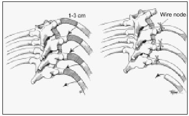
Figure 2. Schematic depiction of thoracoplasty.
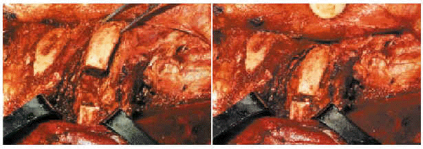
Figure 3. Thoracoplasty, surgical image.
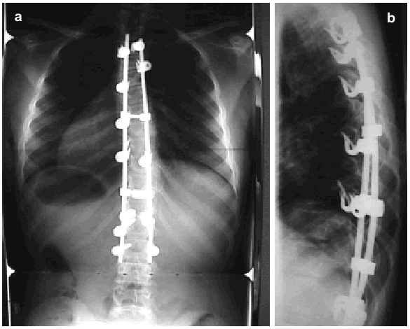
Figure 4. X-ray of a scoliotic thoracic spine after surgical treatment, in anteroposterior (a) and lateral (b) views.
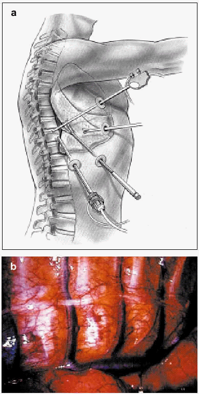
Figure 5. An artist's depiction (a) and a surgical image (b) of thoracoscopy.
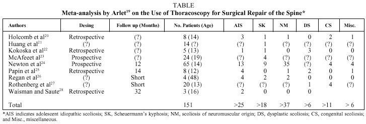
TABLE. Meta-analysis by Arlet19 on the Use of Thoracoscopy for Surgical Repair of the Spine*
Show moreShow less
Full Text

Introduction

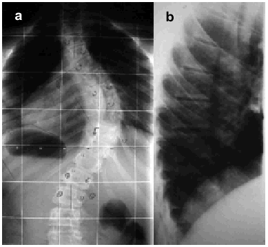
Scoliosis is defined as lateral curvature of the spine with vertebral rotation, meaning that it is a 3-dimensional deformity. Scoliosis leads to thoracic deformity if the spinal alterations occur in that zone (Figure 1).

Figure. 1. Thoracoabdominal x-ray showing posteroanterior (a) and lateral (b) views of a spine with scoliosis.

Scoliosis is classified as either idiopathic or secondary, regardless of what part of the spine is affected. Idiopathic scoliosis is currently thought almost certainly to arise from multiple factors. The most common causes are disordered development of the central nervous system or genetically determined familial disorders.1,2

The prevalence of idiopathic scoliosis in the population at risk (individuals between 10 and 16 years of age) is approximately 2% to 3%. As the magnitude of the curve increases, the incidence decreases, and it is estimated that the percentage of patients who require surgery does not exceed 0.1%.3

The decision of how to treat patients with spinal deformity should be based on an understanding of the natural history of the process. Any decisions taken should have as their objective the modification of that natural history. The main factors influencing therapeutic decisions are the type and magnitude of the curve, age, skeletal maturity, and sex. The most important factors at the time of diagnosis are age and curve magnitude.4,5 Age is especially important because it is related to elasticity3 and on elasticity will depend the possibility of reducing the angle of curvature and the patient's muscular response to it.

Respiratory Function and Deformity

Respiratory function is mainly affected in cases of thoracic scoliosis. The development of the pulmonary parenchyma ends when a child is 8 years old, and if the onset of deformity occurs at that time or earlier, the process may be accompanied by decreased vital capacity (VC) and total lung capacity (TLC).6

A direct relation has been found between lung function changes and magnitude of the thoracic curve. The type of alteration seen most often is restriction, which means less lung volume, greater work of breathing, higher energy expenditure, hypoxemia, respiratory acidosis due to hypoventilation, inadequate ventilatory response to hypoxic stimulus, pulmonary artery hypertension, underdevelopment of the pulmonary parenchyma, and an altered relationship between lung function and respiratory muscle mechanics.

Individuals with deformity of this type have reduced VC and, to a lesser degree, reduced forced expiratory volume in 1 second (FEV1) even if they are asymptomatic. A higher prevalence of preoperative and postoperative pulmonary problems in these patients has been mentioned in numerous articles.

The most marked alterations in lung function have been reported to occur when the Cobb angle of the curve exceeds 70º-80º. In such cases, diaphragm function changes and respiratory function comes to depend more on accessory muscles. Given that accessory muscle activity decreases at night, hypoxemia and, as a consequence, higher red cell counts may develop. Other factors to remember are the location and number of thoracic vertebrae involved.7 In the long run, only patients with curves exceeding 100º concomitant with other factors develop chronic respiratory insufficiency and cor pulmonale. According to some authors, when VC is less than 40% of the reference value, the risk of pulmonary function impairment is great.8 When the deformity is no greater than 50º but is associated with hypokyphosis (loss of the natural thoracic kyphosis), the prognosis worsens. Smoking also worsens the prognosis. Scoliosis in the middle range (40º-60º) in the absence of changes in the normal kyphosis is unlikely to cause severe respiratory problems. In cases of mild thoracic scoliosis (<35º), lung function can be improved by exercise. Nevertheless, the overall mortality of individuals with idiopathic thoracic scoliosis is comparable to that of the general population.

Little is known of the situation in adulthood of patients who had juvenile scoliosis, whether treated surgically or not.6 Such individuals usually experience certain limitations when engaging in intense physical activity, such as lifting or carrying heavy objects.

Treatment

Orthopedic approaches (braces) and surgery seek to reduce spinal curvature in order to improve appearance and prevent the likely deterioration of respiratory function. The outcome of treatment depends on the spine's elasticity. Therefore, the timing of treatment is essential. As will be discussed later, the course of respiratory function is directly related to patient age--whether the patient is an adolescent, considered "more elastic," or an adult, considered more "rigid."3

Orthopedic treatment with rigid braces has been shown to be effective in curvatures of up to 40º and in individuals with elastic deformities. However, recurrence has been observed in some patients. When this treatment is considered, 3 basic questions must be answered. What deformity is going to be treated? Why must it be treated? Can it be successfully corrected by conservative treatment?

Orthopedic treatment is used in adolescents, who usually have a remarkable ability to adapt. Although continuous application of such braces causes problems for thoracic expansion, long-term studies seem to indicate that respiratory function deterioration does not persist.9

Surgery is used to reduce 3-dimensional vertebral deformity by manipulating vertebrae to achieve a stable arthrodesis that prevents progression.4 Surgery is indicated when deformity is greater than 60º and symptomatic (pain in the adult, esthetic problems in the child or adolescent), or greater than 40º during periods of growth, given that curvature has been shown to progress in such cases in spite of orthopedic treatment. Finally, surgery is appropriate if there is a deformity that is unacceptable for the patient.4,5 No evidence supports surgical intervention solely to treat deterioration in lung function.

Thoracic deformities can be corrected using an anterior approach (by thoracotomy or thoracoscopy), a posterior approach, or a combination. All approaches enlist the spine's own elastic properties to reduce curvature. Thus, the approach to the spine will depend not only on technical considerations, but also on the type of curve, elasticity, and number of vertebrae that must be fused. When the effect on appearance is very evident owing to the presence of a gibbus (hump), thoracoplasty might also be performed for esthetic reasons, with partial resection of the ribs in the most prominent zone. This technique is usually seen in posterior orthopedic interventions. The aims of surgical alteration of the thoracic curve are to correct the defect (preserving the sagittal balance of the spine), to preserve or improve lung function, to exercise a positive influence on the lumbar spine, to achieve minimum morbidity and pain for the patient, and to maximize return to full function.

Nevertheless, the effect of surgical correction on the lung function of patients with idiopathic scoliosis is not entirely clear. Although several studies have demonstrated that such surgical correction improves some lung function variables,10 others have observed no such improvement11 or have even seen functional decline.12 Such discrepancies may arise from the fact that many studies have enrolled populations that are heterogenous as to age, sex, type and intensity of curvature, and etiology of the scoliosis. Moreover, the techniques used for surgical correction have differed, as have the criteria used to evaluate respiratory function.

The study performed by Rizzi et al,7 in 35 adult patients undergoing spinal surgery between 1978 and 1994 gave mixed outcomes. Seven patients died during the first year, while the rest experienced considerably improved respiratory function and quality of life (72-month follow-up). The authors concluded that the outcome of spinal surgery may range from "extremely good" (a high percentage of improvement) to "extremely unsatisfactory" (high mortality).

Surgical Technique

During the last decade, surgical approaches to treating thoracic deformities have changed markedly in terms of indications and pre-, intra-, and postoperative management of the patient.4 Currently, the objective is to halt the progression of deformity and maintain or even increase respiratory capacity in these patients.

As has been mentioned, surgery may involve either posterior or anterior approaches, the latter involving thoracotomy (usually with section of the diaphragm) or thoracoplasty. The two techniques may also be combined, in what is called a "double approach" which includes: a) the anterior thoracic approach to perform diskectomy and insert an implant in the space vacated by the intervertebral disk, and b) the posterior route, with spinal fixation and placement of an implant if necessary. Some specialists refer to this technique as "circumferential arthrodesis."

Thoracoplasty

Thoracoplasty consists in a partial resection of the ribs of the convexity of the curve (Figures 2 and 3). The main aim is to improve appearance. Thoracoplasty has also been described in the context of instrumentation performed through a posterior route. When this technique is used as a complement to vertebral fixation, a high number of peri- and postoperative complications (pneumothorax) occur, with altered respiratory mechanics during the first 2 months (decline in lung volume of 12% to 16%). Some authors attribute part of this decline to postoperative pain.13 However, 2 years after surgery, respiratory function has returned to its preoperative status.

Figure 2. Schematic depiction of thoracoplasty.

Posterior Approach

A posterior approach is chosen if the elasticity of the spine allows the deformity to be modified in this way. The main positive feature of this approach is that it does not alter the structure of muscles that act directly on ventilatory mechanics. The first instruments used were Harrington rods,6,8,9,14 which did not allow for a natural thoracic kyphosis. As a result, esthetic defects (a flattened spine) and, probably, functional impairment developed. Some authors report that this technique produces a slight gain in VC and static lung volumes, although they do not usually become normal. However, other authors have observed a slight decrease in VC, which they have attributed to the loss of kyphosis. Nevertheless, a very high percentage of such patients had also undergone prolonged immobilization with braces, and that treatment may also have influenced the outcome.

The implants used currently with the same approach have undergone marked changes (third generation implants). The changes introduced have had the aim of producing a repositioning of the thoracic cage that is as anatomically normal as possible by way of a distraction-rotation maneuver. With this technique, applied to elastic thoracic deformities, correction has taken place in the coronal and frontal planes, in the deformity of the rib cage, and in the asymmetry of the thoracic cage (normal kyphosis in the lateral plane, the normal axis in the frontal plane and derotations in the coronal plane) (Figure 4).

Figure 4. X-ray of a scoliotic thoracic spine after surgical treatment, in anteroposterior (a) and lateral (b) views.

Adolescent patients treated by this approach and with third generation implants have experienced improved VC and forced expiratory volume in 1 second. Some authors report that this improvement endures, whereas others have seen it disappear after 1 year.12

Anterior Approach: Thoracotomy

Thoracotomy is considered an alternative to the posterior approach.15 The objective is to obtain greater reduction of spinal curvature, to involve the least number of vertebrae in the fixation procedure and prevent pseudoarthrosis. Nevertheless, this approach leads to marked changes in elements directly involved in ventilatory mechanics, such as the large dorsal muscles, the diaphragm, and the intercostal muscles. Moreover, it is difficult to correct the loss of thoracic kyphosis. The main drawback to the anterior approach is that the lung on the side through which the spine is approached may collapse, probably leading to posterior expansion. The most frequent complications of this type of intervention are atelectasis and decreased lung volume. In general the patients experience impaired lung function for 3 postoperative months, although function has recovered by 2 years later.

A retrospective study by Grossfeld et al16 in 599 children operated on between 1967 and 1991 identified a set of risk factors in spinal interventions: use of thoracotomy, age over 14 years, male sex, presence of a kyphotic curve, a Cobb angle greater than 100º and a VC less than 40% of the reference value. The most important risk factors for "serious" postoperative complications were age and marked angulation. However, a study by Rawlins et al17 in 32 patients aged between 7 and 17 years found that the results of using thoracotomy to access the spine can be good, even when the patient's VC is less than 40% of the predicted value, provided interdisciplinary medical support is effective (supervision by pneumologists and internists).

Anterior Approach: Thoracoscopy

Minimal access spinal surgery has been moving forward in recent years and will therefore be covered in more detail in this review. Thoracoscopy (Figure 5) has been introduced as a new therapeutic approach for treating spinal pathology. The endoscope was first used in surgery on the spine in 1993, when it was considered an alternative to "open surgery." At first it was used mainly in patients considered to be at high risk from thoracotomy, given that the minimal procedure is less traumatic for the chest wall.18 Time has shown, however, that the deformity must be elastic and the angle greater than 75º if maximum benefit is to be derived from thoracoscopy. The technique should not be used in patients in whom a lung must be collapsed, who have severe respiratory insufficiency, who require high positive pressures to maintain adequate ventilation, or who have pleural adhesions. For the past 7 years, an anterior endoscopic approach has been used for spinal release surgery (with the purpose of increasing flexibility), for anterior arthrodesis, for epiphysiodesis (to prevent a flat back or the crankshaft problem), and for disk resections. It has also been used to treat certain primary tumors, spinal metastases, and fractured or herniated disks.

Figure 5. An artist's depiction (a) and a surgical image (b) of thoracoscopy.

Thoracoscopy is preferred from the patient's point of view because it is less invasive and causes less pain and morbidity. However, these advantages are less evident to the spinal surgeon. The minimal access approach requires a longer training period, more time in the operating room and often the help of a thoracic or abdominal surgeon. An argument in its favor, however, is the growing interest in shortening hospital stays and decreasing costs.

Arlet19 published a meta-analysis of 151 studies that attempted to answer prevailing questions about the indications for thoracoscopy in spinal deformities. Only 57 of the articles found, however, were relatively acceptable. Twenty-one were about spinal fusion, 11 on spinal release, and the rest on complications of thoracoscopy. Of the 57, only 9 were considered optimal sources of evidence (Table).20-28 Most cases involved children, in whom the spine is much more flexible than it is in the adult. It is interesting to note, moreover, that the mean angle of curvature was 65º for the cases published, although the anterior approach is widely held to be indicated only for patients with curvatures greater than 75º and with radiologically evident lateral bending of less than 50º. However, thoracoscopy might be justified for preventing the development of the flat-back syndrome, performing epiphysiodesis with the aim of eliminating growth and/or performing disk removal to afford flexibility to the spine. From a technical standpoint, all the reports reviewed approached the spine from the convex side of the curvature, with the patient in lateral decubitus position, with selective collapse of the ipsilateral lung (as is the case in open thoracotomy). The learning curve rose very gradually for this technique, given that training is needed for the approach phase and for the performance of disk removal itself. An additional problem is the difficulty of evaluating the amount of intervertebral disk removed. The duration of interventions (mean 2.5 hours) was unrelated to type of deformity. When an operation included a period in which a posterior approach was taken, the duration of surgery was much longer (10 hours). Successful repair of the deformity was achieved in 56% to 63% of cases. The cost of thoracoscopy was 30% higher than that of open thoracotomy,14 when the learning curve, material, equipment depreciation, hours in the operating room, and the need for immediate availability for conversion to open thoracotomy were all taken into account. The mean duration of hospitalization was 9 days. Complications developed in 20% of the patients (6% being serious), although they did not affect the final outcomes. The duration of assisted ventilation was seen to be high, more so in patients with neuromuscular problems.

Newton and Wenger,29 following a study comparing thoracotomy and thoracoscopy in children and adolescents with spinal deformity, reported that the benefits of one procedure over the other were not significant, and they even found that patients who underwent thoracoscopy needed longer pleural drainage.

From these reports we can conclude that endoscopic surgery on the thoracic spine has important limitations related to the infrastructure required and the ratio between training and yield. For that reason, the indications for thoracoscopy and the hospitals where it is performed should be strictly rationalized.

Indications for Thoracoscopy in Scoliotic Deformity. Endoscopy can be effective for pediatric patients, but the results of repair are similar to those achieved by open procedures. When the deformity is severe or flexibility of the spine is reduced (as in adults), outcome is less satisfactory.

Indications for Thoracoscopy in Kyphotic Deformity. The consensus is that only kyphoses exceeding 75º should be reduced. The fixation systems currently available allow us to reduce the deformity by facetectomy (posterior resection of articulations), making an anterior approach unnecessary. This surgical procedure is easier in kyphotic patients because they have a "larger" thoracic cavity.

Conclusion

The magnitude of the thoracic curve determines how respiratory function is affected, creating the possibility that a restrictive ventilatory pattern might develop. The presence of a loss of normal kyphosis also influences lung function negatively. Thus, in scoliotic deformities of equal magnitude, those in whom kyphosis is reduced are more likely to develop altered respiratory function. Smoking also aggravates functional impairment (a nonsmoker only develops functional alterations when deformity is between 100º and 120º).

Scoliotic deformity can be resolved using an anterior (endoscopy or thoracotomy), posterior, or combined approach. Moreover, thoracoplasty can be considered as a complementary procedure for improving the appearance of the patient. The application of each technique will depend on the indications explained in this review.

Although thoracoplasty initially raised hopes in surgery to repair thoracic spinal deformity, important limitations on its application exist (specific indications, a prolonged learning curve, and moderately positive outcomes).


Correspondence: Dr. A. Molina Ros.

Servicio de COT. Hospital del Mar.

Pg. Marítim, 27. 08003 Barcelona. España.

E-mail: 20017@imas.imim.es

Manuscript received April 30, 2003.

Accepted for publication May 13, 2003.

Bibliography
[1]
Machida M..
Cause of idiopathic scoliosis..
Spine, 24 (1999), pp. 2576-83
[2]
Winter R..
Classification and terminology. In: Lippincot-Ravent, editor. Textbook of scoliosis and other spinal deformities. Philadelphia: W.B. Saunders Co..
Classification and terminology. In: Lippincot-Ravent, editor. Textbook of scoliosis and other spinal deformities. Philadelphia: W.B. Saunders Co., (1995), pp. 39-43
[3]
Upadhyay SS, Mullaji AB, Luk KD, Leong JC..
Relation of spinal and thoracic cage deformities and their flexibilities with altered pulmonary functions in adolescent idiopathic scoliosis..
Spine, 20 (1995), pp. 2415-20
[4]
Bunell W..
The natural history of idiopathic scoliosis..
Clin Orthop, 229 (1988), pp. 20-5
[5]
Duval-Beaupere G, Lamireau T..
Scoliosis at less than 30 degrees. Properties of the evolutivity (risk of progression)..
Spine, 10 (1985), pp. 421-4
[6]
Pehrsson K, Danielsson A, Nachemson A..
Pulmonary function in adolescent idiopathic scoliosis: a 25 year follow up after surgery or start of brace treatment..
Thorax, 56 (2001), pp. 388-93
[7]
Rizzi PE, Winter RB, Lonstein JE, Denis F, Perra JH..
Adult spinal deformity and respiratory failure. Surgical results in 35 patients..
Spine, 22 (1997), pp. 2517-30
[8]
Vedantam R, Lenke LG, Bridwell KH, Linville DL, Blanke K..
A prospective evaluation of pulmonary function in patients with adolescent idiopathic scoliosis relative to the surgical approach for spinal arthrodesis..
Spine, 25 (2000), pp. 82-90
[9]
Pehrsson K, Bake B, Larssson S, Nachmenson A..
Lung function in adults with idiopathic scoliosis: a 20 year follow-up..
Thorax, 46 (1991), pp. 474-8
[10]
Borowitz D, Armstrong D, Cerny F..
Relief of central airways obstruction following spinal release in a patient with idiopathic scoliosis..
Pediatric Pulmonology, 31 (2001), pp. 86-8
[11]
Estenne M, Derom E, De Troyer A..
Neck and abdominal muscle activity in patients with severe thoracic scoliosis..
Am J Resp Crit Care Med, 158 (1998), pp. 452-7
[12]
Vedantam R, Lente LG, Bridwell KH, Haas J..
A proposed evaluation of pulmonary function in patients with adolescent idiopathic scoliosis relative to the surgical approach used for spinal arthrodesis..
Spine, 25 (2000), pp. 82-90
[13]
Lenke LG, Bridwel KH, Blanke K, Baldus C..
Analysis of pulmonary function and chest cage dimension changes after thoracoplasty in idiopathic scoliosis..
Spine, 20 (1995), pp. 1343-50
[14]
Ordiales JJ, Fernández JC, López L, Colubi J..
Estudio de la función ventilatoria pre y pos cirugía de la escoliosis torácica por el método de Harrington..
Rev Clin Esp, 198 (1998), pp. 356-9
[15]
Freixinet J, Hussein M, Mhaidli H, Rodríguez Suárez P, Robaina F, Rodríguez de Castro F..
Abordaje transtorácico en raquis..
Arch Bronconeumol, 34 (1998), pp. 492-5
[16]
Grossfeld S, Winter RB, Lonstein JE, Denis F, Leonard A, Johnson L..
Complications of anterior spinal surgery in children..
J Pediatr Orthop, 17 (1997), pp. 89-95
[17]
Rawlins BA, Winter RB, Lonstein JE, Denis F, Kubic PT, Wheeler WB, et al..
Reconstructive spine surgery in pediatric patients with major loss in vital capacity..
J Pediatr Orthop, 16 (1996), pp. 284-92
[18]
Burgos J, Rapariz JM, González-Herranz P..
Anterior endoscopic approach to the thoracolumbar spine..
Spine, 23 (1998), pp. 2427-31
[19]
Arlet V..
Anterior thoracoscopic spine release in deformity surgery: a meta-analysis and review..
Eur Spine J, 9 (2000), pp. 17-23
[20]
Holcomb GW II.I, Mencio GA, Green NE..
Video-assisted thoracoscopic diskectomy and fusion..
J Pediatr Surg, 32 (1997), pp. 1120-2
[21]
Huang TJ, Hsu RW, Sum CW, Liu HP..
Complications in thoracoscopic spinal surgery: a study of 90 consecutive patients..
Surg Endosc, 13 (1999), pp. 346-50
[22]
Kokoska ER, Gabriel KR, Silen ML..
Minimally invasive anterior spinal exposure and release in children with scoliosis..
J Bone Joint Sung, 2 (1998), pp. 255-8
[23]
McAfee PC, Regan JR, Zdeblick T, Zuckerman J, Picetti GD II.I, Heim S, et al..
The incidence of complications in endoscopic anterior thoracolumbar spinal reconstructive surgery. A prospective multicenter study comprising the first 100 consecutive cases..
Spine, 20 (1995), pp. 1624-32
[24]
Newton PO, Wenger DR, Mubarak SJ, Meyer RS..
Anterior release and fusion in pediatric spinal deformity. A comparison of early outcome and cost of thoracoscopic and open thoracotomy approaches..
Spine, 22 (1997), pp. 1398-406
[25]
Papin P, Arlet V, Marchesi D, Laberge JM, Aebi M..
Treatment of scoliosis in the adolescent by anterior release and vertebral arthrodesis under thoracoscopy..
Rev Chir Orthop Reparatrice Appar Mot, 84 (1998), pp. 231-8
[26]
Regan JJ, Mack MJ, Picetti GD III..
A technical report on video-assisted thoracoscopy in thoracic spinal surgery. Preliminary description..
Spine, 20 (1995), pp. 831-7
[27]
Rothenberg S, Erickson M, Eilert R, Fitzpatrick J, Chang F, Glancy G, et al..
Thoracoscopic anterior spinal procedures in children..
J Pediatr Surg, 33 (1998), pp. 1168-70
[28]
Waisman M, Saute M..
Thoracoscopic spine release before posterior instrumentation in scoliosis..
Clin Orthop, 336 (1997), pp. 130-6
[29]
Newton PO, Wenger DR..
Anterior release and fusion in pediatric spinal deformity. A comparison of early outcome and cost of thoracoscopic and open thoracotomy approaches..
Spine, 22 (1997), pp. 1398-406
Archivos de Bronconeumología
Article options
Tools

Are you a health professional able to prescribe or dispense drugs?