
We report the case of a 56-year-old man, a smoker who consulted due to respiratory infection with nodularity of the pleural surface in the right hemithorax detected on chest radiograph. The patient had been treated with talc pleurodesis (TP) for recurrent pneumothorax 12 years previously. A chest CT confirmed the existence of a partially calcified nodular thickening of the pleural surface in the right hemithorax, as well as a solid indeterminate pulmonary nodule measuring 1cm in the left lung. On PET-CT, the left pulmonary nodule showed no FDG uptake, but the right pleural lesions showed high metabolism (SUVmax 14) (Fig. 1). A repeat chest CT performed 3 months later showed no changes in the pleural lesions. In view of these findings, a diagnosis was given of benign pleural granulomatous inflammation after TP.

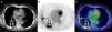
(A) Axial image of the CT component of the PET-CT scan showing partially calcified pseudonodular thickening of the pleural surface in the right hemithorax, particularly the posterior aspect (arrows). (B) Axial image of the PET component of the PET-CT scan (same level as (A) showing hypermetabolic foci in the pleural surface in the right hemithorax (arrows). (C) Axial PET-CT fusion image (same level as A and B) showing more clearly how the hypermetabolic foci correspond to the pleural thickening in the right hemithorax (arrows).
TP is an effective treatment of persistent pleural effusion or recurrent pneumothorax in which the inflammatory pleural response (visceral and parietal) induced by the talc causes fibrosis with secondary pleurodesis. TP is a cause of false positives in PET-CT, since FDG is captured by the pleural granulomas, and hypermetabolism of these lesions can persist for years after pleurodesis is performed.1 This case serves as a reminder that benign pleural uptake may be observed on PET-CT, even if pleurodesis was performed several years earlier.
Please cite this article as: Gorospe Sarasúa L, Jover-Díaz R, Arrieta P. Captación pleural intensa de FDG 12 años después de una pleurodesis con talco: hallazgos en PET/TC. Arch Bronconeumol. 2017;53:638.


