


A 58-year-old man with history of coronary artery bypass operation was admitted to our hospital with atypical chest pain. For atypical angina, coronary computed tomography (CT) angiography was performed. CT angiography showed aorto coronary artery bypass graft and atherosclerotic plaques of the coronary arteries. CT scans also revealed a calcified hydatid cyst in the interventricular septum (Fig. 1A) and a hyperdense lesion in the left ventricular base (Fig. 1B and C). The ventricular base lesion had the same density as the intraventricular contrast material. For differential diagnosis of ventricular aneurysm and calcified hydatid disease, non-contrast CT was performed. CT scan revealed a calcified hydatid cyst in the left ventricular base (Fig. 1D). The cyst was mimicking contrast filling of a ventricular aneurysm in coronary CT angiography.
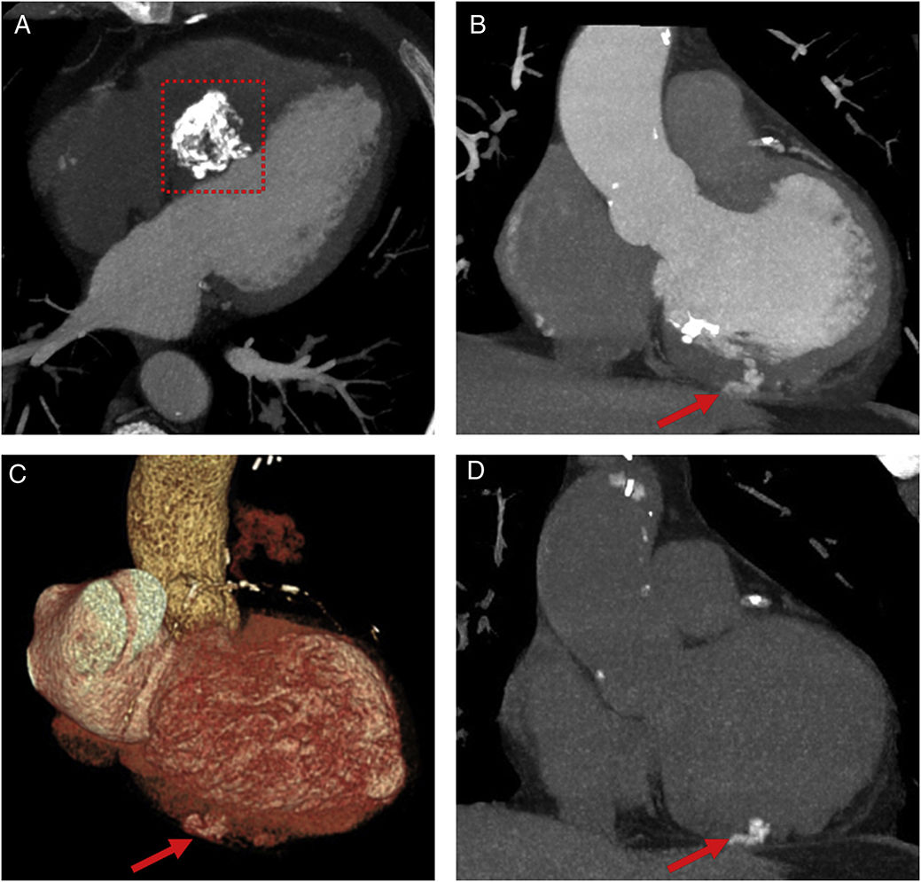
Contrast enhanced axial and coronal CT scans (A and B) and coronal 3D reformat CT angiography (C) show a calcified hydatid cyst (frame) in the interventricular septum and hyperdense lesion (arrows) in the left ventricular base. Non-contrast coronal CT scan (D) reveals a calcified hydatid cyst (arrow) in the left ventricular base.
Hydatic disease is a zoonotic disease that is caused by Echinococcus granulosus. Liver and lungs are affected most common by the disease. Heart is involved rarely by the hydatid disease (0.02–2% of all cases). Left ventricle is the most affected chamber with a 50–60% ratio of all hydatid disease followed by interventricular septum (10–20%), right ventricle (5–15%), pericardium (10–15%) and atriums (5–8%).1 Although serological test for hydatic disease is a very important diagnostic method, false negative findings may lead to a missed diagnosis. Calcified hydatid cyst of the heart can be rarely mistaken for ventricular aneurysm.2 Cardiac calcified hydatid cyst should be considered in the differential diagnosis of hyperdense masses on CT scans in endemic areas. Pre- and post-contrast CT scans can give valuable information about cardiac masses or their mimickers.


