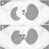
A 45-year-old man, current smoker came to the emergency department after a self-limited episode of mild hemoptysis (less than 30ml). No other symptoms were associated. Chest tomography showed right paratracheal opacity with bilateral adenopathies. As neoplasm and infection was suspected, bronchoscopy was later performed with negative cytology. All microbiological studies were also negative. A prolonged empirical antibiotic treatment was then maintained with satisfactory radiological progression (Fig. 1). The final diagnosis was infected azygos lobe bulla.
The azygos lobe is usually identified as a paratracheal convex line containing normal parenchyma, although 10–15% of cases can be identified as opaque images, mainly because superposition of structures1,2. Nevertheless, there is no reported incidence of azygos lobe opacity due to total occupation of its parenchyma. Moreover, we did not find previous cases of infected and occupied bullas in this anatomical variant causing atelectasis of the azygos lobe as described in this case.
Physicians should be aware of the anatomical and clinical implications of the azygos lobe to insure the differential approaches of paratracheal opacities in imaging studies.


