
We report the case of a young adult who was diagnosed with cystic lung disease at the age of 16, although no specific diagnosis was made until resection of a single lung 14 years later. Thick-walled cystic lesions and associated bilateral nodular opacities were observed and the patient was admitted for pneumothorax up to 6 times. The only relevant extrapulmonary findings were several benign subcutaneous tumors on the back and armpit, that were resected. Histological evaluation of one of the lung cysts showed histologically benign fibrohistiocytic proliferations associated with the formation of cysts lined by metaplastic bronchiolar, squamous or type II alveolar epithelium, and intracystic bleeding. The histology of the lung lesions was similar to that of the last resected subcutaneous nodular lesion. These unusual multiple bilateral fibrohistiocytic tumors of the lung should be included in the differential diagnosis of bilateral lung cystic lesions in young adults.
Our patient was a 30-year-old man with no toxic habits, who was an educator in a juvenile center. He reported no significant family history or environmental exposure. Pathological background of interest included an incidental finding of spina bifida occulta and a renal cyst in the right lower pole measuring 17mm on abdominal ultrasound, with no other changes. A left periaxillary mass was resected and found to be consistent with angiomatoid fibrous histiocytoma (Fig. 1A).

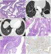
(A) Skin lesion: fibrohistiocytic strain showing vascular structures of varying calibers and pseudocystic cavities with no epithelial or endothelial lining. No pleomorphism or necrosis observed. Suggestive findings of fibrous histiocytoma. (B and C) High-resolution computed tomography of the chest: multiple diffuse and bilateral cystic lesions. Cysts predominantly in upper lobes, random distributed (subpleural and peribronchovascular) with thin and well-defined walls, polylobulated and of irregular size and shape. Bulky lesion containing air–fluid component. (D) Pulmonary cyst biopsy: cyst wall showing congestive thick vessels accompanied by fusocellular proliferation with no atypia, mitosis or necrosis. (E) High magnification image (400×) showing previously described cellular characteristics consistent with fibrous histiocytoma. (F) Immunohistochemical staining corresponding to a fibroblastic–fibrohistiocytic strain, expressing CD10, factor XIII, and CD68 with a Ki67 index <5%.
At the age of 16, the patient was diagnosed by the respiratory medicine department with bilateral cystic disease following a chest CT performed for a fractured rib (Fig. 1B and C). Multiple bilateral pneumothoraces required several chest drains and surgical interventions (bilateral apical plication and bilateral talcage). Cystic fibrosis screening was performed and no CFTR gene mutation was identified. A genetic study for Birt–Hogg–Dubé syndrome (FLCN gene) was negative. Fiberoptic bronchoscopy was performed twice showing no changes, and microbiological cultures and cytology studies were negative.
Massive hemoptysis at the age of 25 years required angiography and bronchial embolization. Embolization was performed in the right bronchial artery emerging from the intercostobronchial trunk with associated distal pulmonary fistula, and the common bronchial trunk, both of which were pathological in appearance. The patient was referred by the thoracic surgery department to the pulmonary interstitial functional unit for evaluation.
During the evaluation period, the patient was admitted to the pulmonology department for hemoptysis associated with Pseudomonas aeruginosa and Hafnia alvei respiratory superinfection. Antibiotic treatment was started according to sensitivity testing results. He also developed new episodes of pneumothorax requiring chest drains. After these events, we finally decided to use video-assisted thoracoscopy and talcage to resect a cyst in the left upper lobe that was adhered to the mediastinum and pericardium, causing compression of these structures. The pathology study found that the lung tissue (Fig. 1F) had the same characteristics as the previously extracted subcutaneous lesion. Histological evaluation is conclusive in pulmonary and cutaneous angiomatoid fibrous histiocytomas.
Angiomatoid fibrous histiocytoma of the lung is extremely rare,1–3 especially when presentation is bilateral and involves concomitant extrapulmonary tumors of the same cell strain. Its origin is unknown and treatment includes tumor resection. Some groups have attempted chemotherapy (with subsequent infectious complications), but no effective medical treatment is yet available. Cyst morphology on chest high-resolution computed tomography and the identification of concomitant subcutaneous tumors should prompt a diagnostic suspicion. Possible complications that may be associated with lung cysts should be taken into account, for example, infections, pneumothorax, hemoptysis, or mediastinal compression.
Conflict of interestsThe authors state that they have no conflict of interests.
The following are the supplementary data to this article:


