Journal Information
Share
Share
Download PDF
More article options
Visits
349
Original Article
Full text access
Available online 17 February 2025
Persistent Blood Eosinophilia and Eosinopenia: Relationship with Outcomes in Bronchiectasis
Visits
349
Grace Osculloa,n, Amina Bekkia,n, Casilda Olveirac, Rosa Girónd, Marta García-Clementee, Luis Máizf, Oriol Sibilag, Rafael Golpeh, Juan Luis Rodríguez-Hermosai, Esther Barreirob,j, Raúl Méndeza,b, Concepción Pradosk, Juan Rodríguez-Lópezl, David de la Rosam, Miguel Ángel Martínez-Garcíaa,b,n,
Corresponding author
mianmartinezgarcia@gmail.com

Corresponding author.
a Servicio de Neumología, Hospital Universitario y Politécnico La Fe, Valencia, Spain
b CIBERES de Enfermedades Respiratorias, ISCIII, Madrid, Spain
c Servicio de Neumología, Hospital Regional Universitario de Málaga, Instituto de Investigación Biomédica de Málaga (IBIMA)/Universidad de Málaga, Málaga, Spain
d Servicio de Neumología, Instituto de Investigación Sanitaria, Hospital Universitario de la Princesa, Madrid, Spain
e Servicio de Neumología, Hospital Universitario Central de Asturias, Oviedo, Asturias, Spain
f Servicio de Neumología, Hospital Ramón y Cajal, Madrid, Spain
g Servicio de Neumología, Hospital Clínico, Barcelona, Spain
h Servicio de Neumología, Hospital Lucus Augusti, Lugo, Spain
i Servicio de Neumología, Hospital San Carlos, Madrid, Spain
j Servicio de Neumología, Hospital del Mar-IMIM, UPF, CIBERES, Barcelona, Spain
k Servicio de Neumología, Hospital La Paz, Madrid, Spain
l Servicio de Neumología, Hospital San Agustín, Avilés, Asturias, Spain
m Servicio de Neumología, Hospital Santa Creu i Sant Pau, Barcelona, Spain
n Instituto de Investigaciones Salud La Fe de Valencia, Spain
Ver más
This item has received
Received 19 November 2024. Accepted 28 January 2025
Article information
Abstract
Full Text
Bibliography
Download PDF
Statistics
Figures (4)
Show moreShow less
Tables (4)
Table 1. Absolute Numbers (Eosinophils/μL) in Peripheral Blood Samples in Three Consecutive Measures in the Persistent and Non-persistent Eosinophilic/Eosinopenic Groups.
Table 2. Comparison Between the Groups With and Without Persistent Eosinophilia.
Table 3. Comparison Between the Groups With and Without Persistent Eosinopenia.
Table 4. Comparison Between the Groups With Persistent Eosinophilia and Persistent Eosinopenia.
Show moreShow less
Abstract
Introduction

Blood eosinophil counts (BEC) have been related to the severity of bronchiectasis and its response to inhaled corticosteroids. However, only the baseline BEC has been used to assess this relationship and it is known that BEC could change over time. The objective of this study is to analyse the association of persistent eosinophilia or eosinopenia with outcomes in bronchiectasis.

Methods

Multicentre, prospective and observational study from 43 centres in Spain derived from the Spanish Bronchiectasis Registry (RIBRON). Asthma and anti-eosinophil treatments were excluded. Patients with at least two yearly BEC measures (including the baseline measure) were included. Persistent eosinophilia (at least 300cells/μL) or persistent eosinopenia (less than 100cells/μL) were defined as the persistence in the same eosinophil group after three yearly measures (being the baseline the first measure).
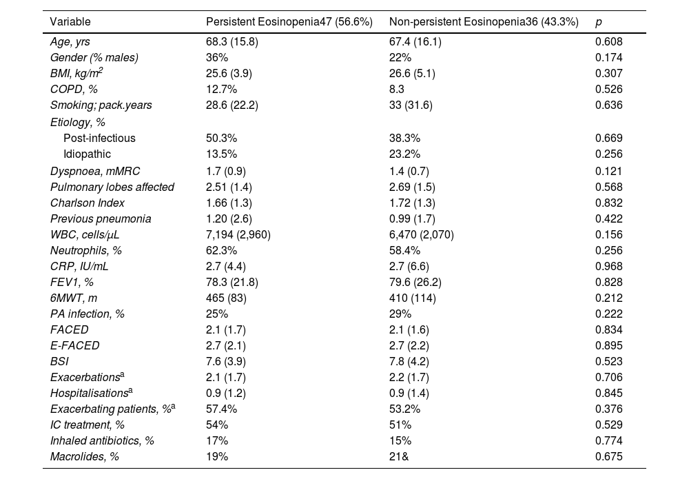
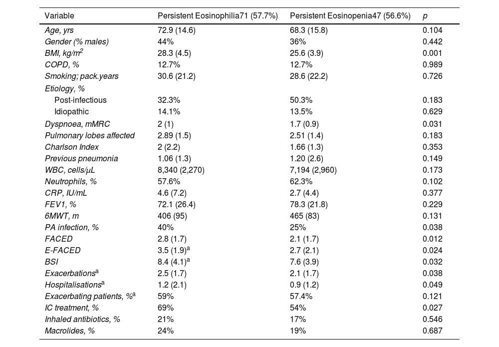
Results

Five hundred two patients with at least three BEC measures were included; 24.5% and 16.6% presented baseline eosinophilia or eosinopenia, respectively. Of these, 57.7% and 56.6% presented persistent eosinophilia and eosinopenia, respectively. Patients with persistent eosinophilia presented greater severity and a higher number/greater severity of exacerbations than those with non-persistent eosinophilia and those with persistent or non-persistent eosinopenia. Finally, patients with non-persistent eosinopenia presented more severity and a higher number/greater severity of exacerbations than those with non-persistent eosinophilia.

Conclusion

When only the baseline BEC was taken into account, patients with eosinopenia presented greater severity than those with eosinophilia. However, patients with persistent eosinophilia presented greater severity than those with persistent eosinopenia. Monitoring the BEC seems to be important in bronchiectasis.

Keywords:
Eosinophils
Bronchiectasis
Severity
Exacerbations
Graphical abstract
Full Text
Introduction

Bronchiectasis is a heterogeneous disease in its clinical presentation, aetiology and prognosis.1–4 The pathophysiological substrate necessary for its formation is the existence of bronchial inflammation, sometimes complemented by a bronchial infection by pathogenic microorganisms that amplifies the inflammation and causes disease progression,2,5 and, in turn, clinical deterioration and exacerbations.3,4 Although in most cases the predominant inflammation in patients with bronchiectasis is neutrophilic,2,6–9 the eosinophil count may also be elevated in respiratory samples from individuals with bronchiectasis.10–14

Given that counting the relative number of eosinophils from sputum samples or bronchial biopsies can be expensive, time-consuming or invasive, in recent years blood eosinophil counts (BEC) have been used to assess the severity, prognosis or response to treatment of bronchiectasis,10–14 as in the cases of other diseases such as COPD,15–17 although the correlation between the BEC and bronchial eosinophil numbers seems to be relatively modest.16,17 Thus, a low (less than 50–100cells/μL) or high (>300cells/μL) BEC have been related to more severe forms of bronchiectasis, a greater number of exacerbations and even, in patients with peripheral eosinophilia, a better response to inhaled corticosteroid (IC) treatment (even after asthma and other eosinophilic diseases have been ruled out).10–12 Nevertheless, only baseline BEC is taken into account to assess its correlation with outcomes or when making therapeutic decisions. It is known that BEC can change over time due to a number of intrinsic and extrinsic factors16 so the situation should be reconsidered after a given time with a new determination of BEC.

In some patients, however, eosinophilia or eosinopenia may persist over time,17 but it is not known whether this persistence is associated with any specific clinical phenotype of patients or with outcomes of interest in bronchiectasis. Therefore, our objective with the present study was to assess the characteristics of patients with bronchiectasis and persistent eosinophilia or eosinopenia, defined as their presence in at least three measures, with one separation of at least one year, taken consecutively from the baseline value as well as to analyse the association of persistent eosinophilia or eosinopenia with important outcomes in bronchiectasis.

MethodsStudy Design

This was a multicentre, prospective and observational study from 43 centres in Spain derived from the Spanish Bronchiectasis Registry (RIBRON).18,19 Patients were recruited from February 2015 to December 2019. All the patients signed their informed written consent to participate in the registry. Ethical approval was obtained from the Ethics Committee at the Hospital Josep Trueta in Girona (reference number: 001-2012), in the coordinating centre and in the local participating centres.

Patients

Inclusion criteria were adult patients (at least 18 years old) diagnosed with bronchiectasis by means of high-resolution computerised tomography in conditions of clinical stability (defined as at least 4 weeks free of an exacerbation period) with three BEC measures available on inclusion in the registry (one baseline and two more yearly during at least 2 years). Exclusion criteria included asthma, allergic bronchopulmonary aspergillosis (ABPA) and treatment with systemic corticosteroid or anti-eosinophil biological treatments. Asthma was excluded following the recommendation of international guidelines, mainly based on lack of typical symptoms and negative complementary tests in case of reasonable doubt (negative reversibility test, IgE levels or another complementary test to rule out ABPA).20 All blood extractions for the measurement of BEC were performed in a stable state (at least four weeks away from a period of exacerbation).

Variables and Definitions

The following variables were used for the purposes of this study: baseline general and anthropometric data, aetiology, severity scores (FACED,21 E-FACED,22 and Bronchiectasis Severity Index [BSI]23), lung function, clinical, analytical and microbiological data and the number and severity of exacerbations prospectively recorded in the year after inclusion in the registry, and their treatments.

An exacerbation was defined (when the registry was created) as a worsening of the typical symptoms of bronchiectasis: cough, dyspnoea, haemoptisis, increase in the volume or purulence of the sputum, chest pain and sibilance with an evolution of more than 24h requiring antibiotic treatment. An exacerbation was considered mild-moderate when the patient needed oral antibiotics, and severe in cases of hospital admission or when intravenous antibiotic treatment was required.24 Exacerbating patients were defined as those with at least three exacerbations per year or two mild-to-moderate exacerbations plus at least one hospitalization.25,26 Exacerbations occurring in the first year of follow-up were considered for analysis.

For the purposes of this study, we defined eosinophilic bronchiectasis as a BEC of at least 300eosinophils/μL and eosinopenic bronchiectasis as a BEC of less than 100eosinophils/μL. There is no established definition of persistent eosinophilia or eosinopenia, but in the present study we used the one presented by Casanova et al for patients with COPD, whereby persistent eosinophilia is defined as the presence of at least 300eosinophils/μL in three consecutive BEC measures, the first being the baseline, with a separation of at least one year between two of the measures.17 Similarly, persistent eosinopenia was defined as the presence of less than 100eosinophils/μL in three consecutive BEC measures, the first being the baseline, with a separation of at least one year between two of the measures.

Statistical Analysis

The data were tabulated using the mean (standard deviation [SD]) or median (interquartile range) for quantitative data, depending on the distribution of the variables. The normality of the distribution was analysed using the Kolmogorov–Smirnov test and the variance homogeneity by the Levene's test.

The qualitative data were tabulated according to the percentage with respect to the total value. Four comparisons were made: 1. Within the patients with baseline eosinophilia, the group of patients with persistent eosinophilia versus those without it; 2. Within the patients with baseline eosinopenia, the group with persistent eosinopenia versus those without it; 3. The group with persistent eosinophilia versus the group with persistent eosinopenia. In all cases, a T-student, chi-square or U-Mann Whitney test was used, depending on the distribution of the variables; 4. Multiple relationship between the four groups (persistent eosinophilia, non-persistent eosinophilia, persistent eosinopenia and non-persistent eosinopenia) was made using one-way ANOVA test with Bonferroni's correction. A p value of less than 0.05 were considered as significant. The statistical packages SPSS Inc. 20 and R software were used.

Results

Of the 2642 patients with non-cystic fibrosis bronchiectasis and an age of at least 18 years, 582 individuals had complete baseline data and at least two additional valid yearly measures of BEC. Of these, 69 patients with asthma (6 with ABPA and 6 taking anti-eosinophil biological treatment) were excluded, along with 11 patients taking systemic corticosteroids. Therefore, 502 subjects were finally included in the analysis (Fig. 1).

Fig. 1.

Flow-chart of the study. BEC: blood eosinophils counts; ABPA: allergic bronchopulmonary aspergillosis.

(0.26MB).

Of these 502 included patients, 123 (24.5%) presented baseline eosinophilia (414.2 [117.5]cells/μL) and 83 (16.6%) eosinopenia (49.3 [22.1]cells/μL). Of those patients with baseline eosinophilia, 71 (57.7%) presented persistent eosinophilia, whereas, of those patients with baseline eosinopenia, 47 (56.6%) presented persistent eosinopenia. Table 1 shows the BEC values according to the presence of eosinophilia or eosinopenia and their persistence.

Table 1.

Absolute Numbers (Eosinophils/μL) in Peripheral Blood Samples in Three Consecutive Measures in the Persistent and Non-persistent Eosinophilic/Eosinopenic Groups.

  Baseline Measure  2nd Measure  3rd Measure 
Persistent eosinophilia  418.5 (125)  439 (128)  463.8 (136) 
No persistent eosinophilia  401.1 (97)  236.9 (126)  210.1 (86) 
Persistent eosinopenia  48.8 (21)  42.8 (18.3)  44.6 (17.5) 
No persistent eosinopenia  51.2 (22.3)  138.9 (78)  143.2 (88) 

Patients with persistent eosinophilia presented more dyspnoea, comorbidities, bronchial infection by Pseudomonas aeruginosa (PA), a greater number and severity of exacerbations, higher use of ICs and greater global disease severity compared with those without persistent eosinophilia (Table 2). However, there were no differences between patients with persistent and non-persistent eosinopenia (Table 3). Moreover, those patients with persistent eosinophilia also presented greater disease severity, more PA bronchial infections and a greater number/severity of exacerbations than those with persistent eosinopenia (Table 4). However, as can be seen in Fig. 2A–C, although the highest mean value in all three multidimensional severity scores (FACED, E-FACED and BSI) was seen in the persistent eosinophilic group, both the eosinopenic groups (persistent and non-persistent) had greater severity values than the group with non-persistent eosinophilia, with very similar results regarding exacerbation (Fig. 3A) and hospitalisations (Fig. 3B).

Table 2.

Comparison Between the Groups With and Without Persistent Eosinophilia.

Variable  Persistent Eosinophilia71 (57.7%)  Non-persistent Eosinophilia52 (42.3%)  p 
Age, yrs  72.9 (14.6)  72.3 (13.7)  0.798 
Gender (% males)  44%  37%  0.431 
BMI, kg/m2  28.3 (4.5)  26.8 (4.1)  0.061 
COPD, %  12.7%  7.7%  0.379 
Smoking; pack.years  30.6 (21.2)  23.3 (23.1)  0.254 
Aetiology, %
Post-infectious  32.3%  44.2%  0.183 
Idiopathic  14.1%  17.3%  0.629 
Dyspnoea, mMRC  1.95 (1)  1.53 (0.9)  0.031 
Pulmonary lobes affected  2.89 (1.5)  2.98 (1.4)  0.731 
Charlson Index  2 (2.2)  1.6 (1.3)  0.023 
Previous pneumonia  1.06 (1.3)  0.76 (1.9)  0.073 
WBC, cells/μL  8,340 (2,270)  7,433 (2,460)  0.061 
Neutrophils, %  57.6%  55.9%  0.493 
CRP, IU/mL  4.6 (7.2)  2.9 (6.9)  0.042 
FEV1, %  72.1 (26.4)  70.2 (26.1)  0.699 
6MWT, m  406 (95)  425 (144)  0.734 
PA infection, %  40%  21%  0.038 
FACED  2.8 (1.7)  1.7 (1.6)  0.001 
E-FACED  3.5 (1.9)a  2.3 (1.9)  0.004 
BSI  8.4 (4.1)a  7.2 (4.3)  0.003 
Exacerbationsa  2.5 (1.7)  1.3 (1.8)  0.014 
Hospitalisationsa  1.2 (2.5)  0.7 (1.1)  0.025 
Exacerbating patients, %a  59%  42%  0.032 
IC treatment, %  69%  52%  0.027 
Inhaled antibiotics, %  21%  19%  0.546 
Macrolides, %  24%  21%  0.687 

BMI: body mass index; COPD: chronic obstructive pulmonary disease; WBC: white blood count; CRP: C-reactive protein; PA: Pseudomonas aeruginosa; IC: inhaled corticosteroids; 6MWT: 6minute walk test; mMRC: Modified Medical Research Council.

a

First year from inclusion.

Table 3.

Comparison Between the Groups With and Without Persistent Eosinopenia.

Variable  Persistent Eosinopenia47 (56.6%)  Non-persistent Eosinopenia36 (43.3%)  p 
Age, yrs  68.3 (15.8)  67.4 (16.1)  0.608 
Gender (% males)  36%  22%  0.174 
BMI, kg/m2  25.6 (3.9)  26.6 (5.1)  0.307 
COPD, %  12.7%  8.3  0.526 
Smoking; pack.years  28.6 (22.2)  33 (31.6)  0.636 
Etiology, %
Post-infectious  50.3%  38.3%  0.669 
Idiopathic  13.5%  23.2%  0.256 
Dyspnoea, mMRC  1.7 (0.9)  1.4 (0.7)  0.121 
Pulmonary lobes affected  2.51 (1.4)  2.69 (1.5)  0.568 
Charlson Index  1.66 (1.3)  1.72 (1.3)  0.832 
Previous pneumonia  1.20 (2.6)  0.99 (1.7)  0.422 
WBC, cells/μL  7,194 (2,960)  6,470 (2,070)  0.156 
Neutrophils, %  62.3%  58.4%  0.256 
CRP, IU/mL  2.7 (4.4)  2.7 (6.6)  0.968 
FEV1, %  78.3 (21.8)  79.6 (26.2)  0.828 
6MWT, m  465 (83)  410 (114)  0.212 
PA infection, %  25%  29%  0.222 
FACED  2.1 (1.7)  2.1 (1.6)  0.834 
E-FACED  2.7 (2.1)  2.7 (2.2)  0.895 
BSI  7.6 (3.9)  7.8 (4.2)  0.523 
Exacerbationsa  2.1 (1.7)  2.2 (1.7)  0.706 
Hospitalisationsa  0.9 (1.2)  0.9 (1.4)  0.845 
Exacerbating patients, %a  57.4%  53.2%  0.376 
IC treatment, %  54%  51%  0.529 
Inhaled antibiotics, %  17%  15%  0.774 
Macrolides, %  19%  21&  0.675 

BMI: body mass index; COPD: chronic obstructive pulmonary disease; WBC: white blood count; CRP: C-reactive protein; PA: Pseudomonas aeruginosa; IC: inhaled corticosteroids; 6MWT: 6minute walk test; mMRC: Modified Medical Research Council.

a

First year from inclusion.

Table 4.

Comparison Between the Groups With Persistent Eosinophilia and Persistent Eosinopenia.

Variable  Persistent Eosinophilia71 (57.7%)  Persistent Eosinopenia47 (56.6%)  p 
Age, yrs  72.9 (14.6)  68.3 (15.8)  0.104 
Gender (% males)  44%  36%  0.442 
BMI, kg/m2  28.3 (4.5)  25.6 (3.9)  0.001 
COPD, %  12.7%  12.7%  0.989 
Smoking; pack.years  30.6 (21.2)  28.6 (22.2)  0.726 
Etiology, %
Post-infectious  32.3%  50.3%  0.183 
Idiopathic  14.1%  13.5%  0.629 
Dyspnoea, mMRC  2 (1)  1.7 (0.9)  0.031 
Pulmonary lobes affected  2.89 (1.5)  2.51 (1.4)  0.183 
Charlson Index  2 (2.2)  1.66 (1.3)  0.353 
Previous pneumonia  1.06 (1.3)  1.20 (2.6)  0.149 
WBC, cells/μL  8,340 (2,270)  7,194 (2,960)  0.173 
Neutrophils, %  57.6%  62.3%  0.102 
CRP, IU/mL  4.6 (7.2)  2.7 (4.4)  0.377 
FEV1, %  72.1 (26.4)  78.3 (21.8)  0.229 
6MWT, m  406 (95)  465 (83)  0.131 
PA infection, %  40%  25%  0.038 
FACED  2.8 (1.7)  2.1 (1.7)  0.012 
E-FACED  3.5 (1.9)a  2.7 (2.1)  0.024 
BSI  8.4 (4.1)a  7.6 (3.9)  0.032 
Exacerbationsa  2.5 (1.7)  2.1 (1.7)  0.038 
Hospitalisationsa  1.2 (2.1)  0.9 (1.2)  0.049 
Exacerbating patients, %a  59%  57.4%  0.121 
IC treatment, %  69%  54%  0.027 
Inhaled antibiotics, %  21%  17%  0.546 
Macrolides, %  24%  19%  0.687 

BMI: body mass index; COPD: chronic obstructive pulmonary disease; WBC: white blood count; CRP: C-reactive protein; PA: Pseudomonas aeruginosa; IC: inhaled corticosteroids; 6MWT: 6minute walk test; mMRC: Modified Medical Research Council.

a

First year from inclusion.

Fig. 2.

Comparison between the FACED value (A), E-FACED value (B) and BSI (C) in the four studied groups. Data expressed as media and standard deviation.

(0.3MB).
Fig. 3.

Comparison between the number of exacerbations (A) and hospitalisations (B) in the four studied groups. Data expressed as media and standard deviation.

(0.2MB).
Discussion

According to our results, 57.7% and 56.6% of the patients with bronchiectasis who presented eosinophilia and eosinopenia at baseline, respectively, persisted in the same group after three measures in a space of two years. Although no significant differences were found in terms of clinical characteristics, exacerbations or severity of patients based on persistence of eosinopenia, those patients with persistent eosinophilia presented greater severity and a higher number of exacerbations than those with non-persistent eosinophilia. Moreover, although patients with non-persistent eosinopenia presented greater severity than those with non-persistent eosinophilia at baseline, those with persistent eosinophilia presented greater severity than those with persistent eosinopenia.

Recent studies have shown that both high (at least 300eosinophils/μL) and low (less than 50–100eosinophils/μL) basal values of BEC were associated with a more pronounced clinical picture, greater severity of bronchiectasis (according to the multidimensional scales used), a higher number of exacerbations and a poor prognosis.10–12 We have used in our study the same cutoff points of eosinophils in order to make the studies comparable. However, these observations were made by assessing only the baseline value of BEG, without taking into account that these values may vary over time as a result of various circumstances.16,17 In fact, a recent study showed how the degree of correlations between the baseline BEG values and subsequent measures did not remain robust after six months, forcing us to reassess, since this variation could imply changes in the classification of the severity of the disease and even have therapeutic implications.27 Therefore, it is not known whether the persistence of elevated or decreased eosinophil counts in bronchiectasis patients may have different associations with significant disease outcomes beyond any elevated or decreased of baseline BEC measurement. This analysis has, however, been carried out in patients with COPD. Casanova et al observed in patients with COPD from two large series (424 patients from the CHAIN series and 308 patients from the BODE series) that the persistence of eosinophilia defined as at least 300cells/μL in at least three separate determinations within a period of two years was not related to the number of exacerbations, although it was related to a higher mortality (15.8% vs 33.7%; p=0.026) than that of the baseline measure.17 In contrast, there was no analysis of patients with eosinopenia (also important, since in COPD it has been observed that a decreased BEC is associated with a lack of response to ICs and more adverse effects28 from this treatment, while in bronchiectasis it has been associated with a greater severity and also lack of response to ICs).10,12

Our group recently reported a U-shaped relationship between the baseline BEC and the severity and number of exacerbations in bronchiectasis. In fact, both eosinophilic (at least 300cells/μL) and, more clearly, eosinopenic (less than 50cells/μL) groups were associated with more severe presentation of bronchiectasis.10

The present study shows some novel results: 1. Persistent eosinophilic bronchiectasis patients had the greatest severity of bronchiectasis compared with non-persistent eosinophilia and even with eosinopenia (both persistent and non-persistent); 2. The severity in the eosinopenic group did not increase over time; 3. In spite of the lack of the change in the severity of the eosinopenic group, this group presented more severity than the non-persistent eosinophilic group (but not more severity than the eosinopenic group).

Therefore, according to our results, it would be important to monitor the BEC of those bronchiectasis patients with baseline eosinophilia in order to detect those moving towards persistent eosinophilia, since this group of patients will present a more severe form of bronchiectasis. However, after two years the severity of bronchiectasis patients with baseline eosinophilia remains constant. In other words, those patients with persistent eosinophilia and baseline eosinopenia are the patient with more severe forms of bronchiectasis and a greater number and severity of exacerbations.

One of the prominent aspects of the present study that could be most controversial is the exclusion of patients with asthma (with a higher probability of persistent eosinophilia) and those with systemic corticosteroids or biological treatments with a higher probability of persistent eosinopenia. Our objective was to assess whether bronchiectasis per se could present a persistent eosinophilic or eosinopenic endotype associated with a particular phenotype that was not influenced by other diseases. Patients being treated with ICs were not excluded as these have not been shown to influence peripheral eosinophil levels.

Among the strengths of this study is that it is the first to be carried out on the value of the persistence of eosinophilia and the persistence of eosinopenia in patients with bronchiectasis and that it has been carried out using the RIBRON Registry, which brings together more than 40 centres throughout Spain.18,19 Among the limitations is the lack of an external validation of the results, since eosinophil values can be influenced by various geographical factors. Furthermore, not all our patients presented repeated eosinophil measurements, so we cannot rule out the existence of some selection bias and a subsequent reduction in statistical power. Finally, we do not have information on some important outcomes, such as mortality, so it is important for future studies to address this issue by trying to consolidate “persistent eosinophilia” as a special endotype related to a characteristic phenotype. Similarly, it is important to assess whether this condition could be a “treatable trait” the role of inhaled corticosteroids,29–31 biologic treatment32 and the impact on chronic bronchial infection and its treatment in bronchiectasis.5

In conclusion, taking into account only the baseline BEC, patients with eosinopenia showed more severe bronchiectasis and a greater number/severity of exacerbations than those with eosinophilia, although patients with persistent eosinophilia showed more severity and a greater number/severity of exacerbations than those with persistent eosinopenia. Therefore, it seems necessary to monitor the values of peripheral eosinophils in steady-state bronchiectasis, especially in those patients with baseline eosinophilia.

CRediT Authorship Contribution Statement

Study design: MAMG; Data acquisition: MAMG, CO, AB, RG, MGC, LM, OS, RG, RM, JRL, EB, CP, JLR, GO and DR; Data analysis: GL and MAMG; Data interpretation and writing of the manuscript: All authors. All authors critically reviewed the manuscript and approved its final submitted version.

Declaration of Generative AI and AI-assisted Technologies in the Writing Process

None declared.

Funding

None declared.

Conflict of Interests

The authors state that they have no conflict of interests.

Acknowledgements

The corresponding author is guarantor of the data included in the manuscript.

References
[1]
P.A. Flume, J.D. Chalmers, K.N. Olivier.
Advances in bronchiectasis: endotyping, genetics, microbiome, and disease heterogeneity.
[2]
D.J. Maselli, A.A. Diaz.
Mortality risk in bronchiectasis.
Arch Bronconeumol, 60 (2024), pp. 333-335
[3]
S. Fuschillo, A. De Felice, G. Balzano.
Mucosal inflammation in idiopathic bronchiectasis: cellular and molecular mechanisms.
Eur Respir J, 31 (2008), pp. 396-406
[4]
B. Urrutia-Royo, I. Garcia-Olivé, M. Compte, C. Folgado, A. Rosell, J.A. Capa.
Effect of comorbidities and gender differences in survival in patients with bronchiectasis.
Arch Bronconeumol, 60 (2024), pp. 388-390
[5]
B. Solarat, L. Perea, R. Faner, D. de La Rosa, M.Á. Martínez-García, O. Sibila.
Pathophysiology of chronic bronchial infection in bronchiectasis.
Arch Bronconeumol, 59 (2023), pp. 101-108
[6]
J.D. Chalmers, S.H. Chotirmall.
Bronchiectasis: new therapies and new perspectives.
Lancet Respir Med, 6 (2018), pp. 715-726
[7]
B. Urrutia-Royo, I. Garcia-Olivé, M. Compte, C. Folgado, A. Rosell, J. Abad Capa.
Impact of comorbidities in clinical outcomes in patients admitted for exacerbation of bronchiectasis.
Arch Bronconeumol, 59 (2023), pp. 762-764
[8]
X. Wang, C. Olveira, R. Girón, M. García-Clemente, L. Máiz, O. Sibila, et al.
Blood neutrophil counts define specific clusters of bronchiectasis patients: a hint to differential clinical phenotypes.
Biomedicines, 10 (2022), pp. 1044
[9]
M.B. Long, J.D. Chalmers.
Treating neutrophilic inflammation in airways diseases.
Arch Bronconeumol, 58 (2022), pp. 463-465
[10]
A. Shoemark, M. Shteinberg, A. De Soyza, C. Haworth, H. Richardson, Y. Gao, et al.
Characterisation of eosinophilic bronchiectasis: a European multicohort study.
Am J Respir Crit Care Med, 205 (2022), pp. 894-902
[11]
X. Wang, C. Villa, Y. Dobarganes, C. Olveira, R. Girón, M. García-Clemente, et al.
Phenotypic clustering in non-cystic fibrosis bronchiectasis patients: the role of eosinophils in disease severity.
Int J Environ Res Public Health, 18 (2021), pp. 8431
[12]
M.Á. Martínez-García, R. Méndez, C. Olveira, R. Girón, M. García-Clemente, L. Máiz, et al.
The U-shaped relationship between eosinophil count and bronchiectasis severity: the effect of inhaled corticosteroids.
Chest, (2023),
[13]
E. Fireman Klein, Z. Abboud, W. Saliba, M. Humbert, Y. Adir.
Association between peripheral blood eosinophils count and mortality in respiratory infections.
Arch Bronconeumol, 60 (2024), pp. 590-592
[14]
W.J. Guan, G. Oscullo, M.Z. He, D.Y. Xu, J.D. Gómez-Olivas, M.A. Martinez-Garcia.
Significance and potential role of eosinophils in non-cystic fibrosis bronchiectasis.
J Allergy Clin Immunol Pract, (2022),
[15]
D. Singh, A. Agusti, F.J. Martinez, A. Papi, I.D. Pavord, J.A. Wedzicha, et al.
Blood eosinophils and chronic obstructive pulmonary disease: a global initiative for chronic obstructive lung disease science committee 2022 review.
Am J Respir Crit Care Med, 206 (2022), pp. 17-24
[16]
C. Cai, J. Wang.
Factors influencing the stability of blood eosinophils counts in chronic obstructive pulmonary disease patients.
Can Respir J, 2022 (2022), pp. 8369521
[17]
C. Casanova, B. Celli, J.P. de Torres, C. Martinez, B. Cosio, V. Punto-Plara, et al.
Prevalence of persistent blood eosinophilia: relation to outcomes in patients with COPD.
Eur Respir J, 50 (2017), pp. 1701162
[18]
M.Á. Martínez-García, G. Oscullo, J.D. Gómez-Olivas, C. Olveira, R. Girón, M. García-Clemente, et al.
Bronchiectasis: changes in the characterization of patients during 20 years of follow-up. data from the Spanish bronchiectasis registries.
Arch Bronconeumol, 59 (2023), pp. 688-690
[19]
D. de la Rosa-Carrillo, L. Máiz-Carro, M.Á. Martínez-García.
What have we learned about bronchiectasis from RIBRON (Spanish Bronchiectasis Registry)?.
Arch Bronconeumol, 59 (2023), pp. 625-626
[20]
V. Plaza, I. Alobid, C. Alvarez, M. Blanco, J. Ferreira, G. García, et al.
Spanish Asthma Management Guidelines (GEMA) VERSION 5.1. Highlights and controversies.
Arch Bronconeumol, 58 (2022), pp. 150-158
[21]
M.A. Martinez-Garcia, J. de Gracia, M. Vendrell, R.M. Girón, L. Maiz, D. de la Rosa, et al.
Multidimensional approach to non-cystic fibrosis bronchiectasis: the FACED score.
Eur Respir J, 43 (2014), pp. 1357-1367
[22]
M.A. Martinez-Garcia, R.A. Athanazio, R. Girón, L. Máiz-Carro, D. de la Rosa, C. Olveira, et al.
Predicting high risk of exacerbations in bronchiectasis: the E-FACED score.
Int J Chron Obstruct Pulmon Dis, 12 (2017), pp. 275-284
[23]
J.D. Chalmers, P. Goeminne, S. Aliberti, M.J. McDonnell, S. Lonni, J. Davidson, et al.
The bronchiectasis severity index. An international derivation and validation study.
Am J Respir Crit Care Med, 189 (2014), pp. 576-8516
[24]
A.T. Hill, C.S. Haworth, S. Aliberti, A. Barker, F. Blasi, W. Boersma, et al.
Pulmonary exacerbation in adults with bronchiectasis: a consensus definition for clinical research.
Eur Respir J, 49 (2017), pp. 1700051
[25]
J.D. Chalmers, S. Aliberti, A. Filonenko, M. Shteinberg, P.C. Goeminne, A.T. Hill, et al.
Characterization of the “frequent exacerbator phenotype” in bronchiectasis.
Am J Respir Crit Care Med, 197 (2018), pp. 1410-1420
[26]
Martinez-Garcia MÁ, R. Athanazio, G. Gramblicka, M. Corso, F. Cavalcanti Lundgren, M. Fernandes de Figueiredo, et al.
Prognostic value of frequent exacerbations in bronchiectasis: the relationship with disease severity.
Arch Bronconeumol, 55 (2019), pp. 81-87
[27]
M.A. Martinez-Garcia, T. Posadas, G. Sotgiu, F. Blasi, L. Saderi, S. Aliberti.
Repeteability of circulating eosinophil measures and inhaled corticosteroids effect in bronchiectasis. A post hoc analysis of a randomized clinical trial.
Arch Bronconeumol, 56 (2020), pp. 681-683
[28]
A. Agustí, B.R. Celli, G.J. Criner, D. Halpin, A. Anzueto, P. Barnes, et al.
Global initiative for chronic obstructive lung disease 2023 report: GOLD executive summary.
Arch Bronconeumol, 59 (2023), pp. 232-248
[29]
M.A. Martinez-Garcia, T. Posadas, G. Sotgiu, F. Blasi, L. Saderi, S. Aliberti.
Role of inhaled corticosteroids in reducing exacerbations in bronchiectasis patients with blood eosinophilia pooled post-hoc analysis of 2 randomized clinical trials.
Respir Med, 173 (2020), pp. 106127
[30]
S. Aliberti, G. Sotgiu, F. Blasi, L. Saderi, T. Posadas, M.A. Martinez-García.
Blood eosinophils predict inhaled fluticasone response in bronchiectasis.
Eur Respir J, 56 (2020), pp. 2000453
[31]
M. Cazzola, M.G. Matera, M.Á. Martínez-García.
Dual broncodilator and triple therapy in bronchiectasis. Clinical trials are urgently needed.
Arch Bronconeumol, 59 (2023), pp. 787-788
[32]
M. Nigro, E. Simonetta, M.Á. Martínez-García, S. Aliberti.
Biologics in bronchiectasis: a future treatment?.
Arch Bronconeumol, 59 (2023), pp. 139-141
Copyright © 2025. SEPAR
Archivos de Bronconeumología
Article options
Tools

Are you a health professional able to prescribe or dispense drugs?