
Severe wheezing episodes (i.e., wheezing illnesses requiring hospitalisation) in early life are associated with an increased risk of childhood asthma.1,2 In terms of pathophysiology, this association is primarily ascribed to an interplay between environmental factors (e.g., respiratory viruses) and host innate immune responses,3 while interferons (IFN) and IFN-stimulated genes (ISG) are considered to play a key role in the latter.4 These mediators limit respiratory virus replication early on, thereby preventing chronic airway inflammation, a key feature of asthma.4 There are more than one thousand ISGs, but attention has focused on the myxovirus resistance (Mx) genes, since the expression of these genes and their products (e.g., IFNs) have a broad inhibitory role in terms of respiratory virus replication and may act prior to genome replication at an early post-entry step of the respiratory virus life cycle.5,6 Specifically, some structural analyses have shown that Mx proteins interact with viral ribonucleoprotein complexes. The affinity of this interaction may be low, possibly explaining the broad target specificity of Mx proteins. However, the interaction between low-affinity Mx protein and viral ribonucleoprotein can trigger the assembly of Mx proteins into oligomers and the subsequent disassembly of viral target structures.7 The current understanding is that in infants and older children with severe respiratory viral infections, high peripheral blood MxA (i.e., the protein encoded by the MX1 gene) levels indicate viral rather than bacterial pathogens.8 This increase in MX1 expression has also been confirmed in an age-dependent manner in children presenting with a wheezing exacerbation.9 However, no evidence is yet available to suggest a link between peripheral blood MxA levels in early-life wheezing illnesses and the risk of developing asthma.
To identify the evidence, we analysed biological data from the Viral Inception of Asthma (VINKU2) study, the design of which is described elsewhere.10 The VINKU2 cohort inclusion criteria were age 3–23 months, delivery at 36 weeks of gestation or older, first wheezing episode (based on parental report and confirmed from medical records), ongoing signs of lower respiratory tract symptoms (cough, noisy breathing, or wheezing), and written informed consent from a caregiver. Since the exposure of interest for this paper was peripheral blood MxA levels, our analytical cohort consisted of 106 infants with available peripheral blood MxA data. MxA protein was detected in diluted 1:20 blood samples using ELISA. A multiplex polymerase chain reaction (PCR) test was used for the detection of rhinovirus (RV) A and B, respiratory syncytial virus (RSV) A and B, parainfluenza virus types 1–3, human metapneumovirus, adenovirus, coronavirus (229E, NL63, OC43, and HKU1), and influenza A and B virus from frozen samples. Human bocavirus-1 was analysed by using PCR and serology, as previously described.10 The primary outcome was the development of asthma by the age of 8 years, documented in either a physician report or healthcare insurance registries. These data were successfully obtained from 86 (81%) of 106 infants. As an exploratory outcome, we investigated whether peripheral blood MxA was secreted at different levels in children with only RV or only RSV illness. Potential confounders (i.e., age, history of eczema, parental asthma, and treatment with prednisolone) were accounted for in the model. The role of smoking in the home was investigated as a possible effect modifier.11 Recruited patients were screened for respiratory viral infections, as described previously.10 We examined the between-groups differences in the infants’ baseline characteristics and virology, using the Mann–Whitney U test and Chi-square tests as appropriate.
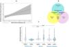
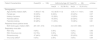
Of 106 infants hospitalized with a first wheezing episode, the median age was 12 months and 33% were girls (Table 1). After adjusting for confounders, we observed that a 1 unit (μg/ml) increase in MxA levels was associated with a 4.7-fold chance of developing asthma by the age of 8 years (adjusted OR 4.7, 95% CI 1.2–18.9; p=0.042) (Fig. 1A). This association was not modified by smoking in the home (p-interaction=0.99). The frequency of respiratory viruses detected in this analytical cohort can be seen in Fig. 1B. The exploratory analysis showed that infants with RV infection only had significantly lower peripheral blood MxA levels than infants with RSV-only and with RSV-RV coinfections (both p<0.05, respectively) (Fig. 1C). The statistical analysis and figures were produced using R Version 4.2.3 and STATA/BE 17.0 software.
Demographic Characteristics and Respiratory Virus Detection in the VINKU2 Analytical Sub-cohort (i.e., N=106 in total) and Participants With Complete Asthma Diagnostic Data Asthma Diagnosis by the Age of 8 Years (i.e., N=86 in Total).
| Patient Characteristics | Overall(N=106) | Asthma by Age of 8 Yearsa(N=86) | p-Value | |
|---|---|---|---|---|
| Yes(N=33; 38.4%) | No(N=53; 61.6%) | |||
| Demographics | ||||
| Age (months) median (IQR) | 11.96 (6.7–16) | 10.3 (8.55–11.9) | 12.8 (11.1–14.01) | 0.06 |
| Female sex | 35 (33%) | 8 (24%) | 23 (43%) | 0.07 |
| History of eczema | 30 (28%) | 12 (36%) | 13 (25%) | 0.24 |
| Parental asthma | 47 (44%) | 18 (55%) | 22 (42%) | 0.24 |
| Parental smoking | 30 (28%) | 11 (33%) | 19 (36%) | 0.81 |
| Peripheral blood cytokine levels | ||||
| MxA levels (μg/L) median (IQR) | 281 (181–474) | 314 (242–684) | 212 (157–383) | 0.015 |
| Respiratory virus data | ||||
| RSV-only | 9 (8%) | 3 (9%) | 5 (9%) | 0.96 |
| RSV-rhinovirus only | 8 (7.5%) | 3 (9%) | 3 (6%) | 0.54 |
| Rhinovirus-only | 47 (44%) | 16 (48%) | 23 (43%) | 0.65 |
| Virus-only (other)b | 5 (5%) | 2 (6%) | 3 (6%) | 0.94 |
Note: Data are N (%) of children unless otherwise indicated.
MxA: myxovirus resistance protein 1; RSV: respiratory syncytial virus.

A multi-panel graphical representation illustrating the key findings of the study.
Panel (A): Relationships of peripheral blood MxA levels with the development of asthma in infants hospitalized with a first wheezing episode. The fitted line represents a locally estimated scatterplot smoothed (loess) curve. The grey bar represents the range in which 95% of corresponding data are present.
Panel (B): This Venn diagram describes the frequency of respiratory viruses detected in nasopharyngeal aspirates from infants in the VINKU2 analytical sub-cohort (i.e., N=106 in total). The left circle (i.e., yellow) includes RSV-positive cases. The middle circle (i.e., light blue) includes cases positive as solo or a coinfection for human metapneumovirus, influenza, parainfluenza, adenovirus, or bocavirus, but not RSV or rhinovirus (RV). The right circle (i.e., pink) includes RV-positive cases. The intersections between the left, middle, and right circles include three groups; the RSV-other viruses coinfections, the RSV-RV-double positive cases, and the RSV-RV-other viruses coinfections. Similarly, the intersections between the right, middle, and left circles include three groups; the RSV-other viruses coinfections, the RSV-RV-double positive cases, and the RSV-RV-other viruses coinfections.
Panel (C): The violin plots depict statistically significant differences in mean peripheral blood MxA levels (i.e., in microgram/ml) in infants hospitalized with an RV-only (N=47) as compared to an RSV-only (N=9) first wheezing episode (p<0.05), as well as in infants hospitalized with an RV-only (N=47) as compared to RV-coinfections (N=21) first wheezing episode (p<0.05). The RV-coinfections group is consistent with the description followed in the Venn diagram in Panel (B). Comparisons have been made with the Mann–Whitney U test.
MxA, myxovirus resistance protein 1; RSV, respiratory syncytial virus; RV, rhinovirus.
To the best of our knowledge, this is the first study of infants hospitalised with a first wheezing episode that suggests that peripheral blood MxA levels are associated with a high risk of childhood asthma. This observation is interesting, particularly following evidence from birth cohort studies showing that MX1 polymorphisms may be associated with an increased risk of asthma exacerbations at school age.7,12 If these data can be confirmed in a larger cohort providing additional evidence of specificity, sensitivity and positive predictive value, peripheral blood MxA could be proposed as a biomarker for childhood asthma. This could prove beneficial, given that peripheral blood MxA levels can be readily identified in point-of-care testing.
We also analysed the available respiratory virus data for RSV-only and RV-only positive groups, and for groups with RSV-RV coinfection. Interestingly, despite the limited sample size, our results in RSV- and RV-only positive groups are in line with reported observational and mechanistic evidence.8,13 To be more precise, we noted among infants hospitalised with a first wheezing episode that those who tested positive for RSV alone showed the highest MxA levels. A similar trend in RSV-positive children was also observed in a Swedish study.8 Furthermore, mechanistic data have shown that in RV infection, ciliated epithelial cells express lower levels of ISGs, possibly through a mechanism that facilitates RV replication.13 This reported mechanism could possibly explain our findings in infants positive for RV only. While we would expect an association between reduced ISG expression and increased asthma risk, this association seems to be more complex than expected. For instance, data from a multicentre bronchiolitis study reveal that infants with RSV-positive bronchiolitis and increased IFN transcription (e.g., linked to probable increased ISG expression) in their nasal airway samples were actually at a greater risk of developing asthma during their school years than infants with RSV-bronchiolitis and reduced IFN transcription in nasal airway samples.3 Further study is needed to clarify if this is related to the respiratory virus type. Another factor that needs to be considered is the timing (i.e., how many days since the onset of symptoms) in the context of the wheezing illness hospitalisation. For example, we know that in adult patients with asthma, IFN expression in response to respiratory virus stimulation is lower than in control patients.14 Nevertheless, recent evidence from an in vitro model showed that IFN deficiency may be associated with increased respiratory virus replication and a subsequent increase in IFN and ISG expression.15 Furthermore, infection with different respiratory viruses may be associated with diverse immune responses in the context of a wheezing illness.16 In this study, we have taken a different approach to the definition of respiratory virus aetiology for analytical purposes. For example, in the statistical analysis, we opted to analyse data by “virus-only-positive” groups based on emerging evidence indicating that respiratory viral coinfections, within the scope of a lower respiratory viral illness, may define groups with unique pathophysiological characteristics.17
Despite its strengths, our study also has several potential limitations. Firstly, the study size in terms of the primary outcome is limited (N=86), although we did observe a significant association between peripheral blood MxA and asthma development up to the age of 8 years. Our study findings provide preliminary evidence that warrants further testing in a larger cohort. Secondly, this study does not have data on asthma phenotypes. The sample size was too small to derive a firm conclusion as to whether peripheral blood MxA could be a more specific marker of childhood asthma development in children with RSV-positive wheezing illnesses. The initial findings from our analysis, however, indicate that this concept should be examined in a larger group of patients.
To summarise, this is the first study to demonstrate a link between peripheral blood MxA levels in infants hospitalised with a first wheezing episode and the development of childhood asthma. The findings are preliminary but significant and warrant further investigation in a larger group of patients. This evidence, when validated, may help design a crucial strategy for the prevention of childhood asthma, which remains a public health priority.
Conflict of InterestsThe authors state that they have no conflict of interests.


