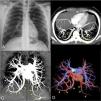
A 22-year-old asymptomatic female was referred to our hospital due to an abnormal finding on a chest radiograph during a routine check-up (Fig. 1A). Physical examination was unremarkable. She underwent enhanced chest computed tomography (CT) examination (Fig. 1B–D), which demonstrated bilateral pulmonary veins with anomalous routes in the lower pulmonary regions, and normal drainage into the left atrium. The diagnosis of bilateral meandering pulmonary veins (MPV) was made based on the CT findings, and the patient was discharged with no further investigation.
Posteroanterior chest radiograph (A) showing bilateral anomalous curvilinear vessels in the lower pulmonary regions. Enhanced chest computed tomography (CT) examination with axial (B) and coronal (C) maximum intensity projection imaging, which demonstrated bilateral pulmonary veins with anomalous routes in the lower pulmonary regions (yellow arrowheads), but draining normally into the left atrium. Coronal volume-rendering 3D reconstruction (D) shows the anomalous veins (red) and normal pulmonary arteries (blue).
It is important to distinguish between pulmonary venous return malformation (PVRM) and MPV. PVRM (e.g., scimitar syndrome) results in a left-to-right shunt, which can lead to cyanosis and may require surgical correction. Consequently, patients are often symptomatic and present at a young age. In contrast, MPV involves no left-to-right shunt.1,2 As in our case, patients with MPV are usually asymptomatic, with the diagnosis made incidentally. Recognition of these variations is important for the clinician to avoid unnecessary investigation when the differential diagnosis includes vascular malformations requiring surgical or interventional treatment. Furthermore, when patients with MPV require surgery for other reasons, thoracic surgeons should be aware of the anomaly to avoid injury to the anomalous vein. Treatment has not been required in any reported case of MPV.


