Journal Information
Share
Share
Download PDF
More article options
Visits
2486
Original Article
Full text access
Available online 16 September 2024
The Effect of Obstructive Sleep Apnea on Subclinical Target Organ Damage in Patients With Resistant Hypertension
Visits
2486
Mireia Dalmasesa,b, Manuel Sánchez-de-la-Torreb,c, Dolores Martinezd, Olga Minguezd, Rafaela Vacad, Lydia Pascuald, Maria Aguilád, Esther Gracia-Lavedand, Ivan D. Benitezd, Lucía Pinillab,d, Anunciación Cortijod, Clara Gort-Paniellod, Ramon Bascompte Clarete, Miguel Ángel Martinez-Garciaf, Olga Medianob,g, Sofía Romero Peraltag, Ana Maria Fortuna-Gutierrezb,h, Paola Ponte Marquezi, Luciano F. Dragerj, Mayara Cabrinij..., Juan Fernando Masab,k, Jaime Corral Peñafielb,k, Susana Vázquezl, Jorge Abadb,m, Francisco García-Riob,n, Raquel Casitasb,n, Chi-Hang Leeo, Ferran Barbéb,d, Gerard Torresb,d,
Corresponding author
gtorres@gss.cat

Corresponding author.
Ver más
a Sleep Unit, Department of Pulmonary Medicine, Hospital Clínic de Barcelona, Institut d’Investigacions Biomèdiques August Pi i Sunyer (IDIBAPS), Barcelona, Spain
b Centro de Investigación Biomédica en Red de Enfermedades Respiratorias (CIBERES), Instituto de Salud Carlos III, Madrid, Spain
c Group of Precision Medicine in Chronic Diseases, Hospital Nacional de Parapléjicos, IDISCAM, Department of Nursing, Physiotherapy and Occupational Therapy, Faculty of Physiotherapy and Nursing, University of Castilla-La Mancha, Toledo, Spain
d Hospital Universitari Arnau de Vilanova and Santa Maria, Group of Translational Research in Respiratory Medicine, IRB Lleida, Lleida, Spain
e Cardiology Department, Hospital Arnau de Vilanova, Centro de Investigación en Red en Enfermedades Cardiovasculares (CIBERCV), Lleida, Spain
f Hospital Universitario y Politécnico La Fe, Respiratory Department, Valencia, Spain
g Sleep Unit, Pneumology Department, Hospital Universitario de Guadalajara, Guadalajara, Spain
h Sleep Unit, Respiratory Department, Hospital de la Santa Creu i Sant Pau, Biomedical Research Institute Sant Pau, Universitat Autònoma de Barcelona, Barcelona, Spain
i Internal Medicine, Emergency Department, Hypertension and Cardiovascular Risk Unit, Hospital de la Santa Creu i Sant Pau, Universitat Autónoma de Barcelona, Bellaterra, Spain
j Unidades de Hipertensão, Instituto do Coração (InCor) e Divisão de Nefrologia, Hospital das Clínicas HCFMUSP, Faculdade de Medicina, Universidade de Sao Paulo, Sao Paulo, SP, Brazil
k Hospital San Pedro de Alcantara, Respiratory Dept, Cáceres, Spain
l Hypertension and Vascular Risk Unit, Nephrology Department, Hospital del Mar, Parc de Salut Mar, IMIM, UAB-UPF, Barcelona, Spain
m Respiratory Department, Hospital Universitari Germans Trias i Pujol, Badalona, Spain
n Respiratory Department, Hospital Universitario La Paz, Instituto de Investigación Hospital Universitario La Paz, Madrid, Spain
o Department of Medicine, Yong Loo Lin School of Medicine, National University of Singapore, Singapore
Ver más
Highlights

  • OSA is associated to worse renal function in patients with RH.

  • There is a dose–response association between glomerular filtration rate and OSA severity.

  • Renal function could be ameliorated with adherent CPAP treatment.

This item has received
Article information
Abstract
Full Text
Bibliography
Download PDF
Statistics
Figures (5)
Show moreShow less
Tables (2)
Table 1. Characteristics of the Cohort Stratified by Obstructive Sleep Apnea Presence.
Table 2. Subclinical Organ Parameters in the OSA and Non-OSA Groups at Baseline.
Show moreShow less
Abstract
Introduction

Among all patients with hypertension, those with resistant hypertension (RH) have the highest rates of subclinical organ damage (SOD). The prevalence of obstructive sleep apnea (OSA) is high in RH patients, and it could contribute to SOD. We aimed to investigate how OSA and its treatment are related to SOD in a large cohort of RH patients.

Methods

This is an ancillary analysis to the SARAH study, a multicentre observational cohort aiming to evaluate the impact of OSA on RH. Individuals with RH who were undergoing a sleep study and have information on at least one of the SOD variables (vascular, cardiac or renal damage) were selected. Patients were followed-up for three years.
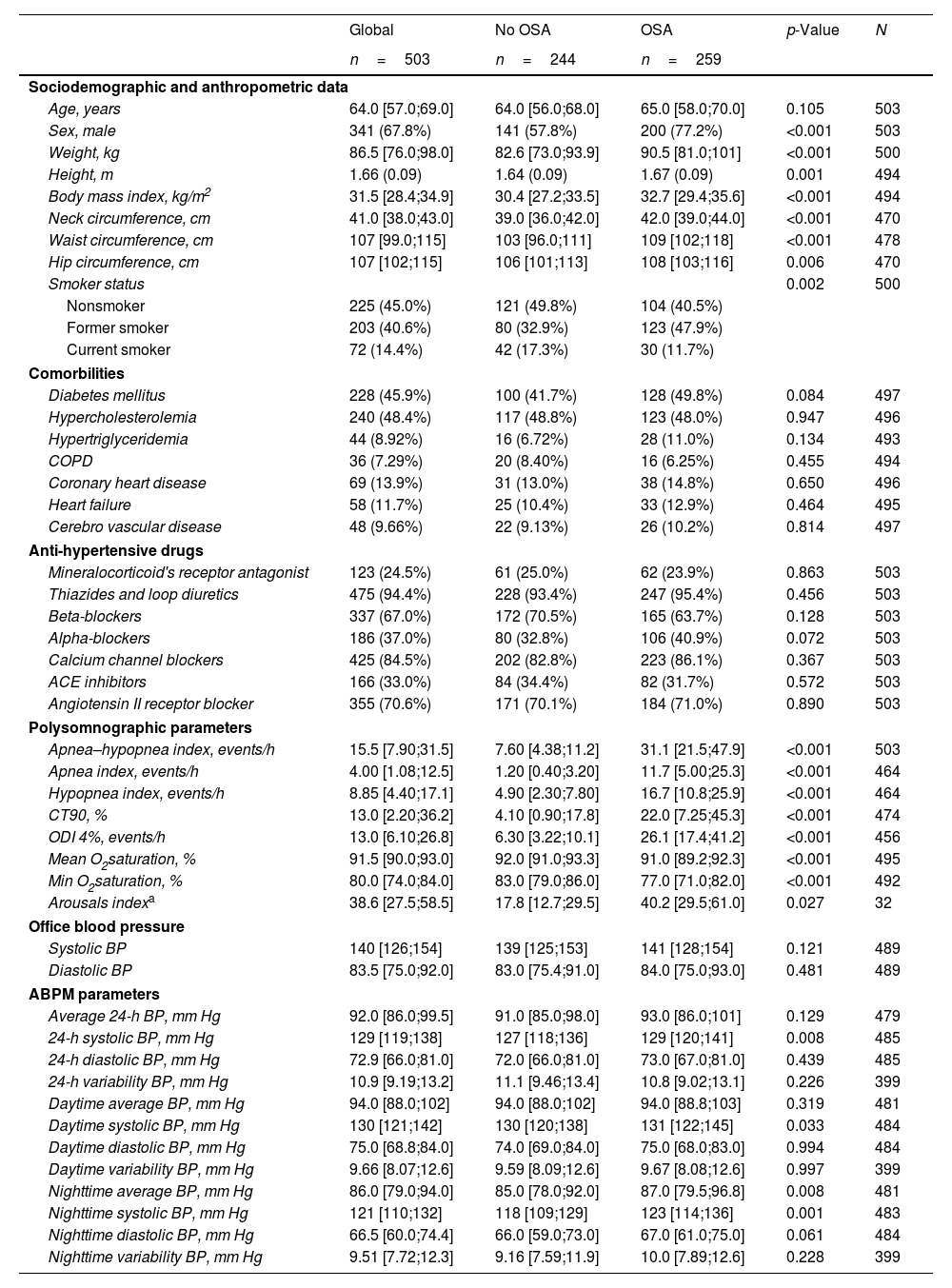
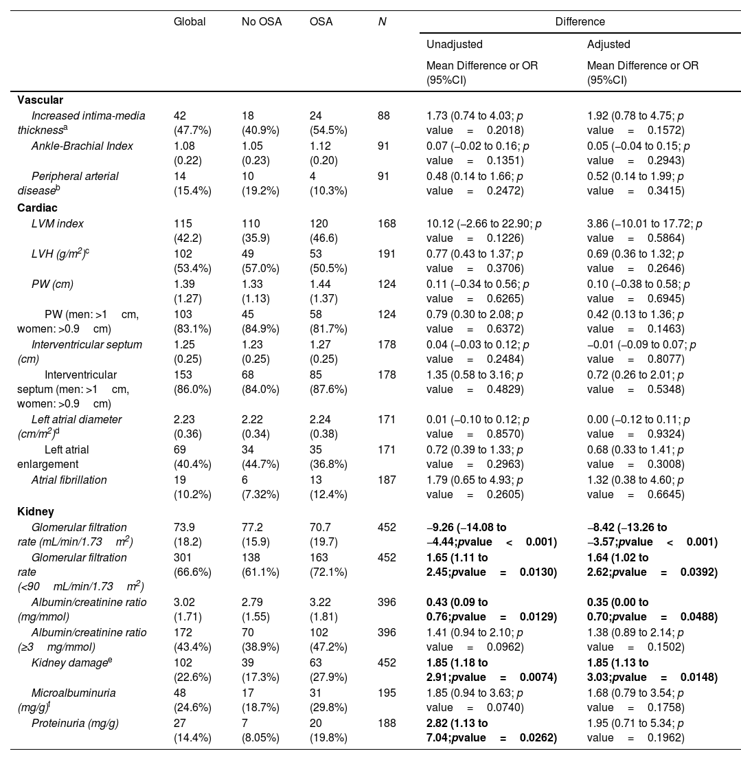
Results

In total, 503 subjects were included. The participants were predominantly male, obese, and the median (IQR) apnea–hypopnea index (AHI) was 15.5 (7.90–31.5)events/h. No differences in the presence of vascular or cardiac damage were observed between OSA and non-OSA patients. A lower estimated glomerular filtration rate (eGFR) was observed in participants with OSA than in those without OSA, with an adjusted effect of −8.69mL/min/1.73m2 (−13.59, −3.79; p value<0.001). Kidney damage was also greater in subjects with OSA, with an adjusted OR (95% CI) of 1.77 (1.09, 2.87; p value=0.02). The eGFR showed a linear dose–response relationship with OSA severity. Among patients treated with CPAP, lower eGFR values were observed in noncompliant subjects.

Conclusions

OSA could contribute to worsening renal function in patients with RH. No compliance with CPAP was associated with lower values of eGFR.

Keywords:
Continuous positive airway pressure
Obstructive sleep apnea
Resistant hypertension
Subclinical organ damage
Renal function
Glomerular filtration rate
Abbreviations:
ABI
ABPM
AF
AHI
BMI
BP
CPAP
CT90
eGFR
ESS
IMT
LAD
LAE
LVH
LVM
ODI
OSA
RH
SOD
Full Text
Introduction

Resistant hypertension (RH) is defined as blood pressure (BP) that remains above the goal despite the use of three different antihypertensive drugs, including a diuretic, prescribed at the optimal dose or as those that require four or more drugs to achieve BP control.1 Among all hypertensive patients, RH patients have the worst prognosis and have estimated rates of subclinical organ damage (SOD) and cardiovascular events 50% greater than those of patients with controlled hypertension.2,3

There is a high prevalence of obstructive sleep apnea (OSA) in patients with RH.4 OSA is associated with daytime symptoms, a decrease in quality of life and morbidity and mortality, mainly from cardiovascular alterations.5 It is well known that OSA produces hemodynamic changes, inflammation, oxidative stress, endothelial damage and atherosclerosis5,6; therefore, it can potentially produce SOD. Moreover, a dose–response association between OSA severity and BP measurements has been described, especially at night, which could also have an impact on the generation of SOD.7

SOD represents an intermediate stage in the continuum of cardiovascular disease, and it is considered a powerful marker of increased cardiovascular risk that can even predict the recurrence of cardiovascular events.8 Early detection of SOD in patients with RH who are at high cardiovascular risk is crucial for implementing therapeutic strategies to stop disease progression and possibly favor its regression. Therefore, knowing whether other comorbid conditions frequent in RH patients, such as OSA, could have a synergistic effect on the induction of SOD could be important in the management of these patients, especially when this condition is potentially treatable.

Nevertheless, few studies have evaluated the prevalence of SOD in patients with RH and OSA and they are single-center studies with a cross-sectional design. The aim of this study was to assess the presence of SOD in a large cohort of RH patients with and without OSA, evaluate whether there is a dose–response association with OSA severity, identify the OSA profile associated with the presence of SOD and evaluate the effect of CPAP treatment.

Material and MethodsStudy Design and Population

This is an ancillary study to the SARAH study (Long-Term Cardiovascular Outcomes in Patients with RH and OSA with or without Treatment with continuous positive airway pressure (CPAP)), a multicenter, international, prospective, observational cohort study (NCT03002558) aimed at evaluating the impact of OSA and CPAP treatment on cardiovascular outcomes in patients with RH.

The study included subjects aged 18–75 years diagnosed with RH based on the American Heart Association guidelines1 and confirmed by 24-h ambulatory blood pressure monitoring (ABPM). The exclusion criteria were RH secondary to other causes (endocrinological, treatment with nonsteroidal anti-inflammatory agents, immunosuppressants or cortisone, renal artery stenosis, intracranial tumors or aortic coarctation), life expectancy<1 year and current treatment with CPAP. The methodology of the SARAH trial is published elsewhere.9 The ethics committee of each participating center approved the study protocol (Ethics Committee number CEIC-1547), and all participants provided informed consent.

For the current study, we selected 503 participants in the SARAH study who had information on at least one of the SOD variables. See the study flow chart in Fig. 1. Patients were followed once a year for three years.

Fig. 1.

Study flow chart.

(0.05MB).
ProceduresBaseline and Follow-Up Visits

At the initial visit, data regarding sociodemographic characteristics, habits, comorbidities, medication, anthropometric measures and self-reported sleepiness (evaluated with the Epworth Sleepiness Scale (ESS)) were collected. Diabetes and dyslipidemia were determined by self-report, abnormal values in the blood sample analysis or medication use and confirmed by a doctor. Chronic obstructive pulmonary disease, coronary heart disease, heart failure and cerebrovascular diseases were identified by self-reports or medication and confirmed by a doctor or a hospital report. At baseline, we performed a sleep study, 24-h ABPM and an evaluation of SOD. At the follow-up visits, anthropometric measurements, 24-h ABPM and blood samples were collected.

Sleep Evaluation

A sleep test consisting of either respiratory polygraphy or polysomnography was performed on all included subjects. All the studies were performed in accredited sleep units with certified technicians and somnologists. Sleep studies were performed using different types of devices according to their availability at each center. The majority of the studies were performed using Embletta sleep monitor. The rest of the studies were performed using Compumedics E-Profusion 3.4, Sibelmed Exea Serie 5, Philips Respironics Alice 6 LDx, Somnomedics Somnoscreen plus Version 2.7.0 or ApneaLink Resmed. Apnea was defined as an absence or reduction of airflow ≥90% lasting ≥10s. Hypopnea was defined as a reduction in airflow (30–90%) lasting10s associated with an oxygen desaturation index (ODI)4% or arousal. The apnea–hypopnea index (AHI) was defined as the number of apnea and hypopnea events per hour of recording or sleep, depending on the study (polygraphy or polysomnography, respectively). CT90 was defined as the percentage of time with an oxygen saturation<90%. According to the AHI, participants were classified as non-OSA (AHI<15/h) or OSA (AHI15/h). The OSA diagnosis and treatment recommendations were based on guidelines.10 CPAP titration was conducted by means of automated equipment following previously described methods.11 After titration, patients continued treatment with fixed CPAP. Good compliance with CPAP treatment was defined as the use of ≥4h/night.

Blood Pressure Monitoring

Office BP and 24-h ABPM measurements were performed according to the guidelines.12 Based on the 24-h ABPM results, those subjects with a 24-h mean BP that remained above the target (average SBP130mmHg, average DBP80mmHg or both) despite the use of three antihypertensive drugs at full dose (one of which should be a diuretic) or those patients treated with ≥4 antihypertensive medications regardless of the BP values obtained during the 24-h ABPM were included. The devices used to perform the 24-h ABPM were Spacelabs 90207/90217A devices (Spacelabs Inc.), Mortara Ambulo 2400, Microlife WatchBP (Microlife AG), and Dyna-MAPA (Cardios Sistemas Coml. Indl. Ltda.).

All participants maintained their prescribed antihypertensive treatment during the measurements. Compliance with antihypertensive treatment was considered when subjects retrieved from the pharmacy>80% of their prescribed medications.13 In addition, all patients underwent Morisky–Green and Hayness–Sackect test at each follow-up visit.

Subclinical Organ Damage Assessment

The evaluation of SOD at baseline was not compulsory in the SARAH study; therefore, it was performed at the discretion of the physician. During the follow-up, only blood analyses were performed; therefore, only data on the estimated glomerular filtration rate (eGFR) were available. SOD was evaluated as follows:

  • Cardiac damage: Left ventricular mass (LVM), left atrial diameter (LAD), posterior wall and interventricular septum were evaluated following the American Society of Echocardiography recommendations.14 Left ventricular hypertrophy (LVH) was defined as an LVM>115g/m2 in men and >95g/m2 in women. Left atrial enlargement (LAE) was considered when the LAD was >2.3cm/m2. Atrial fibrillation (AF) was diagnosed when the patient had a previous history, received antiarrhythmic treatment or was observed via electrocardiography.

  • Renal damage: Kidney function was evaluated based on eGFR and/or albuminuria in accordance with the International Kidney Disease guidelines.15 Microalbuminuria was defined as albuminuria between 30 and 300mg/g creatinine, and proteinuria was defined as albuminuria>300mg/g in 24h. Alterations should be confirmed in two blood or urine analyses separated by at least 6 months. Kidney damage was defined as an eGFR<60mL/min/1.73m2 and/or an albumin/creatinine ratio3mg/mmol.

  • Vascular disease: The intima-media thickness (IMT) was evaluated by carotid ultrasound following the American Society of Echocardiography recommendations.16 An increased IMT was defined as an IMT>0.9mm protuberant in the lumen. Peripheral arterial disease was defined by an ankle-brachial index (ABI)<0.9.17

Statistical Analyses

Descriptive statistics were used to determine the characteristics of the study population. The normality of the distributions was assessed by the Shapiro–Wilk test. Normally distributed continuous data were summarized using the mean (standard deviation), and nonnormally distributed continuous data using the median (25th percentile; 75th percentile). Categorical data were summarized using frequencies (percentages). The clinical and sociodemographic characteristics of the patients were compared between the OSA and non-OSA groups using t tests (or equivalent nonparametric tests) or Chi-square tests for quantitative and categorical variables, respectively. SOD parameters were compared among the OSA and non-OSA groups using linear models for continuous variables, tobit regression for truncated variables and logistic regression for dichotomous variables. Unadjusted models and adjusted for confounding factors (age, sex, BMI, alcohol consumption and smoking status) were constructed. The dose–response relationship between the outcomes and the severity of OSA was evaluated using generalized additive models for parameters that showed significant differences between study groups after adjusting for confounding factors. Furthermore, a linear mixed model was fitted to compare the eGFR during follow-up visits (baseline, one year and three years) between OSA patients and non-OSA patients. Patients with OSA who were receiving CPAP treatment were excluded. Patient status was included in the model as a random effect, and study group, visit and their interaction were included as fixed effects. Finally, the eGFR during follow-up visits was assessed according to CPAP adherence (<4 or ≥4h/day) in patients receiving CPAP treatment. Patient status was included in the model as a random effect, and study group, visit and their interaction were included as fixed effects. The level of statistical significance was set at 0.05. All analyses were performed using the statistical software R-project (version 4.1.1).

ResultsCohort Characteristics

In total, 503 subjects with RH were included. The main sociodemographic and clinical characteristics of the population are shown in Table 1. Briefly, the median (IQR) age was 64.0 (57.0–69.0) years, the participants were predominantly male (67.8%), and the median body mass index (BMI) was 31.5kg/m2 (28.4–34.9). The most prevalent comorbidities were hypercholesterolemia (48.4%) and diabetes mellitus (45.9%). A total of 87.6% of the included subjects took ≥4 antihypertensive drugs. Related to the type of antihypertensive drugs, 94.4% take thiazides and loop diuretics, 84.5% calcium channel blockers and 70.6% angiotensin II receptor blockers. No significant differences in antihypertensive treatment were observed between the groups (Table 1).

Table 1.

Characteristics of the Cohort Stratified by Obstructive Sleep Apnea Presence.

  Global  No OSA  OSA  p-Value  N 
  n=503  n=244  n=259     
Sociodemographic and anthropometric data
Age, years  64.0 [57.0;69.0]  64.0 [56.0;68.0]  65.0 [58.0;70.0]  0.105  503 
Sex, male  341 (67.8%)  141 (57.8%)  200 (77.2%)  <0.001  503 
Weight, kg  86.5 [76.0;98.0]  82.6 [73.0;93.9]  90.5 [81.0;101]  <0.001  500 
Height, m  1.66 (0.09)  1.64 (0.09)  1.67 (0.09)  0.001  494 
Body mass index, kg/m2  31.5 [28.4;34.9]  30.4 [27.2;33.5]  32.7 [29.4;35.6]  <0.001  494 
Neck circumference, cm  41.0 [38.0;43.0]  39.0 [36.0;42.0]  42.0 [39.0;44.0]  <0.001  470 
Waist circumference, cm  107 [99.0;115]  103 [96.0;111]  109 [102;118]  <0.001  478 
Hip circumference, cm  107 [102;115]  106 [101;113]  108 [103;116]  0.006  470 
Smoker status        0.002  500 
Nonsmoker  225 (45.0%)  121 (49.8%)  104 (40.5%)     
Former smoker  203 (40.6%)  80 (32.9%)  123 (47.9%)     
Current smoker  72 (14.4%)  42 (17.3%)  30 (11.7%)     
Comorbilities
Diabetes mellitus  228 (45.9%)  100 (41.7%)  128 (49.8%)  0.084  497 
Hypercholesterolemia  240 (48.4%)  117 (48.8%)  123 (48.0%)  0.947  496 
Hypertriglyceridemia  44 (8.92%)  16 (6.72%)  28 (11.0%)  0.134  493 
COPD  36 (7.29%)  20 (8.40%)  16 (6.25%)  0.455  494 
Coronary heart disease  69 (13.9%)  31 (13.0%)  38 (14.8%)  0.650  496 
Heart failure  58 (11.7%)  25 (10.4%)  33 (12.9%)  0.464  495 
Cerebro vascular disease  48 (9.66%)  22 (9.13%)  26 (10.2%)  0.814  497 
Anti-hypertensive drugs
Mineralocorticoid's receptor antagonist  123 (24.5%)  61 (25.0%)  62 (23.9%)  0.863  503 
Thiazides and loop diuretics  475 (94.4%)  228 (93.4%)  247 (95.4%)  0.456  503 
Beta-blockers  337 (67.0%)  172 (70.5%)  165 (63.7%)  0.128  503 
Alpha-blockers  186 (37.0%)  80 (32.8%)  106 (40.9%)  0.072  503 
Calcium channel blockers  425 (84.5%)  202 (82.8%)  223 (86.1%)  0.367  503 
ACE inhibitors  166 (33.0%)  84 (34.4%)  82 (31.7%)  0.572  503 
Angiotensin II receptor blocker  355 (70.6%)  171 (70.1%)  184 (71.0%)  0.890  503 
Polysomnographic parameters
Apnea–hypopnea index, events/h  15.5 [7.90;31.5]  7.60 [4.38;11.2]  31.1 [21.5;47.9]  <0.001  503 
Apnea index, events/h  4.00 [1.08;12.5]  1.20 [0.40;3.20]  11.7 [5.00;25.3]  <0.001  464 
Hypopnea index, events/h  8.85 [4.40;17.1]  4.90 [2.30;7.80]  16.7 [10.8;25.9]  <0.001  464 
CT90, %  13.0 [2.20;36.2]  4.10 [0.90;17.8]  22.0 [7.25;45.3]  <0.001  474 
ODI 4%, events/h  13.0 [6.10;26.8]  6.30 [3.22;10.1]  26.1 [17.4;41.2]  <0.001  456 
Mean O2saturation, %  91.5 [90.0;93.0]  92.0 [91.0;93.3]  91.0 [89.2;92.3]  <0.001  495 
Min O2saturation, %  80.0 [74.0;84.0]  83.0 [79.0;86.0]  77.0 [71.0;82.0]  <0.001  492 
Arousals indexa  38.6 [27.5;58.5]  17.8 [12.7;29.5]  40.2 [29.5;61.0]  0.027  32 
Office blood pressure
Systolic BP  140 [126;154]  139 [125;153]  141 [128;154]  0.121  489 
Diastolic BP  83.5 [75.0;92.0]  83.0 [75.4;91.0]  84.0 [75.0;93.0]  0.481  489 
ABPM parameters
Average 24-h BP, mm Hg  92.0 [86.0;99.5]  91.0 [85.0;98.0]  93.0 [86.0;101]  0.129  479 
24-h systolic BP, mm Hg  129 [119;138]  127 [118;136]  129 [120;141]  0.008  485 
24-h diastolic BP, mm Hg  72.9 [66.0;81.0]  72.0 [66.0;81.0]  73.0 [67.0;81.0]  0.439  485 
24-h variability BP, mm Hg  10.9 [9.19;13.2]  11.1 [9.46;13.4]  10.8 [9.02;13.1]  0.226  399 
Daytime average BP, mm Hg  94.0 [88.0;102]  94.0 [88.0;102]  94.0 [88.8;103]  0.319  481 
Daytime systolic BP, mm Hg  130 [121;142]  130 [120;138]  131 [122;145]  0.033  484 
Daytime diastolic BP, mm Hg  75.0 [68.8;84.0]  74.0 [69.0;84.0]  75.0 [68.0;83.0]  0.994  484 
Daytime variability BP, mm Hg  9.66 [8.07;12.6]  9.59 [8.09;12.6]  9.67 [8.08;12.6]  0.997  399 
Nighttime average BP, mm Hg  86.0 [79.0;94.0]  85.0 [78.0;92.0]  87.0 [79.5;96.8]  0.008  481 
Nighttime systolic BP, mm Hg  121 [110;132]  118 [109;129]  123 [114;136]  0.001  483 
Nighttime diastolic BP, mm Hg  66.5 [60.0;74.4]  66.0 [59.0;73.0]  67.0 [61.0;75.0]  0.061  484 
Nighttime variability BP, mm Hg  9.51 [7.72;12.3]  9.16 [7.59;11.9]  10.0 [7.89;12.6]  0.228  399 
a

Only for patients diagnosed with PSG.

Abbreviations: ACE=angiotensin-converting enzyme; AHI=apnea–hypopnea index; COPD=chronic obstructive pulmonary disease; CT90=percentage of time with oxygen saturation 90%; ODI=oxygen desaturation index.

Notes: Quantitative variables are described by median [25th percentile; 75th percentile], and qualitative variables are described by absolute and relative frequencies.

Regarding OSA parameters, the median (IQR) AHI was 15.5 (7.90–31.5)events/h, and the median 4%-ODI was 13.0 (6.10–26.8)events/h. The median CT90 was 13.0% (2.20–36.2), and the median ESS was 6 (4–10). The prevalence of OSA (defined as an AHI15events/h) was 51.4%. With regard to OSA severity, 24.2% had moderate OSA, and 27.2% had severe OSA. CPAP treatment was indicated in 193 subjects and the mean CPAP compliance at 1-year follow-up was 5.28 (3.95–6.71)h/night, without significant changes during the follow-up period.

Male sex, BMI and ABPM parameters, especially nighttime BP values, were greater in the OSA group than in the non-OSA group. Conversely, there were no differences in the number or type of drugs used between the groups.

Subclinical Organ Damage at Baseline

The SOD parameters were compared between the OSA and non-OSA groups. The evaluation of vascular disease (n=91) revealed that 47.7% of the participants presented increased IMT in the carotid territory, and 15.4% had peripheral arterial disease. There were no statistically significant differences in vascular diseases (neither in carotid nor in peripheral arterial territories) between the groups (Table 2).

Table 2.

Subclinical Organ Parameters in the OSA and Non-OSA Groups at Baseline.

  Global  No OSA  OSA  N  Difference
          Unadjusted  Adjusted 
          Mean Difference or OR (95%CI)  Mean Difference or OR (95%CI) 
Vascular
Increased intima-media thicknessa  42 (47.7%)  18 (40.9%)  24 (54.5%)  88  1.73 (0.74 to 4.03; p value=0.2018)  1.92 (0.78 to 4.75; p value=0.1572) 
Ankle-Brachial Index  1.08 (0.22)  1.05 (0.23)  1.12 (0.20)  91  0.07 (−0.02 to 0.16; p value=0.1351)  0.05 (−0.04 to 0.15; p value=0.2943) 
Peripheral arterial diseaseb  14 (15.4%)  10 (19.2%)  4 (10.3%)  91  0.48 (0.14 to 1.66; p value=0.2472)  0.52 (0.14 to 1.99; p value=0.3415) 
Cardiac
LVM index  115 (42.2)  110 (35.9)  120 (46.6)  168  10.12 (−2.66 to 22.90; p value=0.1226)  3.86 (−10.01 to 17.72; p value=0.5864) 
LVH (g/m2)c  102 (53.4%)  49 (57.0%)  53 (50.5%)  191  0.77 (0.43 to 1.37; p value=0.3706)  0.69 (0.36 to 1.32; p value=0.2646) 
PW (cm)  1.39 (1.27)  1.33 (1.13)  1.44 (1.37)  124  0.11 (−0.34 to 0.56; p value=0.6265)  0.10 (−0.38 to 0.58; p value=0.6945) 
PW (men: >1cm, women: >0.9cm)  103 (83.1%)  45 (84.9%)  58 (81.7%)  124  0.79 (0.30 to 2.08; p value=0.6372)  0.42 (0.13 to 1.36; p value=0.1463) 
Interventricular septum (cm)  1.25 (0.25)  1.23 (0.25)  1.27 (0.25)  178  0.04 (−0.03 to 0.12; p value=0.2484)  −0.01 (−0.09 to 0.07; p value=0.8077) 
Interventricular septum (men: >1cm, women: >0.9cm)  153 (86.0%)  68 (84.0%)  85 (87.6%)  178  1.35 (0.58 to 3.16; p value=0.4829)  0.72 (0.26 to 2.01; p value=0.5348) 
Left atrial diameter (cm/m2)d  2.23 (0.36)  2.22 (0.34)  2.24 (0.38)  171  0.01 (−0.10 to 0.12; p value=0.8570)  0.00 (−0.12 to 0.11; p value=0.9324) 
Left atrial enlargement  69 (40.4%)  34 (44.7%)  35 (36.8%)  171  0.72 (0.39 to 1.33; p value=0.2963)  0.68 (0.33 to 1.41; p value=0.3008) 
Atrial fibrillation  19 (10.2%)  6 (7.32%)  13 (12.4%)  187  1.79 (0.65 to 4.93; p value=0.2605)  1.32 (0.38 to 4.60; p value=0.6645) 
Kidney
Glomerular filtration rate (mL/min/1.73m2)  73.9 (18.2)  77.2 (15.9)  70.7 (19.7)  452  9.26 (14.08 to4.44;pvalue<0.001)  8.42 (13.26 to3.57;pvalue<0.001) 
Glomerular filtration rate (<90mL/min/1.73m2)  301 (66.6%)  138 (61.1%)  163 (72.1%)  452  1.65 (1.11 to 2.45;pvalue=0.0130)  1.64 (1.02 to 2.62;pvalue=0.0392) 
Albumin/creatinine ratio (mg/mmol)  3.02 (1.71)  2.79 (1.55)  3.22 (1.81)  396  0.43 (0.09 to 0.76;pvalue=0.0129)  0.35 (0.00 to 0.70;pvalue=0.0488) 
Albumin/creatinine ratio (≥3mg/mmol)  172 (43.4%)  70 (38.9%)  102 (47.2%)  396  1.41 (0.94 to 2.10; p value=0.0962)  1.38 (0.89 to 2.14; p value=0.1502) 
Kidney damagee  102 (22.6%)  39 (17.3%)  63 (27.9%)  452  1.85 (1.18 to 2.91;pvalue=0.0074)  1.85 (1.13 to 3.03;pvalue=0.0148) 
Microalbuminuria (mg/g)f  48 (24.6%)  17 (18.7%)  31 (29.8%)  195  1.85 (0.94 to 3.63; p value=0.0740)  1.68 (0.79 to 3.54; p value=0.1758) 
Proteinuria (mg/g)  27 (14.4%)  7 (8.05%)  20 (19.8%)  188  2.82 (1.13 to 7.04;pvalue=0.0262)  1.95 (0.71 to 5.34; p value=0.1962) 

Notes: Descriptive results are shown as the mean (SD) or n (%) for quantitative or categorical variables, respectively.

a

Increased intima-media thickness (IMT) is defined as an intima-media thickness greater than 0.9mm.

b

Peripheral arterial disease is defined by an ankle-brachial index (ABI)<0.9.

c

Left ventricular hypertrophy (LVH) is defined as a left ventricular mass (LVM) greater than 115g/m2 in men and greater than 95g/m2 in women.

d

Left atrial enlargement is defined as a left atrial diameter greater than 2.3cm/m2.

e

Kidney damage is defined as an eGFR<60mL/min/1.73m2 and/or an albumin/creatinine ratio3mg/mmol.

f

Microalbuminuria was defined as albuminuria between 30 and 300mg/g creatinine, and proteinuria was defined as albuminuria greater than 300mg/g in 24h.

With regard to cardiac damage (n=191), the mean (SD) of LVM was 119.5 (47.7)g/m2 in men and 108.4 (30.3)g/m2 in women, and LVH was observed in 47.2% of men and 65.2% of women. The mean LAD was 2.23 (0.36)cm/m2, and 40.4% of patients presented with LAE. Moreover, 10% of the participants had AF. There were no statistically significant differences in cardiac parameters between OSA and non-OSA patients (Table 2).

Regarding kidney parameters (n=452), patients had a mean (SD) eGFR of 73.9 (18.2)mL/min/1.73m2. We observed lower values of eGFR in participants with OSA than in those without OSA, with an adjusted effect of −8.42mL/min/1.73m2 (−13.26, −3.57; p value<0.001). The albumin–creatinine ratio was also greater in participants with OSA than in those without OSA, with an adjusted OR (95% CI) of 0.35 (0.00, 0.70; p value=0.05). The presence of kidney damage was greater in subjects with OSA than in those without OSA, with an adjusted OR (95% CI) of 1.85 (1.13, 3.03; p value=0.01). Proteinuria was also slightly greater in the OSA group, although differences did not reach statistical significance after adjusting for confounding factors. For microalbuminuria, no significant differences were detected between the non-OSA and OSA groups after adjusting for confounding factors (Table 2).

The eGFR showed a linear dose–response relationship with OSA severity measured by the AHI (Fig. 2). Specifically, the ODI was the respiratory parameter that was strongly associated with the eGFR (p value=0.0026) (Fig. 3).

Fig. 2.

Glomerular filtration according to OSA severity measured by the AHI. The relationships between the AHI and eGFR were evaluated using generalized additive models adjusted for confounding factors (age, sex, BMI, alcohol consumption and smoking status).

(0.07MB).
Fig. 3.

Associations between different sleep parameters and the eGFR. The relationships between the OSA parameters and eGFR were evaluated using generalized additive models adjusted for confounding factors (age, sex, BMI, alcohol consumption and smoking status).

(0.16MB).
Evolution of the Glomerular Filtration Rate

The change in eGFR over time according to the presence of OSA was analyzed after excluding patients who underwent CPAP treatment. A longitudinal model showed that the eGFR decreased slightly at the three-year follow-up, with a change (95% CI) from the baseline of −3.12 (−7.58; 1.33)mL/min/1.73m2. Compared with the non-OSA group, the OSA group had a mean difference (95% CI) in the eGFR of −7.07mL/min/1.73m2 (−11.40, −2.79, p value=0.0013) at baseline. These differences were sustained throughout the follow-up, but there were no differences in the evolution trend (Fig. 4A). In patients with CPAP, adherence to CPAP treatment was significantly associated with the eGFR at the one-year follow-up, which persisted at three years and was lower in noncompliers (mean differences of −8.21mL/min/1.73m2 [−13.51, −2.90, p value<0.01) (Fig. 4B).

Fig. 4.

Longitudinal changes in the eGFR during the follow-up period. (A) The values of eGFR at the follow-up visits in the OSA and non-OSA groups. The OSA patients with CPAP were excluded from this analysis. The points represent the estimated marginal means of eGFR per OSA group derived from the linear mixed-effects model. The model included confounding factors (age, sex and BMI), OSA group, visit and OSA-visit interaction as fixed effects and patient as a random effect. (B) The values of eGFR at the one- and three-year follow-ups in OSA patients receiving CPAP treatment according to adherence group. Only patients with CPAP were included. Linear mixed-effects model included confounding factors (AHI, age, sex, and BMI), adherence group, visit and adherence-visit interaction as fixed effects and patient as a random effect.

(0.09MB).
Discussion

The study showed that patients with RH and OSA do not present differences in vascular or cardiac damage but present worse kidney function than subjects without OSA. Lower eGFR values were observed in OSA patients, with a dose–response relationship with OSA severity. Moreover, the study showed that the ODI is the OSA parameter that has a greater association with the eGFR and indicated that there is an association between adherence to CPAP treatment and kidney function, with lower eGFR in noncompliant subjects.

These results are in line with studies describing a decrease in the eGFR in patients with OSA and an increased risk of kidney disease.18 OSA is a risk factor for decreased eGFR in patients with hypertension, hypertrophic cardiomyopathy, diabetic kidney disease and in patients without comorbidities,19–21 with an effect similar to simple hypertension and an additive effect in RH patients.22 We observed a mild reduction in the eGFR, which is consistent with previous studies demonstrating discrete reductions in kidney function.23 The eGFR found in our study, even in the group without OSA, was slightly lower than that described in elderly subjects with OSA from the general population,24 suggesting that RH patients may be especially vulnerable to renal damage.25 The relationship between renal damage and OSA could be bidirectional, and we cannot exclude that a decrease in eGFR could produce fluid overload and favor OSA.26 The albumin–creatinine ratio was also greater in subjects with OSA than in those without OSA and proteinuria was slightly greater in the OSA group than in the control group, but these differences did not reach statistical significance after adjusting for confounding factors. Our results are in line with previous studies describing increases in albuminuria in patients with OSA27 and relating them to OSA severity28 and as far as we are concerned, these increases have not been previously described in subjects with RH.

The ODI was the sleep parameter that was strongly associated with the eGFR in patients with RH. The kidney medulla is known to be particularly sensitive to hypoxia,29 and OSA-related hypoxia has been associated with kidney disease30 and glomerular filtrate loss.31 Hypertension produces renal damage through glomerular hypertension and hyperfiltration, and hypoxia mainly affects renal damage at the tubulointerstitial level; therefore, both diseases could have an additive effect.

The eGFR at baseline was lower in OSA patients than in non-OSA patients, and if not treated, it decreased slightly throughout the three-year follow-up, but there were no differences in the evolution trend between the groups. The decline in the eGFR found in our study was greater than previously reported in a healthy young cohort,32 probably related to the vulnerability of the RH itself to deterioration of renal function. Our results are in agreement with those of Canales et al., who did not observe differences in kidney function trajectory during follow-up but contradict those of other studies.33 As the follow-up in our study lasted three years, we cannot exclude the possibility that differences could be found over a longer follow-up period because our cohort included a high-risk group (RH patients with an abnormal eGFR at baseline and comorbidities). In subjects receiving CPAP treatment, the results revealed an association between CPAP adherence and the eGFR at the one-year follow-up that persisted at three years, with lower values in noncompliers. Although the effect of OSA treatment on kidney function is not completely clear,34 our results are in accordance with studies showing that CPAP treatment could improve renal function35,36 and be related to adherence.37

We did not find differences in cardiac or vascular damage between OSA and non-OSA groups. LVH and carotid atherosclerosis have been previously described, while findings related to LAE and arterial stiffness are less conclusive.38 Previous studies included mainly patients without CV diseases or simple hypertension; therefore, differences in the results could be explained, at least in part, by the heterogeneity in the populations in terms of hypertension phenotype and blood pressure control and also related to differences in OSA severity. There are only two studies in RH subjects that reported a greater incidence of LVH in individuals with moderate/severe OSA than in individuals with no/mild OSA.39,40 In contrast with our study, the majority of subjects included were women, and the differences could be related to a differential effect depending on sex, as suggested by the greater proportion of LVH observed in women than in men in our study. Moreover, the incidence of LVH in those studies was slightly greater than in our study, even in patients without OSA, probably related to more severe RH and OSA or longer RH than did patients in our cohort.

The main strength of our study is that this is the first multicentric and international study with a longitudinal design evaluating SOD in different territories in a large and well-characterized cohort of patients with RH and OSA. Second, unlike other studies, OSA diagnosis was based on sleep studies instead of questionnaires; therefore, the accuracy of diagnosis and classification is greater. Third, analyses were adjusted for adequate confounding factors.

This study has some limitations. First, the results should not be generalized to subjects with less severe hypertension. Second, the evaluation of SOD was not compulsory, not all patients had all the parameters available at baseline, and only blood analysis data were available at follow-up. Therefore the vascular and cardiac evaluation may have been underpowered to detect differences in SOD. Third, the assessment of the compliance with the antihypertensive treatment was based on the retrieval of the prescribed medication from pharmacy and the Morisky–Green and Hayness–Sackect test, methods that although being widely accepted, are not so accurate than counting the number of doses taken by the patient in each control visit. Forth, we cannot exclude a healthier user bias in patients with good adherence to CPAP treatment. Fifth, for the study design, cause-effect relationship remains unproven. Sixth, we have no data on residual AHI in treated patients.

Conclusions

Our study suggested that OSA may contribute to worsen kidney function in patients with RH, observing a dose–response association with OSA severity. Moreover, greater values of eGFR were detected in those with good CPAP adherence. Therefore, the results highlight the importance of assessing OSA in RH patients with eGFR reduction, even if the reduction is mild, and indicate that in RH patients diagnosed with OSA, stricter control of renal function may be needed. Due to the exploratory nature of the study, future studies are needed to determine the effect of CPAP treatment on the eGFR in RH patients.

Funding Information

This work was supported by Instituto de Salud Carlos III, grant PI16/00489, PI21/00337, and the Spanish Respiratory Society. F.B is supported by ICREA Academia program.

Authors’ Contributions

M.D., F.B., and G.T. had full access to all the data in the study, and G.T. takes responsibility for (is the guarantor) the content of the manuscript, including the data and analysis. M.S., G.T. and F.B. contributed to the study concept and design, data analysis and interpretation, drafting of the manuscript, and critical revision of the manuscript. M.D. contributed to the study concept, coordination of the data acquisition, data analysis, interpretation and writing of the manuscript. E.G.-L. and I.D.B. participated in the study design, performed the statistical analysis and participated in manuscript preparation. All the authors participated in data acquisition, data interpretation, reviewed the article, and approved its submission to Archivos de Bronoconeumología.

Role of Sponsors

The sponsor had no role in the design of the study, the collection and analysis of the data, or the preparation of the manuscript.

Conflicts of Interest

The authors declare not to have any conflicts of interest that may be considered to influence directly or indirectly the content of the manuscript.

Artificial Intelligence Involvement

None.

References
[1]
D.A. Calhoun, D. Jones, S. Textor, D.C. Goff, T.P. Murphy, R.D. Toto, et al.
Resistant hypertension: diagnosis, evaluation, and treatment: a scientific statement from the American Heart Association Professional Education Committee of the Council for High Blood Pressure Research.
Circulation, 117 (2008), pp. e510-e526
[2]
J.R. Banegas, L.M. Ruilope, A. de la Sierra, E. Vinyoles, M. Gorostidi, J.J. de la Cruz, et al.
Relationship between clinic and ambulatory blood-pressure measurements and mortality.
N Engl J Med, 378 (2018), pp. 1509-1520
[3]
S.L. Daugherty, J.D. Powers, D.J. Magid, H.M. Tavel, F.A. Masoudi, K.L. Margolis, et al.
Incidence and prognosis of resistant hypertension in hypertensive patients.
Circulation, 125 (2012), pp. 1635-1642
[4]
E. Sapinã-Beltran, G. Torres, I. Benitez, A.M. Fortuna-Gutierrez, P.P. Marquez, J.F. Masa, et al.
Prevalence characteristics, and association of obstructive sleep apnea with blood pressure control in patients with resistant hypertension.
Ann Am Thorac Soc, 16 (2019), pp. 1414-1421
[5]
M. Sánchez-de-la-Torre, F. Campos-Rodriguez, F. Barbé.
Obstructive sleep apnoea and cardiovascular disease.
Lancet Respir Med, 1 (2013), pp. 61-72
[6]
E. Sapiña-Beltrán, E. Gracia-Lavedan, G. Torres, A.M. Gaeta, J. Paredes, A. Mayoral, et al.
Prevalence of obstructive sleep apnoea and its association with atherosclerotic plaques in a cohort of subjects with mild–moderate cardiovascular risk.
Arch Bronconeumol, 58 (2022), pp. 490-497
[7]
K. Kario, T.G. Pickering, Y. Umeda, S. Hoshide, Y. Hoshide, M. Morinari, et al.
Morning surge in blood pressure as a predictor of silent and clinical cerebrovascular disease in elderly hypertensives: a prospective study.
Circulation, 107 (2003), pp. 1401-1406
[8]
J.S. Park, J.S. Shin, Y.H. Lee, K.W. Seo, B.J. Choi, S.Y. Choi, et al.
Prognostic impact of left ventricular mass change in patients with ST-elevation myocardial infarction.
[9]
E. Sapiña-Beltrán, G. Torres, M. Martínez-Alonso, M. Sánchez-de-la-Torre, M. Franch, C. Bravo, et al.
Rationale and methodology of the SARAH trial: long-term cardiovascular outcomes in patients with resistant hypertension and obstructive sleep apnea.
Arch Bronconeumol, 54 (2018), pp. 518-523
[10]
P. Lloberes, J. Durán-Cantolla, M.Á. Martínez-García, J.M. Marín, A. Ferrer, J. Corral, et al.
Diagnosis and treatment of sleep apnea–hypopnea syndrome Spanish Society of Pulmonology and Thoracic Surgery.
Arch Bronconeumol, 47 (2011), pp. 143-156
[11]
J.F. Masa, A. Jiménez, J. Durán, F. Capote, C. Monasterio, M. Mayos, et al.
Alternative methods of titrating continuous positive airway pressure: a large multicenter study.
Am J Respir Crit Care Med, 170 (2004), pp. 1218-1224
[12]
G. Mancia, R. Fagard, K. Narkiewicz, J. Redón, A. Zanchetti, M. Böhm, et al.
2013 Practice guidelines for the management of arterial hypertension of the European Society of Hypertension (ESH) and the European Society of Cardiology (ESC): ESH/ESC Task Force for the Management of Arterial Hypertension.
J Hypertens, 31 (2013), pp. 1925-1938
[13]
W.Y. Lam, P. Fresco.
Medication adherence measures: an overview.
Biomed Res Int, 2015 (2015),
[14]
R.M. Lang, L.P. Badano, V. Mor-Avi, J. Afilalo, A. Armstrong, L. Ernande, et al.
Recommendations for cardiac chamber quantification by echocardiography in adults: an update from the American Society of Echocardiography and the European Association of Cardiovascular Imaging.
Eur Heart J Cardiovasc Imaging, 16 (2015), pp. 233-271
[15]
P.E. Stevens, S.B. Ahmed, J.J. Carrero, B. Foster, A. Francis, R.K. Hall, et al.
KDIGO 2024 clinical practice guideline for the evaluation and management of chronic kidney disease.
Kidney Int, 105 (2024), pp. S117-S314
[16]
J.H. Stein, C.E. Korcarz, R.T. Hurst, E. Lonn, C.B. Kendall, E.R. Mohler, et al.
Use of carotid ultrasound to identify subclinical vascular disease and evaluate cardiovascular disease risk: a consensus statement from the American Society of Echocardiography Carotid Intima-Media Thickness Task Force Endorsed by the Society for Vascular Medicine.
J Am Soc Echocardiogr, 21 (2008), pp. 93-111
[17]
M. Anguita Sánchez.
Comentarios a la guía de práctica clínica de la ESC/ESH sobre manejo de la hipertensión arterial de la Sociedad Europea de Cardiología y la Sociedad Europea de Hipertensión 2013 Un informe del Grupo de Trabajo del Comité de Guías de Práctica Clínica de la Sociedad Española de Cardiología.
Rev Esp Cardiol, 66 (2013), pp. 842-847
[18]
A.E. Beaudin, J.K. Raneri, S.B. Ahmed, A.J.M. Hirsch Allen, A. Nocon, T. Gomes, et al.
Risk of chronic kidney disease in patients with obstructive sleep apnea.
[19]
Y.S. Lin, P.H. Liu, S.W. Lin, L.P. Chuang, W.J. Ho, Y.T. Chou, et al.
Simple obstructive sleep apnea patients without hypertension or diabetes accelerate kidney dysfunction: a population follow-up cohort study from Taiwan.
Sleep Breath, 21 (2017), pp. 85-91
[20]
M. Liu, M. Heizhati, N. Li, M. Lin, L. Gan, Q. Zhu, et al.
The relationship between obstructive sleep apnea and risk of renal impairment in patients with hypertension, a longitudinal study.
Sleep Med, 109 (2023), pp. 18-24
[21]
E. Zamarrón, A. Jaureguizar, A. García-Sánchez, T. Díaz-Cambriles, A. Alonso-Fernández, V. Lores, et al.
Obstructive sleep apnea is associated with impaired renal function in patients with diabetic kidney disease.
[22]
C.P. Chang, T.C. Li, L.W. Hang, S.J. Liang, J.J. Lin, C.Y. Chou, et al.
The relationships of sleep apnea, hypertension, and resistant hypertension on chronic kidney disease.
[23]
R.J. Adams, S.L. Appleton, A. Vakulin, P.J. Hanly, S.P. McDonald, S.A. Martin, et al.
Chronic kidney disease and sleep apnea association of kidney disease with obstructive sleep apnea in a population study of men.
[24]
I. Jaussent, J.P. Cristol, B. Stengel, M.L. Ancelin, A.M. Dupuy, A. Besset, et al.
Impact of sleep disturbances on kidney function decline in the elderly.
Eur Respir J, 47 (2016), pp. 860-868
[25]
A. De La Sierra, J.R. Banegas, A. Oliveras, M. Gorostidi, J. Segura, J.J. De La Cruz, et al.
Clinical differences between resistant hypertensives and patients treated and controlled with three or less drugs.
J Hypertens, 30 (2012), pp. 1211-1216
[26]
M. Gleeson, W.T. McNicholas.
Bidirectional relationships of comorbidity with obstructive sleep apnoea.
Eur Respir Rev, (2022), pp. 31
[27]
C. Tsioufis, C. Thomopoulos, K. Dimitriadis, A. Amfilochiou, D. Tsiachris, M. Selima, et al.
Association of obstructive sleep apnea with urinary albumin excretion in essential hypertension: a cross-sectional study.
Am J Kidney Dis, 52 (2008), pp. 285-293
[28]
M.D. Faulx, A. Storfer-Isser, H.L. Kirchner, N.S. Jenny, R.P. Tracy, S. Redline.
Obstructive sleep apnea is associated with increased urinary albumin excretion.
Sleep, 30 (2007), pp. 923-929
[29]
P.J. Hanly, S.B. Ahmed.
Sleep apnea and the kidney: is sleep apnea a risk factor for chronic kidney disease?.
Chest, 146 (2014), pp. 1114-1122
[30]
C.L. Jackson, C. Umesi, S.A. Gaston, A. Azarbarzin, J. Lunyera, J.A. Mcgrath, et al.
Multiple, objectively measured sleep dimensions including hypoxic burden and chronic kidney disease: findings from the Multi-Ethnic Study of Atherosclerosis.
[31]
Y. Sakaguchi, T. Hatta, T. Hayashi, T. Shoji, A. Suzuki, K. Tomida, et al.
Association of nocturnal hypoxemia with progression of CKD.
Clin J Am Soc Nephrol, 8 (2013), pp. 1502-1507
[32]
M.T. Canales, E.W. Hagen, J.H. Barnet, P.E. Peppard, S.F. Derose.
Sleep apnea and kidney function trajectory: results from a 20-year longitudinal study of healthy middle-aged adults.
[33]
M.Z. Molnar, I. Mucsi, M. Novak, Z. Szabo, A.X. Freire, K.M. Huch, et al.
Association of incident obstructive sleep apnoea with outcomes in a large cohort of US veterans.
[34]
Y. Fu, J. Lin, L. Chen, X. Chen, Q. Chen.
Meta-analysis of the effects of CPAP therapy on estimated glomerular filtration rate in patients with obstructive sleep apnea.
[35]
E. Zamarrón, A. Jaureguizar, A. García-Sánchez, T. Díaz-Cambriles, A. Alonso-Fernández, V. Lores, et al.
Continuous positive airway pressure effect on albuminuria progression in patients with obstructive sleep apnea and diabetic kidney disease: a randomized clinical trial.
Am J Respir Crit Care Med, 207 (2023), pp. 757-767
[36]
O. Marrone, F. Cibella, J.L. Pépin, L. Grote, J. Verbraecken, T. Saaresranta, et al.
Fixed but not autoadjusting positive airway pressure attenuates the time-dependent decline in glomerular filtration rate in patients with OSA.
[37]
R. Puckrin, S. Iqbal, A. Zidulka, M. Vasilevsky, P. Barre.
Renoprotective effects of continuous positive airway pressure in chronic kidney disease patients with sleep apnea.
Int Urol Nephrol, 47 (2015), pp. 1839-1845
[38]
C. Cuspidi, M. Tadic, E. Gherbesi, C. Sala, G. Grassi.
Targeting subclinical organ damage in obstructive sleep apnea: a narrative review.
J Hum Hypertens, 35 (2021), pp. 26-36
[39]
E.S. Muxfeldt, V.S. Margallo, G.M. Guimarães, G.F. Salles.
Prevalence and associated factors of obstructive sleep apnea in patients with resistant hypertension.
Am J Hypertens, 27 (2014), pp. 1069-1078
[40]
M.L. Cabrini, T.A. Macedo, E. Castro, S. de Barros, I. Azam, A. Pio-Abreu, et al.
Obstructive sleep apnea and hypertension-mediated organ damage in nonresistant and resistant hypertension.
Hypertens Res, 46 (2023), pp. 2033-2043
Copyright © 2024. SEPAR
Archivos de Bronconeumología
Article options
Tools

Are you a health professional able to prescribe or dispense drugs?