Palliation of malignant tracheobronchial stenosis is challenging. Published experience with self-expanding Y-shaped stents is limited and it seems necessary to evaluate whether they improve clinical results with respect to alternative prostheses.
We present a retrospective case series of 20 consecutive patients with malignant tracheobronchial stenosis that underwent placement of a single-unit, Y-shaped covered metallic stent. Outcomes were: safety of the procedure, palliation of dyspnea, complications, and survival.
All stents were safely and easily placed using a rigid tracheoscope within 24h of admission. Dyspnea was effectively palliated in all patients, and no early or late adverse stent-related events were observed. Thirty-day mortality was 40%. Median survival was 12.2 weeks.
Placement of Y-shaped self-expanding stents is a safe and effective procedure for the palliation of malignant tracheobronchial stenosis, and is currently our stent of choice for this subgroup of patients.
El tratamiento paliativo de la estenosis traqueobronquial maligna es difícil. Las experiencias publicadas con stent en Y autoexpandibles son escasas, por lo que es necesario evaluar si los resultados que ofrecen son mejores que los de otras prótesis alternativas.
Presentamos una serie retrospectiva de 20 pacientes consecutivos con estenosis traqueobronquial maligna, a los que se insertó un stent en Y metálico y recubierto. Las variables analizadas fueron las siguientes: alivio de la disnea, complicaciones y supervivencia.
Los stent se insertaron a través de un traqueoscopio rígido en las 24 h siguientes al ingreso del paciente, de forma segura y sin dificultades. Todos los pacientes lograron un alivio eficaz de la disnea y no se observaron efectos adversos, tempranos o tardíos, relacionados con el stent. La mortalidad a los 30 días fue del 40%, con una mediana de supervivencia de 12,2 semanas.
La inserción de stent en Y autoexpandibles es un procedimiento seguro y eficaz para el tratamiento paliativo de la estenosis traqueobronquial maligna. En la actualidad, este es nuestro stent de elección para este subgrupo de pacientes.
Patients with malignant tracheobronchial obstruction not amenable to treatment with curative intent have poor overall prognosis.1 Appropriate palliative procedures should be adequately combined to obtain maximum benefit for these patients. Ideally, new treatments should be evaluated in the setting of a randomized controlled trial. As this is not possible for this subgroup of patients, and considering the variable level of technical expertise at a given hospital, the evidence for clinical improvements will likely be limited to case series.
Our experience with the treatment of malignant tracheobronchial stenosis began in the 1990s and is related to Hood and Dumon stents.2 We have never used Freitag stents.3 However, both our group and other authors have reported frequent complications with these devices. As an alternative solution, a new type of self-expanding single-unit Y-shaped covered metallic stent was developed.4 We report our experience with this novel stent.
Case ReportWe performed a retrospective review of 20 consecutive patients who underwent placement of self-expanding Y-shaped stent for the treatment of malignant tracheobronchial stenosis between August 2010 and August 2014. The clinicopathological characteristics of our patients are shown in Table 1. All patients underwent rigid bronchoscopy under deep sedation with midazolam and fentanyl. Laser ablation was used selectively to debulk the neoplastic lesions; when necessary, the stricture was also dilated using a balloon dilator to obtain the minimum diameter required by the preloaded stent delivery catheter, or until the rigid bronchoscope could be inserted.
Data From 20 Patients With Tracheobronchial Stent Placement.
| Pt no./age/sex | Histology | Site of obstruction | Stent size diam×length | No. of days in place | Next therapy | Outcome |
|---|---|---|---|---|---|---|
| 1/65/M | Adenoca | Trach, Right MB | 20×50mm | 275 | ES, CHT | DP, died |
| 2/53/M | Esophageal ca | Trach, Left MB (f) | 20×50mm | 65 | ES, CHT; RT CHT, RT | Lost |
| 3/62/M | Squamous Ca | Right MB | 20×50mm | 210 | CHT | Lost |
| 4/71/M | NSCLC | Trach, bilateral (f) | 20×50mm | 70 | CHT | DP, died |
| 5/81/M | SCLC | Trach, bilateral | 20×50mm | 97 | DP | |
| 6/70/M | Squamous Ca | Trach, bilateral | 20×50mm | 5 | Died APE | |
| 7/73/M | Squamous Ca | Trach, Right MB | 20×50mma | 165 | CHT, RT | |
| 8/71/F | Squamous Ca | Trach, Right MB | 20×50mma | 10 | Died AMI | |
| 9/72/M | Esophageal ca | Trach, Right MB | 20×50mm | 285 | CHT, RT | |
| 10/74/F | Esophageal ca | Trach, Left MB | 20×50mm | 80 | RT | Lost |
| 11/57/M | Adenoca | Trach, bilateral | 20×50mm | 150 | DP, died | |
| 12/67/M | Adenoca | Trach, Right MB | 20×50mm | 110 | CHT, RT | Lost |
| 13/73/M | Kidney Mts | Trach, bilateral | 20×50mm | 60 | ||
| 14/67/F | Breast Mts | Trach, bilateral | 18×45mm | 11 | Died AMI | |
| 15/78/M | Squamous Ca | Trach, Right MB | 20×50mm | 1 | Died VCS | |
| 16/74/F | Adenoca | Trach, Right MB | 18×45mm | 18 | Died APE | |
| 17/60/M | Esophageal ca | Trach, Left MB (f) | 20×50mm | 27 | Died | |
| 18/79/F | SCLC | Trach, Right MB | 18×45mm | 7 | DP, died | |
| 19/74/F | Esophageal ca | Trach, bilateral | 18×45mm | 25 | Died | |
| 20/65/M | Squamous Ca | Trach, Right MB | 18×45mma | 40 | CHT, RT |
Ca: cancer; Mts: metastases; NSCLC: non small cell lung cancer; SCLC: small cell lung cancer; MB: main bronchus; ES: esophageal stent; CHT: chemotherapy; RT: radiotherapy; DP: disease progression; AMI: acute myocardial infarction; VCS: vena cava syndrome; APE: acute pulmonary edema.
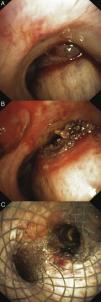
All prostheses were safely and easily placed using a rigid tracheoscope under bronchoscopic or fluoroscopic guidance, within 24h of admission. While the average time of all procedures was 30min, the actual stent insertion procedure required less than 2min (Figure 1). Endoscopic follow-up included revaluation at 15 days, 1, and 3 months and periodically thereafter as long as the stent remained in place.
In 3 cases of right main bronchus involvement (with tumor originating from the right upper bronchus in 2, and from the lobectomy suture line in 1) we placed Y stents with inverted branches. The longer left branch was positioned in the right main bronchus with exclusion of the completely obstructed upper lobar bronchus.
No severe complications occurred during the interventional endoscopic procedures. Patients reported symptomatic relief of dyspnea immediately after stent placement.
Three patients died in the first week due to pulmonary and cerebral edema. The remaining 17 patients were discharged from our hospital. Within 1 month of the procedure, another 5 deaths occurred, bringing overall 30-day mortality to 40% (8/20). Nine out of 12 survivors were subsequently treated by chemotherapy and/or radiotherapy. In 3 cases, following the insertion of a metallic esophageal stent for the treatment of locally advanced esophageal carcinoma infiltrating the carinal region, adding a Y stent provided effective palliation of symptoms of tracheoesophageal fistula. The median survival after stent placement was 12.2 weeks.
DiscussionThe Y-shaped anatomical configuration of the carinal bifurcation represents a real problem in the treatment of neoplastic stenosis. Selection of the best stent is just one step in the treatment process.2–4 Optimal endoscopic management should be minimally invasive, and should permit long-term control of potentially life-threatening obstruction. An ideal stent should be atraumatic with respect to the airway wall, well anchored, thin, biocompatible, easily inserted and removed if necessary, available in different sizes, and inexpensive.
The first experience with the type of stent used in our cohort of patients was published in 2008.4 The authors, who also designed this novel stent, reported that the procedure was safe and effective, with a median survival and median stent patency of 215 days.
We also observed a low rate of stent-related complications. Stent removal, when indicated, was relatively simple in spite of the good radial force applied against the airway wall and the friction exerted.
In the present series, 8 patients (40%) died within 30 days of stent placement for causes unrelated to the procedure. Median survival was poor (12.2 weeks) with respect to Han's series (28 weeks). However, no meaningful comparison is possible without risk-adjustment for comorbidities and tumor staging.
The limitations of this series include the relatively small number of cases and the retrospective nature of the study.
ConclusionPlacement of Y-shaped self-expanding stents is a safe and effective procedure for the palliation of malignant tracheobronchial stenosis, and is currently our stent of choice for this subgroup of patients. Further improvements in overall survival and quality of life are expected from systemic and local anti-tumoral therapies.
The authors are indebted to Prof. Patrick Barron of the Department of International Medical Communications of Tokyo Medical University for his review of this manuscript.
Please cite this article as: Conforti S, Durkovic S, Rinaldo A, Gagliardone MP, Montorsi E, Torre M. Stent en y autoexpandible para el tratamiento de la estenosis traqueobronquial maligna. Estudio retrospectivo. Arch Bronconeumol. 2016;52:e5–e7.


