Journal Information
Visits
80
Scientific Letter
Full text access
Uncorrected Proof. Available online 3 June 2025
Pulmonary Vascular Disease and Pulmonary Exercise Hemodynamics in Patients With Sjögren's Syndrome – A Cross-Sectional Study
Visits
80
Philipp Douschana,1,
Corresponding author
philipp.douschan@medunigraz.at

Corresponding author.
, Teresa Johna,1, Martin Stradnerb, Jutta Horvath-Winterc, Florentine Moazedi-Fürstb, Vasile Forisa, Henrik Abelna, Katarina Zedera, Lisa Cvirna, Adrienn Tornyosd, Jens Thielb, Nikolaus Kneidingera, Horst Olschewskie, Gabor Kovacsa
a Division of Pulmonology, Medical University of Graz, Graz, Austria
b Division of Rheumatology, Medical University of Graz, Graz, Austria
c Department of Ophthalmology, Medical University of Graz, Graz, Austria
d Department of Radiology, Medical University of Graz, Graz, Austria
e Sigmund Freud University, Vienna, Austria
This item has received
Article information
Full Text
Bibliography
Download PDF
Statistics
Figures (1)
Tables (1)
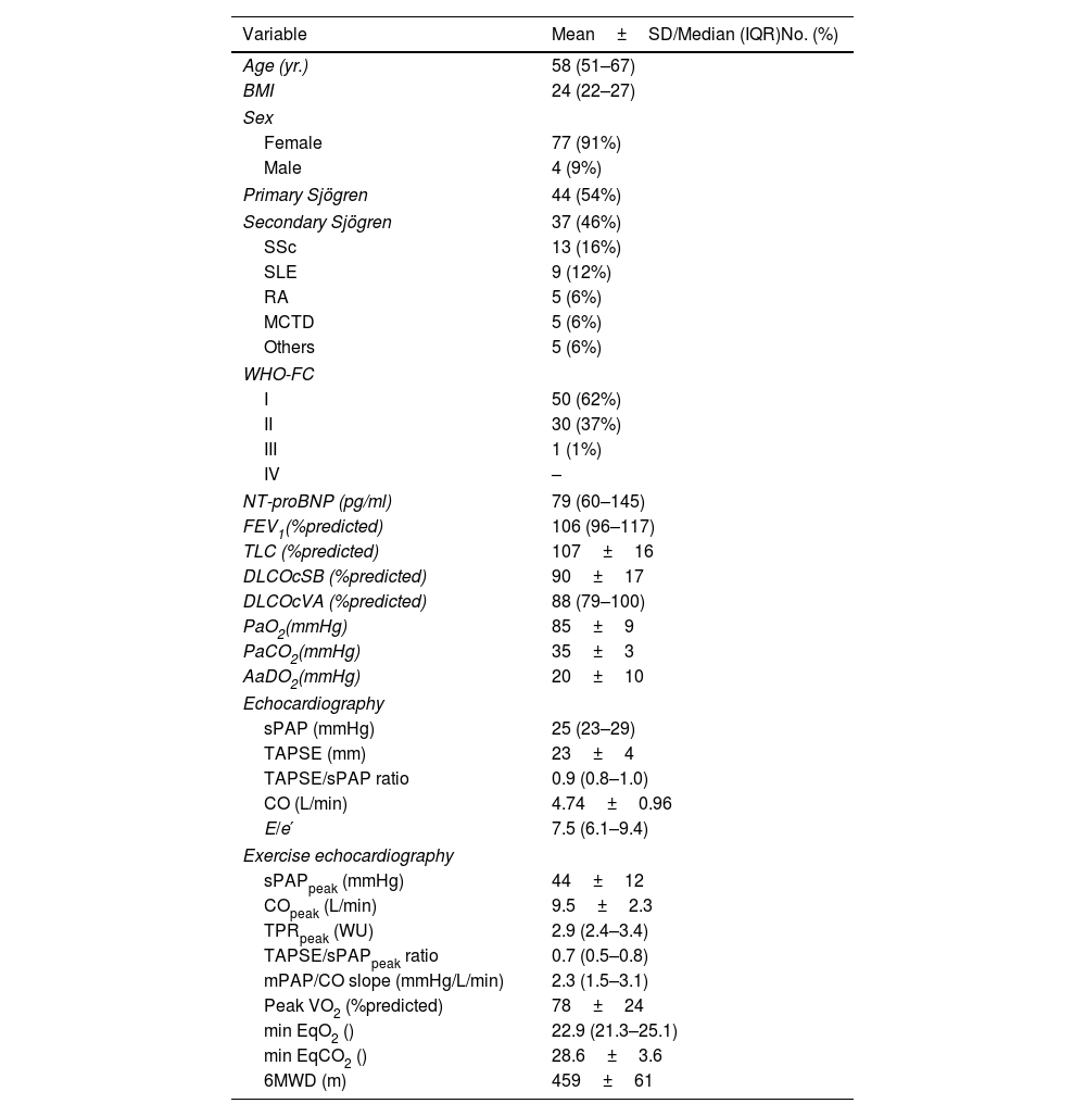
Table 1. Patient Characteristics.
Tables
Additional material (1)
Full Text
To the Director,

Sjögren's syndrome is an autoimmune disease affecting exocrine glands leading to a sicca-syndrome.1 When occurring with other autoimmune diseases it is termed secondary Sjögren's syndrome. Systemic manifestations such as pulmonary vascular disease (PVD) including pulmonary arterial hypertension (PAH) are associated with poor outcome.2 Data regarding the frequency of PAH are mainly limited to Asian registry-collectives.3–5

Of note, due to increasing evidence for the prognostic relevance of mildly elevated mean pulmonary arterial pressure (mPAP) within the range of 21–24mmHg, the definition of pulmonary hypertension (PH) was recently revised.6,7 Furthermore, as pulmonary hemodynamics during exercise are associated with clinical outcome,8,9 the term exercise PH (EPH) was recently reintroduced.7 So far, no data exists regarding the prevalence of EPH and mild PH in Sjögren's syndrome. Hence, this study aimed to assess the frequency of PVD based on most recent guidelines, using a screening algorithm with resting- and exercise echocardiography and right heart catheterization (RHC).

Consecutive patients with diagnosed Sjögren's syndrome from rheumatological and ophthalmological outpatient clinics were invited.10 Exclusion criteria included uncontrolled systemic arterial hypertension, recent pulmonary embolism or myocardial infarction, LV ejection fraction (LVEF)<50%, diastolic dysfunctiongrade 2, significant valvular heart disease or pregnancy/breast-feeding. The study was approved by the Ethics Committee of the Medical University of Graz (26–446 ex 13/14) and registered at clinical trials.gov (NCT02752269). Patients underwent assessment for symptoms, pulmonary function testing, resting- and exercise-echocardiography combined with cardiopulmonary-exercise-testing (CPET) using a cycle ergometer with stepwise increase of workload (25-Watt-increase every 2min). Exercise echocardiography was performed in semi supine left-tilted position. At each step, systolic PAP (sPAP) and velocity-time-integral derived cardiac output (CO) were assessed. SPAP was estimated by using the simplified Bernoulli equation and by adding an estimate of right atrial pressure as previously described.11 Estimated mPAP was calculated from sPAP as 0.61xsPAP+2.12 In patients with resting sPAP38mmHg (estimated invasive mPAP25mmHg), RHC was recommended due to suspected more severe PH and potential indication for treatment. Further, in subjects with suspected mild PH (mPAP: 21–24mmHg) or EPH, resting and exercise RHC was suggested in case of relevant exercise limitation or exercise dyspnea (for decision tree please and hemodynamic criteria please see Fig. 1A). At RHC, CO was assessed using thermodilution. The mPAP/CO-slope was calculated by (peak–resting mPAP)/(peak–resting CO). According to the recent recommendations, PH was defined as RHC-derived mPAP>20mmHg. Pre-capillary PH was defined as mPAP>20mmHg, PAWP15mmHg and PVR>2 WU.13 PAH was defined as pre-capillary PH and exclusion of other PH groups and EPH was defined as mPAP/CO-slope>3mmHg/L/min.13 Data are expressed as means±standard deviation (SD) or median and interquartile range (IQR) for continuous variables. Categorical data are presented as absolute and relative frequencies. For in-between group comparisons, T-Tests and U-Tests were used. Categorical variables were analyzed using Fisher's exact test or Chi2-test. A p-value<0.05 was considered significant. Statistical analysis was performed using IBM SPSS Statistics (Release 20.0.0. 2011. Chicago (IL), USA: SPSS Inc., an IBM Company).

Fig. 1.

(A) Screening algorithm for pulmonary vascular disease. Abbreviations: PVD: pulmonary vascular disease; PH: pulmonary hypertension; EPH: exercise pulmonary hypertension; mPAP: mean pulmonary arterial pressure; CO: cardiac output; sPAP: systolic pulmonary arterial pressure; WHO-FC: World Health Organization functional class; peakVO2: peak oxygen uptake; RHC: right heart catheterization; B: Screening Results; Explanation: The light blue box represents the overall cohort. In 13 (16%) patients echocardiographic mPAP/CO slope could not be calculated due to a lack of adequate mPAP or CO estimates from exercise echocardiography, in these patients echocardiographic sPAP>46mmHg (corresponding to a mPAP>30mmHg) and TPR>3WU was used to rise suspicion for EPH. Green box: no signs of EPH or mild PH according to our screening algorithm; of note, 2/29 patients did not undergo exercise testing. Yellow box left: echocardiographic mPAP/CO slope>3mmHg/l/min or echocardiographic sPAP>46mmHg and echocardiographic TPR>3WU at peak exercise. Yellow box middle: echocardiographic resting sPAP 31–38mmHg. Red box: echocardiographic resting sPAP38mmHg. Abbreviations: PH: pulmonary hypertension; PAH: pulmonary arterial hypertension; EPH: exercise pulmonary hypertension; PVD: pulmonary vascular disease; mPAP: mean pulmonary arterial pressure; sPAP: systolic pulmonary arterial pressure; RHC: right heart catheterization; WHO-FC: World Health Organization functional class; N=number; peakVO2: peak oxygen uptake; TPR=total pulmonary resistance.

(0.61MB).

Two patients were excluded after screening due to newly diagnosed severe exercise induced systemic hypertension (n=1) and moderate to severe mitral insufficiency (n=1). Characteristics of the remaining 81 patients are shown in Table 1. Forty-four (54%) patients had primary and 37 (46%) secondary Sjögren's syndrome. In 4 (5%) cases, sPAP could not be assessed at rest due to missing tricuspid regurgitation signal. In 13 (16%) patients exercise echocardiographic mPAP/CO slope could not be calculated due to inadequate mPAP or CO estimates. No relevant differences were detected between patients with primary- and secondary Sjögren's syndrome (Supplement Table 1). Pulmonary function testing revealed well-preserved lung function (Table 1).

Table 1.

Patient Characteristics.

Variable  Mean±SD/Median (IQR)No. (%) 
Age (yr.)  58 (51–67) 
BMI  24 (22–27) 
Sex
Female  77 (91%) 
Male  4 (9%) 
Primary Sjögren  44 (54%) 
Secondary Sjögren  37 (46%) 
SSc  13 (16%) 
SLE  9 (12%) 
RA  5 (6%) 
MCTD  5 (6%) 
Others  5 (6%) 
WHO-FC
50 (62%) 
II  30 (37%) 
III  1 (1%) 
IV  – 
NT-proBNP (pg/ml)  79 (60–145) 
FEV1(%predicted)  106 (96–117) 
TLC (%predicted)  107±16 
DLCOcSB (%predicted)  90±17 
DLCOcVA (%predicted)  88 (79–100) 
PaO2(mmHg)  85±
PaCO2(mmHg)  35±
AaDO2(mmHg)  20±10 
Echocardiography
sPAP (mmHg)  25 (23–29) 
TAPSE (mm)  23±
TAPSE/sPAP ratio  0.9 (0.8–1.0) 
CO (L/min)  4.74±0.96 
E/e′  7.5 (6.1–9.4) 
Exercise echocardiography
sPAPpeak (mmHg)  44±12 
COpeak (L/min)  9.5±2.3 
TPRpeak (WU)  2.9 (2.4–3.4) 
TAPSE/sPAPpeak ratio  0.7 (0.5–0.8) 
mPAP/CO slope (mmHg/L/min)  2.3 (1.5–3.1) 
Peak VO2 (%predicted)  78±24 
min EqO2 ()  22.9 (21.3–25.1) 
min EqCO2 ()  28.6±3.6 
6MWD (m)  459±61 

Normally distributed variables are expressed as mean±SD, non-normally distributed variables are expressed as median and IQR. Abbreviations: SSc: systemic sclerosis; SLE: systemic lupus erythematodes; RA: rheumatoid arthritis; MCTD: mixed CTD; WHO-FC: World Health Organization functional class; NT-proBNP: N terminal pro brain natriuretic peptide; FEV1: forced expiratory volume at 1second; TLC: total pulmonar capacity; DLCOcSB: single breath diffusion capacity for carbon monoxide; DLCOcVA: DLCO corrected for alveolar volume; PaO2: arterial oxygen partial pressure; PaCO2: arterial carbon dioxide partial pressure; AaDO2: arterial-alveolar oxygen partial pressure difference; E/e′: early diastolic transmitral flow velocity to early diastolic mitral annular tissue velocity; SPAP: systolic pulmonary arterial pressure; TPR: total pulmonary resistance; TAPSE: tricuspid annular plane systolic excursion; mPAP: mean pulmonary arterial pressure; CO: cardiac output; peakVO2: peak oxygen uptake; EqO2: ventilatory equivalent for O2; EqCO2: ventilatory equivalent for CO2; 6MWD: 6minute walking distance.

In the overall collective, 20 (25%) patients had an echocardiographic estimated mPAP/CO-slope>3mmHg/L/min and additional seven patients had abnormal peak exercise sPAP and TPR. In total, 27 patients were classified as possible EPH. N=4/27 patients with suspected EPH additionally showed an echocardiographic resting sPAP38mmHg (suspected severe PH) and 7 had a resting sPAP of 31–38mmHg (suspected mild PH). These 11 patients were not grouped in the “suspected EPH group” but in the group “suspected severe PH” and “suspected mild PH”, respectively. The remaining 16 patients were grouped in the “suspected EPH group”. There was no difference in the frequency of “suspected EPH” in patients with secondary vs. primary Sjögren's syndrome. Patients with abnormal echocardiographic hemodynamics were significantly older (65±10 years vs. 55±10 years, p<0.001), had a higher NT-proBNP (145 (71–343)pg/ml vs. 74 (44–110)pg/ml, p=0.002) and significantly lower TAPSE/sPAP-ratio (0.8±0.2mm/mmHg vs. 1.0±0.2mm/mmHg, p<0.001) (Supplement Table 2). When comparing patients without echocardiographic signs of PVD with those 29 subjects who were at risk for PVD we found no significant differences in the EULAR Sjögren's syndrome disease activity index (ESSDAI) (Supplement Table 2).

Final screening results are presented in Fig. 1B. Overall, out of 10 patients undergoing RHC, PAH was finally confirmed in two patients (2%) and EPH was confirmed in eight (10%) patients. Six patients showed an increase of PAWP>21mmHg during stress testing, indicating possible latent left heart disease. Pulmonary hemodynamics of all patients undergoing RHC are provided in Supplement Table 3. Out of the two patients with PAH, one had primary and the other had secondary Sjögren's syndrome.

PAH is complicating several autoimmune disorders, including Sjögren's syndrome and is associated with increased mortality.2,7 According to recent registry data, the prevalence of PAH in adults is 12–268 in 1,000,000 and approximately 15–30% have PAH due to CTD.14,15 According to the French PAH registry the prevalence of CTD among PAH patients was 15% with systemic sclerosis and systemic lupus erythematosus as leading causes.14 In an echocardiography study 5/47 (11%) patients with primary Sjögren's syndrome had PH.5 However, in this study, only five patients had a sPAP>35mmHg and no RHC was performed to confirm PAH. Prevalence of PAH in Sjögren's has been more frequently reported in Asian patient collectives, representing 16% of all PAH cases in connective tissue disease.16 According to a recent retrospective PH register study from Japan out of 142 patients diagnosed with PAH, only one patient was initially diagnosed with PAH associated with primary Sjögren's syndrome.3 In our study out of four patients with non-invasive SPAP38mmHg, RHC confirmed PAH in only two patients and PAH was only mild, not meeting the indication for targeted PAH therapy.

Besides pulmonary resting hemodynamics we also focused on pathological pulmonary exercise hemodynamics as there is increasing evidence for the clinical relevance of EPH8 and no data have been available so far on pulmonary exercise hemodynamics in Sjögren's syndrome. Assessment of pulmonary exercise hemodynamics may also be possible non-invasively by using exercise echocardiography. Although this may be limited by inadequate estimations of PAP and CO at peak exercise, recent studies supported the prognostic value of the method.17 Accordingly, recent guidelines recommended to consider exercise echocardiography or CPET in symptomatic patients with SSc to aid decisions to perform RHC.7

To best of our knowledge this was the first study to systematically implement exercise-echocardiography in the screening for PVD in Sjögren's syndrome. The underlying mechanisms of EPH were heterogenous in our collective with pre- and post-capillary phenotypes.18 Patients with EPH suspected by echocardiography were significantly older and had higher resting E/e’ suggesting subclinical diastolic dysfunction as an additional contributing factor for pathological exercise hemodynamics. This was also indicated by steeper increase of PAWP during exercise RHC.

Previous publications showed significantly worse prognosis of patients with systemic sclerosis when complicated by secondary Sjögren's syndrome.19 To which extent this is caused by a higher prevalence of PVD is unknown. Accordingly, we decided not only to include patients with primary – but also with secondary Sjögren's syndrome. Interestingly we found a balanced prevalence of PH between primary – secondary Sjögren's syndrome.

Based on our observations, assessment of PVD may be useful in symptomatic patients with Sjögren's Syndrome. So far, no data exist regarding the optimal management of patients diagnosed with EPH or mild PH. However, such patients may be at risk to progress to more severe PAH.20 In addition, even a mild elevation of PAP is associated with poor outcome in patients at risk for PH.6 Current guidelines recommend close follow-up of these patients.7,13 There is no evidence for the safety and efficacy of PAH drugs in patients with exercise PH or mild PH (mPAP<25mmHg) from randomized controls trials.

Our study is limited by its single center character. Due to limited therapeutic consequences and ethical reasons, RHC could not be performed in all patients with suspected EPH. Accordingly, RHC was only offered in case of relevant symptoms. Still, to the best of our knowledge, this is the first prospective cross-sectional study, systematically screening a large number of patients with Sjögren's syndrome for the presence of PVD including mild PH and EPH. The prevalence of PAH in patients with primary- and secondary Sjögren's syndrome in patients of European ancestry was low. Suspected EPH was relatively frequent, warranting follow-up studies concerning the development of severe PVD or relevant left heart disease in this patient collective.

Artificial Intelligence Involvement

None.

Financial/Nonfinancial Disclosures

P.D. reports personal fees from Actelion, Janssen, Ferrer, GSK, Chiesi and non-financial support from Actelion, Astra Zeneca, Boehringer Ingelheim, GSK, MSD, Novartis, Menarini, Ferrer and Chiesi outside, the submitted work; T.J. reports personal fees and non-financial support from Janssen, non-financial support from Ferrer, non-financial support from MSD, non-financial support from Boehringer Ingelheim, personal fees from Roche, outside the submitted work; M.S. has nothing to disclose; J.H.W. has nothing to disclose; A.T. has nothing to disclose; V.F. reports personal fees and non-financial support from Janssen, non-financial support from Boehringer Ingelheim, non-financial support from Astra Zeneca, personal fees and non-financial support from Chiesi, non-financial support from MSD, non-financial support from Ferrer, outside the submitted work; H.A. has nothing to disclose; K.Z. reports grants from Max Kade Foundation, grants from Cardiovascular Medical Research, grants from Education Fund, personal fees from MSD, personal fees from Janssen, personal fees from Ferrer, other from Ferrer, outside the submitted work; L.C. has nothing to disclose; J.T. reports research support from BMS, GSK, fees for lectures and advisory boards: BMS, Novartis, GSK, Janssen, Abbvie, Galapagos, Vifor, Astra-Zeneca, CSL-Behring, UCB; N.K. has nothing to disclose; H.O. reports grants and personal fees from MSD, personal fees from Janssen, personal fees from Ferrer, personal fees from Mondial, personal fees from MedUpdate, grants and personal fees from Iqvia, non-financial support from Ludwig Boltzmann Institute for Lung Vascular Research, outside the submitted work; G.K. reports grants, personal fees and non-financial support from Janssen, grants, personal fees and non-financial support from Boehringer-Ingelheim, personal fees from Bayer, personal fees and non-financial support from MSD, personal fees from Ferrer, personal fees from GSK, personal fees from Astra Zeneca, personal fees and non-financial support from AOP, personal fees from Chiesi, outside the submitted work.

Conflict of Interests

The authors state that they have no conflict of interests.

References
[1]
R.I. Fox.
Sjögren's syndrome.
Lancet Lond Engl, 366 (2005), pp. 321-331
[2]
Z. Liu, X. Yang, Z. Tian, J. Qian, Q. Wang, J. Zhao, et al.
The prognosis of pulmonary arterial hypertension associated with primary Sjögren's syndrome: a cohort study.
Lupus, 27 (2018), pp. 1072-1080
[3]
T. Sato, M. Hatano, Y. Iwasaki, H. Maki, A. Saito, S. Minatsuki, et al.
Prevalence of primary Sjögren's syndrome in patients undergoing evaluation for pulmonary arterial hypertension.
PLOS ONE, 13 (2018), pp. e0197297
[4]
Y. Zhao, Y. Li, L. Wang, X.F. Li, C.B. Huang, G.C. Wang, et al.
Primary Sjögren syndrome in Han Chinese: clinical and immunological characteristics of 483 patients.
Medicine (Baltimore), 94 (2015), pp. e667
[5]
S. Kobak, S. Kalkan, B. Kirilmaz, M. Orman, E. Ercan.
Pulmonary arterial hypertension in patients with primary Sjögren's syndrome.
Autoimmune Dis, 2014 (2014), pp. 710401
[6]
P. Douschan, G. Kovacs, A. Avian, V. Foris, F. Gruber, A. Olschewski, et al.
Mild elevation of pulmonary arterial pressure as a predictor of mortality.
Am J Respir Crit Care Med, (2017),
[7]
M. Humbert, G. Kovacs, M.M. Hoeper, R. Badagliacca, R.M.F. Berger, M. Brida, et al.
2022 ESC/ERS Guidelines for the diagnosis and treatment of pulmonary hypertension.
Eur Respir J, 25 (2022), pp. 2200879
[8]
P. Douschan, A. Avian, V. Foris, T. Sassmann, G. Bachmaier, P. Rosenstock, et al.
Prognostic value of exercise – as compared to resting pulmonary hemodynamics in patients with normal or mildly elevated pulmonary arterial pressure.
Am J Respir Crit Care Med, (2022),
[9]
G. Kovacs, M. Humbert, A. Avian, G.D. Lewis, S. Ulrich, A. Vonk Noordegraaf, et al.
Prognostic relevance of exercise pulmonary hypertension: results of the multicentre PEX-NET Clinical Research Collaboration.
Eur Respir J, 64 (2024), pp. 2400698
[10]
C.H. Shiboski, S.C. Shiboski, R. Seror, L.A. Criswell, M. Labetoulle, T.M. Lietman, et al.
2016 American College of Rheumatology/European League Against Rheumatism Classification Criteria for Primary Sjögren's Syndrome: a consensus and data-driven methodology involving three international patient cohorts.
Arthritis Rheumatol Hoboken NJ, 69 (2017), pp. 35-45
[11]
L.G. Rudski, W.W. Lai, J. Afilalo, L. Hua, M.D. Handschumacher, K. Chandrasekaran, et al.
Guidelines for the echocardiographic assessment of the right heart in adults: a report from the American Society of Echocardiography.
J Am Soc Echocardiogr, 23 (2010), pp. 685-713
[12]
D. Chemla, V. Castelain, M. Humbert, J.L. Hébert, G. Simonneau, Y. Lecarpentier, et al.
New formula for predicting mean pulmonary artery pressure using systolic pulmonary artery pressure.
Chest, 126 (2004), pp. 1313-1317
[13]
G. Kovacs, S. Bartolome, C.P. Denton, M.A. Gatzoulis, S. Gu, D. Khanna, et al.
Definition, classification and diagnosis of pulmonary hypertension.
Eur Respir J, 29 (2024), pp. 2401324
[14]
M. Humbert, O. Sitbon, A. Chaouat, M. Bertocchi, G. Habib, V. Gressin, et al.
Pulmonary arterial hypertension in France: results from a national registry.
Am J Respir Crit Care Med, 173 (2006), pp. 1023-1030
[15]
T. Thenappan, S.J. Shah, S. Rich, M. Gomberg-Maitland.
A USA-based registry for pulmonary arterial hypertension: 1982–2006.
Eur Respir J, 30 (2007), pp. 1103-1110
[16]
Y.J. Hao, X. Jiang, W. Zhou, Y. Wang, L. Gao, Y. Wang, et al.
Connective tissue disease-associated pulmonary arterial hypertension in Chinese patients.
Eur Respir J, 44 (2014), pp. 963-972
[17]
L. Gargani, N.R. Pugliese, N. De Biase, M. Mazzola, G. Agoston, M. Arcopinto, et al.
Exercise stress echocardiography of the right ventricle and pulmonary circulation.
J Am Coll Cardiol, 82 (2023), pp. 1973-1985
[18]
Y.N.V. Reddy, M. Obokata, B. Wiley, K.E. Koepp, C.C. Jorgenson, A. Egbe, et al.
The haemodynamic basis of lung congestion during exercise in heart failure with preserved ejection fraction.
Eur Heart J, 40 (2019), pp. 3721-3730
[19]
M. Scherlinger, J. Lutz, G. Galli, C. Richez, J.E. Gottenberg, J. Sibilia, et al.
Systemic sclerosis overlap and non-overlap syndromes share clinical characteristics but differ in prognosis and treatments.
Semin Arthritis Rheum, 51 (2021), pp. 36-42
[20]
T.R. Assad, B.A. Maron, I.M. Robbins, M. Xu, S. Huang, F.E. Harrell, et al.
Prognostic effect and longitudinal hemodynamic assessment of borderline pulmonary hypertension.
JAMA Cardiol, (2017),

Equally contributed.

Copyright © 2025. The Authors
Archivos de Bronconeumología
Article options
Tools

Are you a health professional able to prescribe or dispense drugs?