


We report the case of a 36-year-old man, former smoker (14 pack-years), candidate for lung transplant due to acquired non-cystic fibrosis bronchiectasis. Standard chest X-ray showed pulmonary hyperinflation and radiolucent images consistent with bronchiectasis. Bronchiectasis was confirmed on computed axial tomography of the chest, which revealed images of air in the mediastinum suggestive of tracheobronchial diverticula (Fig. 1A). Virtual reconstruction shows multiple sac-like images in both main bronchi (Fig. 1B). The neck of the diverticula can been seen in the upper right image. Small depressions in the mucosa corresponding to diverticular perforations were also observed.
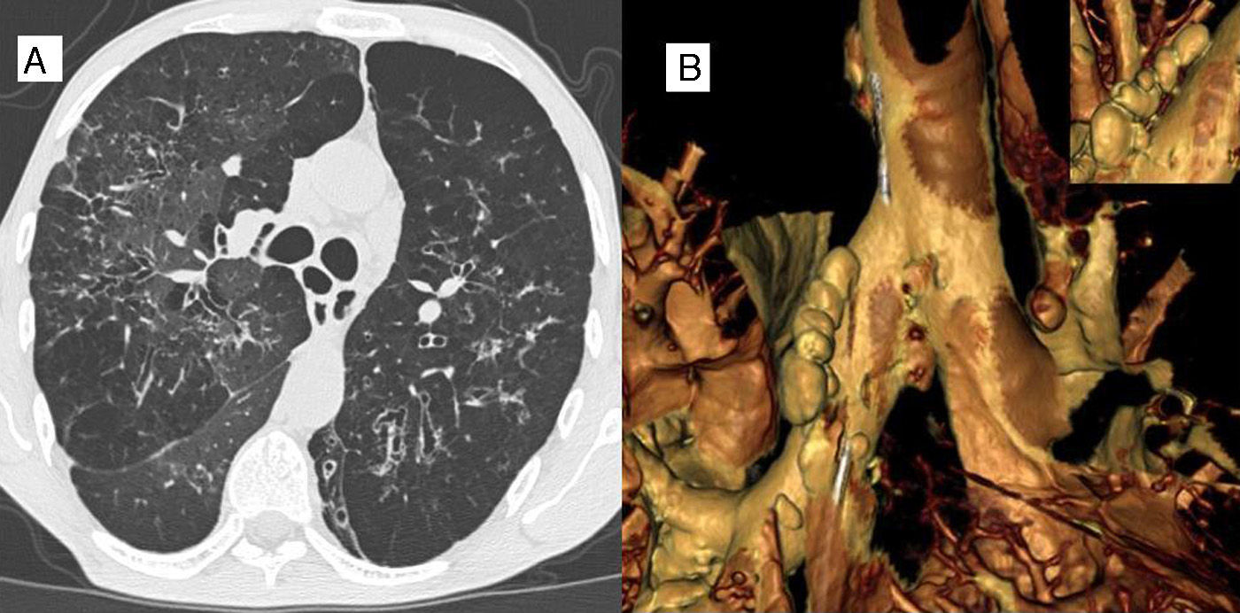
Early reports of this condition suggested a prevalence of 0.09%–0.05%, but a more recent study of 503 smokers and another of 200 adults without lung disease found a prevalence of 45% and 41%, respectively.1,2 The bronchial diverticula were small in size (1–3mm). The interest in our case lies in the large size of the bilateral bronchial diverticula (10–13mm in diameter) observed in tridimensional images reconstructed from computed tomography. These images would not have been observed on standard chest X-ray.
FundingNo funding was received.
Conflict of InterestsNo conflict of interests declared.
Please cite this article as: Orazi ML, Svetliza GN, de Vito EL, Precerutti JA. Divertículos bronquiales gigantes múltiples. Arch Bronconeumol. 2015;51:356.


