Thymomas are the most common primary anterior mediastinal tumors, although a multifocal (synchronous or metastatic) presentation is exceptional: less than 30 cases of synchronous thymomas have been reported in the literature.1–3 There is controversy surrounding the question of whether the presence of more than 1 mass in the anterior mediastinum represents independent multicentric (synchronous) tumors or satellite metastases from a single thymoma.4 Several criteria have been put forward that may help differentiate multicentric thymomas from satellite metastases originating in a primary thymic tumor,5 but they do not factor in information obtained from morphological and metabolic imaging techniques.
We report the case of a patient with 2 mediastinal masses, seen on imaging techniques as 2 synchronous thymomas, and confirmed as such by preoperative percutaneous biopsy of each mass.
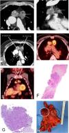
Our patient was a 75-year-old man who consulted for a respiratory infection, and was found to have an abnormal mediastinal contour on chest X-ray. Chest computed tomography (CT) confirmed the existence of 2 solid masses in the anterior mediastinum with different radiological characteristics, suggesting 2 synchronous thymomas (Figs. 1A, B and C). The patient had no symptoms to suggest myasthenia gravis. A positron emission tomography (PET)/CT scan showed that each mediastinal mass had different metabolic activity (the right mass showed a standardized uptake value [SUV] of 3.6 while the left one had a SUV of 6.1), supporting the idea that these were 2 independent lesions (Figs. 1D and E). We decided to carry out radiologically-guided parasternal core needle biopsy of the 2 mediastinal masses, confirming that they were both thymomas (right mass, type B2, and left mass, type B3, according to World Health Organization [WHO] classification) (Fig. 1F and G). The patient underwent successful video-assisted thoracoscopy, confirming that the 2 masses were synchronous thymomas classified as TNM stage I (pT1N0) and Masaoka stage IIB (Fig. 1H).
A) and B) Axial (A) and coronal (B) chest CT images showing two masses in the anterior mediastinum: a right mass (white asterisk) and a left mass (black asterisk). Note the presence of calcification foci in the left mass (arrows). C) Axial CT image of the chest clearly showing the different attenuation characteristics of the mediastinal masses: the right mass (white asterisk) has a mean attenuation of 83 Hounsfield units while the left mass (black asterisk) has a mean attenuation of 54 Hounsfield units, suggesting an independent origin. D) and E) Axial (D) and coronal (E) PET/CT images showing the different metabolic activity of the two mediastinal masses (greater FDG uptake by the left mass [6.1, black asterisk] than by the right mass [3.6, white asterisk]), suggesting two independent tumors. F) Percutaneous biopsy sample of the left mediastinal mass showing neoplastic proliferation of epithelial cells surrounded by fibrous tissue and scant lymphocytes, associated with type B3 thymoma (hematoxylin and eosin). G) Percutaneous biopsy sample of the right mediastinal mass in which a mainly lymphocyte component is identified with some prominent epithelial cell nests, associated with type B2 thymoma. H) Post-surgical macroscopic piece showing both contiguous masses (white circle: right mass; black circle: left mass).
CT: computed tomography; FDG: fluorodeoxyglucose; PET/CT: positron emission tomography.
Multiple thymomas are rare, and there are conflicting opinions in the scientific literature as to whether they correspond to metastases from a single primary thymic tumor or a multicentric/multifocal origin.1–3 In general, multiple thymomas are considered synchronous (independent) if they meet the following criteria: 1) lesions are stage I (an intact capsule theoretically prevents the spread of the tumor outside its margins); 2) there are less than 3 thymomas; 3) the size of the thymomas is relatively similar; and 4) the histology of each tumor is different.4,5 Very few studies recommend radiological or metabolic criteria to differentiate primary (independent) thymic tumors from metastatic (dependent) lesions,6,7 although CT, MRI or PET/CT may all provide information that can help predict the degree of malignancy of thymic epithelial tumors in some cases.8–10 In our patient, it is interesting to note the clearly different morphological characteristics on CT of the two mediastinal masses (attenuation variations and presence of calcification foci in one of the masses) and their different metabolic affinity for fluorodeoxyglucose (FDG) (the right mass showed an SUV of 3.6, and the left mass 6.1). In view of these different radiological and metabolic characteristics, we decided to perform a preoperative percutaneous biopsy of the two masses to characterize them histologically and rule out the possibility of 2 different types of malignancy (e.g., thymoma and germ cell tumor or lymphoma). We have not found any previously published cases of preoperative confirmation by percutaneous core needle biopsy of 2 synchronous thymomas (in a previous paper, 1 of the patient’s 2 tumors was biopsied5). In our case, the mass with the lowest attenuation on CT that contained calcification foci and had the highest metabolism in PET/CT was classified as type B3 thymoma, whereas the mass with the highest density on CT and the lowest metabolic activity on PET/CT was classified as type B2 thymoma. There was therefore a correlation between the WHO histological subtype and PET/CT metabolic activity (type B3 thymomas have a worse prognosis than type B2).11
We believe that in a patient with multiple thymomas, metabolic and radiological characterization of the lesions can help differentiate between multiple synchronous thymomas and satellite metastases of a single thymic tumor and optimize therapeutic management.
Conflict of interestsThe authors state that they have no conflict of interests.
Please cite this article as: Gorospe-Sarasúa L, Ajuria-Illarramendi O, Vicente-Zapata I, Muñoz-Molina GM, Fra-Fernández S, Cabañero-Sánchez A, et al. Diagnóstico de dos timomas sincrónicos mediante técnicas de imagen (TC y PET/TC) y confirmación mediante biopsia percutánea. Arch Bronconeumol. 2021;57:560–562.


![A) and B) Axial (A) and coronal (B) chest CT images showing two masses in the anterior mediastinum: a right mass (white asterisk) and a left mass (black asterisk). Note the presence of calcification foci in the left mass (arrows). C) Axial CT image of the chest clearly showing the different attenuation characteristics of the mediastinal masses: the right mass (white asterisk) has a mean attenuation of 83 Hounsfield units while the left mass (black asterisk) has a mean attenuation of 54 Hounsfield units, suggesting an independent origin. D) and E) Axial (D) and coronal (E) PET/CT images showing the different metabolic activity of the two mediastinal masses (greater FDG uptake by the left mass [6.1, black asterisk] than by the right mass [3.6, white asterisk]), suggesting two independent tumors. F) Percutaneous biopsy sample of the left mediastinal mass showing neoplastic proliferation of epithelial cells surrounded by fibrous tissue and scant lymphocytes, associated with type B3 thymoma (hematoxylin and eosin). G) Percutaneous biopsy sample of the right mediastinal mass in which a mainly lymphocyte component is identified with some prominent epithelial cell nests, associated with type B2 thymoma. H) Post-surgical macroscopic piece showing both contiguous masses (white circle: right mass; black circle: left mass). CT: computed tomography; FDG: fluorodeoxyglucose; PET/CT: positron emission tomography.](https://static.elsevier.es/multimedia/15792129/0000005700000008/v1_202108020523/S1579212921001920/v1_202108020523/en/main.assets/thumbnail/gr1.jpeg?xkr=ue/ImdikoIMrsJoerZ+w98FxLWLw1xoW2PaQDYY7RZU=)


