Introduction
Infection by Strongyloides stercoralis occurs when filarial larvae penetrate the skin and are carried to the lungs through the bloodstream. After burrowing into the alveoli, the larvae migrate through the airways where they are coughed up and swallowed, eventually maturing in the walls of the small intestine. Unlike other nematodes, S stercoralis are capable of autoinfection of the host.
During the migratory cycle, pulmonary infestation may cause abscess, hemorrhaging, acute respiratory distress syndrome, pleural effusion, or asthma-like symptoms.1-3 When asthma-like symptoms present, the patient is likely to be classified as asthmatic and receive inhaled or systemic corticosteroids. Steroid use, as well as immunocompromised status, may lead to the uncontrolled spread of S stercoralis to other organs or to unusually large numbers of them in the organs that are usually involved in the parasite's life cycle (hyperinfection). Such conditions may lead to death.
Although asthma-like symptoms caused by S stercoralis are well described in the literature, their mechanism has not yet been elucidated. The purpose of this study is to report an additional possible obstructive mechanism that causes asthma-like symptoms: the presence of multiple nodules protruding into the lumen of the airways due to massive invasion by S stercoralis larvae.
Clinical Observation
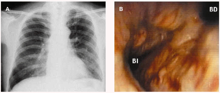
A 55-year-old male farm worker with a 2-year history of illness characterized by cough, dyspnea, and wheezing was treated in our hospital. He had been diagnosed with asthma and received bronchodilators and oral corticosteroids (30 mg/day for the last 5 months). In spite of treatment, his symptoms persisted, and he was referred to our hospital for severe dyspnea. Physical examination showed blood pressure of 110/70 mm Hg, respiratory rate at 24 breaths/min, and wheezing. Chest x-rays revealed air trapping mainly in the right side (Figure 1A). Arterial blood gases were within normal range for the altitude of Mexico City, with pH of 7.42, arterial carbon dioxide pressure of 31 mm Hg, and arterial oxygen pressure of 66 mm Hg. Pulmonary function testing demonstrated moderate obstruction: forced vital capacity (FVC) was 4.7 L (95% of the predicted value), forced expiratory volume in the first second (FEV1) was 2.2 L (62%), the ratio of FEV1/FVC was 45.7%, forced expiratory flow between 25% and 75% of vital capacity was 1.4 L (58%); the bronchodilator test was negative.
Figure 1. Radiographic and bronchoscopic images of the patient. A: initial chest x-ray where slight air trapping was observed, especially in the right lung. B: image observed when the fiberoptic bronchoscope was in the main carina. The main right bronchus can be observed at the top right of the image. Main findings were yellowish mucous, engorged vascular bed, widening of the carina and multiple nodules protruding into the airway lumen and partially obstructing the opening to the left bronchus.
During the first 5 days after admission, the patient was treated as having severe asthma, with intravenous methylprednisolone (125 mg/12 h), intravenous aminophylline (0.7 mg/kg/h), salbutamol, and also ceftazidime (1 g/12 h) due to suspicion of bacterial bronchitis. The patient did not respond to treatment; on the contrary, 3 to 4 days after admission, he presented with pain and diffused abdominal distention, nausea, and vomiting.
Bronchoscopy was performed because pulmonary function tests demonstrated an irreversible obstruction pattern. Yellowish, friable mucous with engorgement of the vascular bed was observed along with irregular contours due to multiple irregular nodules of varying size (Figure 1B). S stercoralis larvae were identified in the bronchoalveolar lavage (Figure 2A). This same nematode was also identified in stool samples. Treatment with albendazole at 400 mg/day was, therefore, started.
Figure 2. Strongyloides stercoralis larvae. A: larva observed in the bronchial brushing (hematoxylin-eosin x400). B: larva (arrow) found in a lymphatic vessel of the bronchial submucosa. The cellular infiltrates are composed primarily of mononuclear cells (hematoxylin-eosin x150).
Three days later, the patient presented with hypotension (90/50 mm Hg), tachypnea (36 breaths/min), heart rate of 80 beats/min, increased dyspnea, and mild hemoptysis and was transferred to the intensive care unit, where he rapidly deteriorated. Hemoptysis increased and mechanical ventilation was started when confusion, hypercapnia, and hypoxemia developed. Six hours later, the patient died.
Necropsy showed pleural adhesion in both lungs, bilateral hemothorax (approximately 300 mL in each side), an abundance of blood clots in the trachea and bronchi. The lung parenchyma was consolidated, reddish with signs of recent pulmonary hemorrhaging, and hyaline membrane formation. S stercoralis larvae were found in the mucosal and submucosal layers of the main bronchi with infiltrates composed primarily of mononuclear cells, but no eosinophils, around the larvae (Figure 2B). Some larvae were observed in the lumen and blood vessel walls with no tissular reaction. The larvae were also found in the duodenum and jejunum, in crypts and lamina propria. Examination of the liver showed centrilobular necrosis as well as other signs of shock. No abnormalities in other organs were noted.
Discussion
It has been well documented that S stercoralis are capable of producing asthma-like symptoms, yet little research about the mechanism has been carried out, and what we do know remains speculative. Infection due to S stercoralis may cause severe exacerbation of asthmatic symptoms in subjects suffering from asthma,2,4-6 and it has been suggested that the cause of such a change in status is increased bronchial inflammation,5 local invasion of the larvae,2 the exacerbation of the allergic process,2 or an increased load of T-helper-2 cytokines.6 Other studies describe relatively sudden onset of asthma-like symptoms in previously healthy people,1-3 but mechanisms have not been clearly established.
In this article we describe another mechanism which may explain the asthma-like symptoms. This mechanism consists of protruding, swollen nodules caused by S stercoralis larval invasion in airway submucosa. In our case, nodules were observed by fiberoptic bronchoscopy, thus, fully explaining the obstruction pattern resistant to salbutamol observed in the respiratory function tests. Migration to the bronchial submucosa indicates that hyperinfection had begun, probably due to the prolonged use of corticosteroids. The route the larvae took was most likely direct penetration into the bronchial tubes.
In at least 2 reported cases, bronchoscopy was performed, and in both cases mucosal inflammation without nodules was observed.7,8 Therefore, it is likely that the development of nodules explains only some cases with refractory symptoms similar to asthma. However, this possibility should always be considered, especially wherever this parasite is endemic.
We conclude that infection by S stercoralis should be taken into account in the differential diagnosis of asthma-like symptoms and, at least in some cases, the production of nodules in the airways may be the cause of the obstruction.
Correspondence: Dr. M.H. Vargas.
Unidad de Investigación Médica en Epidemiología Clínica.
Hospital de Pediatría. Centro Médico Nacional Siglo XXI.
Instituto Mexicano del Seguro Social.
Avda. Cuauhtémoc, 330. 06720 México DF. México.
E-mail: mhvargasb@yahoo.com.mx
Manuscript received March 17, 2003.
Accepted for publication May 6, 2003.


