Thoracic ultrasound (TU) is a complementary tool in respiratory medicine that has many applications in patients with peripheral pulmonary parenchymal and pleural disease. One of these is TU-guided biopsy of peripheral lung lesions (PLL) for the diagnosis of suspected malignancy.1–5
Over the years, the respiratory community has become aware of the importance of conscious sedation in patients undergoing interventional techniques in respiratory medicine. The most commonly used benzodiazepine in sedation is midazolam, given its sedative, anxiolytic, amnesic, and muscle relaxant properties. No studies have yet been published investigating the degree of sedation in TU-guided PLL biopsy and patient satisfaction with or without midazolam.
We report our experience in 2 patient groups undergoing TU-guided PLL biopsy: group A without midazolam; and group B with midazolam.
This was an ambispective, observational case–control study. Data from the control group (A) who did not receive sedation were collected retrospectively, and those from group (B), who received sedation, were collected prospectively.
Control data, including the satisfaction survey, vital signs, complications, and diagnosis, were reviewed retrospectively. For the cases, the satisfaction survey and all other variables were collected prospectively. The satisfaction survey was based on previous publications which reported patient satisfaction with respiratory endoscopic techniques.6–10
The sample size required for group B was calculated from the score obtained on the satisfaction survey of the historical control group.
In total, 39 patients with no contraindications for PLL biopsy or for sedation were included. They were assessed previously by a nurse and a pulmonologist with experience in interventional respiratory medicine. Patients were considered for inclusion if they had PLL in contact with the chest wall, previously visualized on chest computed tomography, suspected to be lung cancer in any disease stage, with an area of contact between the lesion and the chest wall of at least 2cm.
Patients had to meet all inclusion criteria and none of the exclusion criteria (less than 18 years of age, coagulation, liver or kidney disorders, unstable ischemic heart disease, COPD with FEV1 <30%, ASA (American Society of Anesthesiologists) status > III, or hemodynamic instability). At least 2 and at most 3 passes were performed for both fine needle aspirations and biopsies. The patient survey comprised 12 questions, of which 10 required Likert-type responses (A lot, Quite a lot, A bit, A little, Very little). The other 2 questions were multiple-choice. The interventional pulmonologist completed a 3-question survey, 2 of which had multiple numbered choices (0 = None, 1 = A little, and 2 = A lot), and a third question with alternative responses.
Group B received midazolam at a dilution of 1mg/ml, and doses of 1mg were administered during the process at 3-min intervals if required by the patient, to a maximum dose of 5mg. Both groups received local anesthesia with lidocaine 2% applied to the subepidermis and the parietal pleura. No patients in either group used oral anxiolytics before the procedure.
Nineteen patients were included in group A, and 20 in group B.
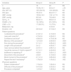
Demographic variables showed no statistically significant differences between the baseline values of both groups, with the exception of age. Nor were differences observed in vital signs or number of biopsies. Procedure duration was shorter, but not statistically, in the sedation group (Table 1).
Study Group Variables.
| Variables | Group A | Group B | P |
|---|---|---|---|
| n | 19 | 20 | NS |
| Age, years | 72.05±10.1 | 65.9±11 | <.05 |
| Sex, M/F (%) | 16/3 (84/16) | 16/4 (80/20) | NS |
| HR, bpm | 76.3±12 | 82.2±11.7 | NS |
| SBP, mmHg | 135.8±14.2 | 134.1±17.9 | NS |
| DBP, mmHg | 80.5±6 | 78.9±9.6 | NS |
| Sat O2, % | 96.8±1.5 | 96.8±1.8 | NS |
| FNAB, n (%) | 19 (100%) | 20 (100%) | NS |
| CNB, n (%) | 17 (89.5%) | 15 (75%) | NS |
| Midazolam, mg | – | 4.5±0.6 | – |
| Duration, min | 24.5±4.2 | 21.9±4.2 | NS |
| Patient questions | |||
| I understood the procedurea | 4.12±1.2 | 4.10±0.9 | NS |
| I was well cared fora | 4.74±0.4 | 4.90±0.3 | NS |
| Confidence and trusta | 4.58±0.5 | 4.75±0.44 | NS |
| Memory of the procedureb | 1.21±0.9 | 4.60±0.9 | .0001 |
| Pain during the procedureb | 3.11±1.1 | 4.85±0.3 | .0001 |
| Length of the procedureb | 3±1.2 | 4.65±0.7 | .0001 |
| I was nervous before the procedureb | 2.47±1.1 | 2.85±1.2 | NS |
| I would be nervous if I had to repeat itb | 3.11±1.1 | 4.40±0.8 | .001 |
| Indifferent to repeating itb | 4.63±1 | 3.05±1.2 | .0001 |
| Discomfort of the procedureb | 3.53±1 | 4.70±0.6 | .0001 |
| Worst moment of the procedurec | 2.11±1.4 | 0.85±0.8 | .009 |
| Repeat the test if necessaryd | 1.79±0.9 | 1.05±0.2 | .002 |
| Physician questions | |||
| Patient collaboratione | 1.74±0.4 | 1.80±0.4 | NS |
| Difficulty of the proceduree | 0.47±0.6 | 0.35±0.5 | NS |
| Procedure completedf | 1.37±0.7 | 1.05±0.2 | NS |
bpm: beats per minute; CNB: core needle biopsy; F: female; FNAB: fine needle aspiration and biopspy; HR: heart rate; NS: not significant; Sat O2: oxygen saturation; DBP: diastolic blood pressure; SBP: systolic blood pressure; M: male.
Scores for each of the Likert-type responses were examined to evaluate the patients’ perception of the procedure. Scores for each question showed a higher feeling of discomfort during the biopsy in group A. Patients who received midazolam were less nervous at the prospect of repeating the procedure. The perception of pain, memory of the procedure, and the perception of the length of the procedure were greater in the group that did not receive midazolam. Patients’ perception of care received and confidence and trust in the staff were similar in both groups.
In the multiple-choice questions, patients in group A reported that the worst moment was receiving the anesthesia, and in group B, the worst moment was going into the procedure room. Group A would probably repeat the biopsy, and group B would definitely repeat it.
No differences were found between groups in the surveys completed by the interventional pulmonologists (Table 1).
The post-procedure stay in the recovery room in both groups was 2h, and no hospital admissions were required. No patients in either group developed complications and a final diagnosis was obtained in all participants.11–14
We conclude that this model of sedation with midazolam may be necessary in patients undergoing this additional test, given the benefits offered by this drug, the absence of side effects and complications, and the lack of impact on the performance of the technique by the pulmonologist.
One of the limitations of this study is the small sample size, and the fact that 1 patient group was analyzed retrospectively. However, this is the first published study which evaluates the opinion of the patient and the physician, with and without midazolam, in this type of respiratory intervention.
For this reason, we recommend that the use of midazolam in this type of procedure be standardized.
Please cite this article as: Wangüemert Pérez AL, González Expósito H, Pascual Fernández L. Valoración de la sedación con midazolam en las punciones pulmonares periféricas dirigidas por ecografía torácica. Arch Bronconeumol. 2018;54:342–343.


